. 2017 Oct 3;7(1):12629.
doi: 10.1038/s41598-017-13015-w.

Neofunctionalization of "Juvenile Hormone Esterase Duplication" in Drosophila as an odorant-degrading enzyme towards food odorants

Affiliations

Neofunctionalization of "Juvenile Hormone Esterase Duplication" in Drosophila as an odorant-degrading enzyme towards food odorants

Claudia Steiner et al. Sci Rep. .

Abstract

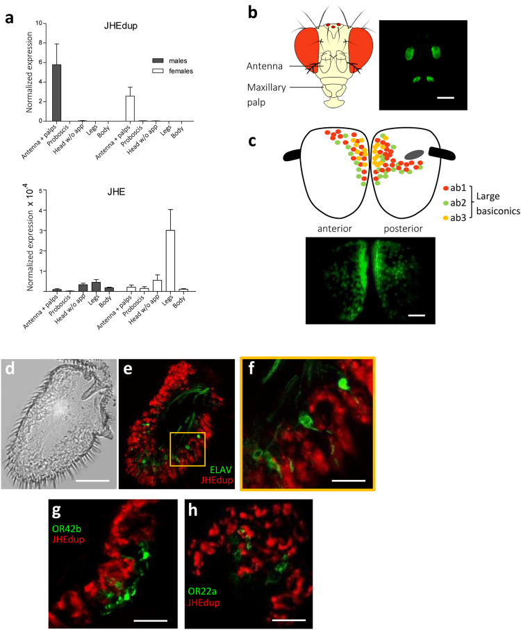
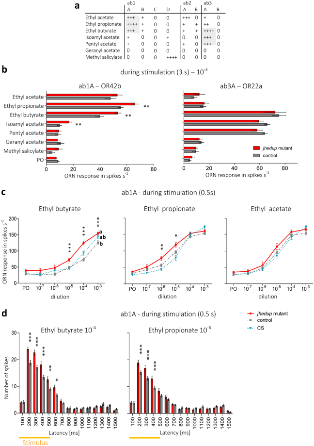
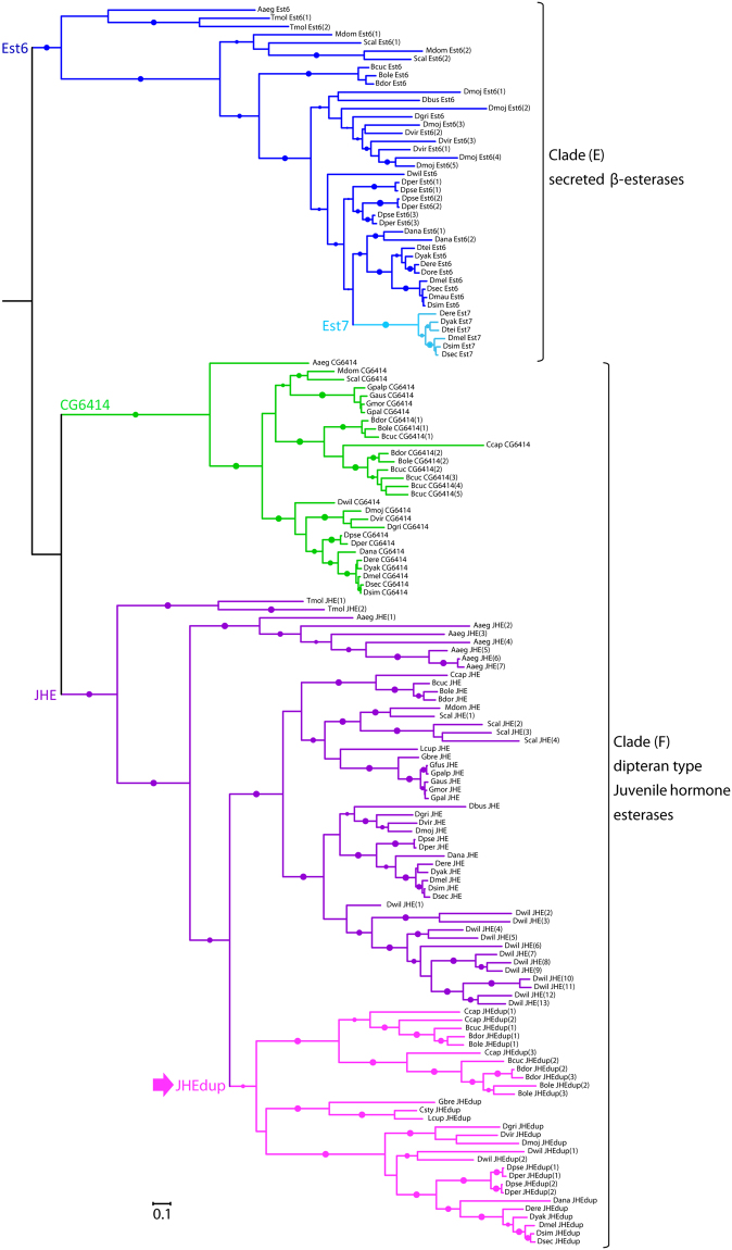
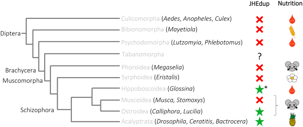
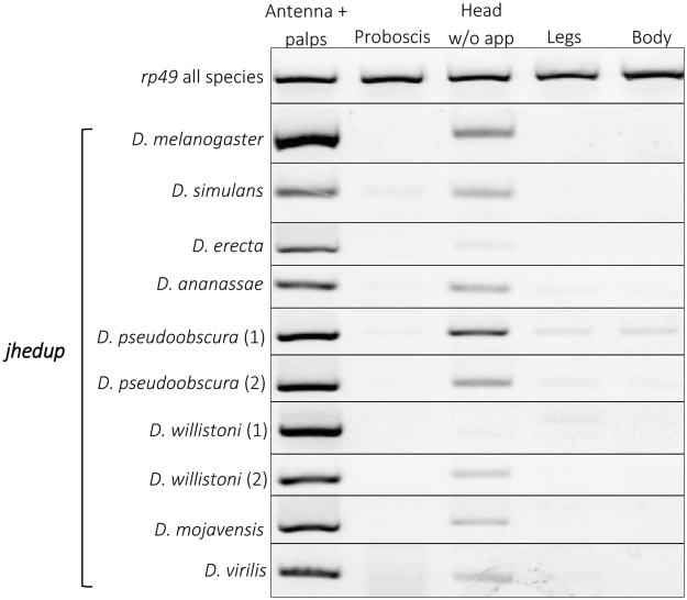
Odorant degrading enzymes (ODEs) are thought to be responsible, at least in part, for olfactory signal termination in the chemosensory system by rapid degradation of odorants in the vicinity of the receptors. A carboxylesterase, specifically expressed in Drosophila antennae, called "juvenile hormone esterase duplication (JHEdup)" has been previously reported to hydrolyse different fruit esters in vitro. Here we functionally characterize JHEdup in vivo. We show that the jhedup gene is highly expressed in large basiconic sensilla that have been reported to detect several food esters. An electrophysiological analysis demonstrates that ab1A olfactory neurons of jhedup mutant flies exhibit an increased response to certain food acetates. Furthermore, mutant flies show a higher sensitivity towards the same odorants in behavioural assays. A phylogenetic analysis reveals that jhedup arose as a duplication of the juvenile hormone esterase gene during the evolution of Diptera, most likely in the ancestor of Schizophora, and has been conserved in all the 12 sequenced Drosophila species. Jhedup exhibits also an olfactory-predominant expression pattern in other Drosophila species. Our results support the implication of JHEdup in the degradation of food odorants in D. melanogaster and propose a neofunctionalization of this enzyme as a bona fide ODE in Drosophilids.


Conflict of interest statement

The authors declare that they have no competing interests.

Figures

Figure 1
Figure 1
Jhedup expression in antennae. (a) Normalized expression of jhedup and jhe in antennae + maxillary palps, proboscises, heads without appendages, legs and bodies of wildtype (Canton-S) males and females using qPCR (reference gene for normalization of expression: pgk). The normalized expression level is indicated as mean ± SE of triplicate biological samples. (b) Expression of jhedup in antennae and maxillary palps of jhedup-GAL4; UAS-mCD8::GFP flies. Scale bar, 150 µm. (c) jhedup expression in the 3rd antennal segment of jhedup-GAL4; UAS-mCD8::GFP flies (anterior and posterior view), including scheme for dispersion of large basiconics on the 3rd antennal segment, scale bar 20 µm. (d) Antennal slice of 3rd antennal segment. Scale bar, 20 µm. (e) Jhedup expression in 3rd antennal segment of jhedup-GAL4; UAS-mCD8::Cherry//elav-LexA; LexAOP-mCD8::GFP flies. Jhedup expression (red), expression of neuronal marker elav (green). (f) Higher magnification of (e), Scale bar, 3 µm. Jhedup expression in 3rd antennal segment of (g) jhedup-GAL4; UAS-mCD8::Cherry//OR42b::GFP and (h) jhedup-GAL4; UAS-mCD8::Cherry//OR22a::GFP flies. Jhedup expression (red), expression of olfactory receptor OR42b (ab1A neuron) or OR22a (ab3A neuron) coupled to GFP (green), detailed view. Scale bars (f/g), 6 µm.
Figure 2
Figure 2
JHEdup’s involvement in physiological responses to food acetates. (a) Response pattern of large basiconic sensilla across a panel of seven acetates, modified from De Bruyne et al.. (b) Single-sensillum responses of ab1 and ab3 sensilla of the jhedup mutant and the control strain to various food acetates; ORN response is shown in number of spikes per sec during the stimulation (3 s), mean ± SE (ab1: N ≥ 14 for each data point; ab3: N ≥ 9 for each data point), Mann Whitney U test. (c) Dose response curves of the three main ligands of OR42b (ethyl butyrate, ethyl propionate, ethyl acetate) of jhedup mutant, control and CS strains; ORN response is shown in number of spikes per second during the stimulation (0.5 s); mean ± SE (N ≥ 13 for each data point); Kruskal-Wallis test followed by Dunn’s multiple comparison post-hoc test (d) Peri-stimulus time histogramfor short stimulation of ethyl butyrate and ethyl propionate, concentrations of high differences between the genotypes were selected for this analysis. 2 way ANOVA, followed by a Bonferroni post-hoc test. PO: Paraffin oil, Data marked with different letters are significantly different; *P ≤ 0.05; **P ≤ 0.01; ***P ≤ 0.001.
Figure 3
Figure 3
JHEdup’s function in behavioural response to certain food acetates. (a) Fly behaviour of jhedup mutant, control and CS was tested in a one trap assay. Food acetates were presented depending on their properties (repellent/attractant) in combination with or without regular food. Behavioural response is shown as number of flies in traps in percent, mean ± SE (N ≥ 10 traps for each data point ≈150 flies). Behavioural responses of non-starved females to (b) the repellent food acetate ethyl butyrate with food and the attractive food acetates (c) ethyl propionate and (d) ethyl acetate without food after 72 h. Kruskal-Wallis test, followed by a Dunn’s multiple comparison post-hoc test; data marked with different letters are significantly different, p < 0.05, ns: not significant. Asterisk indicates significantly different from food only.
Figure 4
Figure 4
Evolutionary relationships of dipteran JHEdups, JHEs and other β-esterases. Phylogeny was created from full-length protein sequences applying the maximum likelihood method. The β-esterases esterase 6 and CG6414 were used as out groups. Dots represent branch support values based on the fast likelihood method, aLRT ≥ 0.9; aLRT < 0.9 were discarded. The branch length corresponds to the number of amino acid substitutions, scale bar indicates the average number of amino acid substitutions per residue.Aaeg, Aedes aegypti; Bcuc, Bactrocera cucurbitae; Bdor, Bactrocera dorsalis; Bole, Bactrocera oleae; Ccap, Ceratitis capitata; Csty, Calliphora stygia; Dana, Drosophila ananassae; Dbus, Drosophila busckii; Dere, Drosophila erecta; Dgri, Drosophila grimshawi; Dmau, Drosophila mauritania; Dmel, Drosophila melanogaster; Dmoj, Drosophila mojavensis; Dore, Drosophila orena; Dper, Drosophila persimilis; Dpse, Drosophila pseudoobscura; Dsec, Drosophila sechellia; Dsim, Drosophila simulans; Dtei, Drosophila teissieri; Dvir, Drosophila virilis; Dwil, Drosophila willistoni; Dyak, Drosophila yakuba; Gaus, Glossina austeni; Gbre, Glossina brevipalpis; Gmor, Glossina morsitans; Gpal, Glossina pallidipes; Gpalp, Glossina palpalis; Lcup, Lucilia cuprina; Mdom, Musca domestica; Scal, Stomoxys calcitrans; Tmol, Tenebrio molitor.
Figure 5
Figure 5
Distribution of JHEdup across the Diptera. Summary of phylogenetic relationships of Diptera adapted from Yeates and Wiegmann. The red cross indicates absence of jhedup, the green star indicates the presence of jhedup in genome or transcriptome of various dipteran species, analyzed genera are indicated in brackets. Genomes and transcriptomes used for analysis come from NCBI database and VectorBase. Jhedup was not present in Culicomorpha, Bibionomorpha, Psychodomorpha, Phoroidea and Syrphoidea. Jhedup first evolved from a duplication of the JHE gene in the Brachycera lineage, before the emergence of the Schizophora. Jhedup was found in blood feeders (G. brevipalpis), carrion feeders (C. stygia, L. cuprina) and flies feeding on fruits (Drosophila, Ceratitis, Bactrocera). *Jhedup was found in one out of six Glossina species, in G. brevipalpis.
Figure 6
Figure 6
Jhedup expression among various Drosophila species. Expression pattern of jhedup in antennae, proboscises, heads without appendages, legs and bodies from adults of several Drosophila species (males and females mixed) using RT-PCR. The reference gene rp49 was used to ensure quality of the cDNAs. Each delimited gel represents a representative view of jhedup expression in the given species. All drosophilid species have only one jhedup in their genome except D. willistoni and D. pseudoobscura with two isoforms.
Figure 7
Figure 7
Comparison of domains and functional sites of JHEdup and JHE proteins. The domains and functional sites ofJHEdup and JHE are highly conserved. JHEdup and JHE differ just in one amino acid in the for carboxylesterases typical GXSXG motif surrounding the serine which is part of the catalytic triad. JHEdup has a GHSAG motif while JHE has a GQSAG motif. JHEdup and JHE sequences of one species have the same color code. Coloured amino acids indicate sequence variations. X represents various amino acids depending on the type of esterase; G: Glycin; S: Serine; Q: Glutamine; A: Alanin; H: Histidin. Species abbreviations see Fig. 4.

Similar articles

Cited by

References

    1. Benton R. On the ORigin of smell: Odorant receptors in insects. Cell. Mol. Life Sci. 2006;63:1579–1585. doi: 10.1007/s00018-006-6130-7. - DOI - PMC - PubMed
    1. Laissue PP, Vosshall LB. Advances in Experimental Medicine and Biology6. 2008;28:102–114. doi: 10.1007/978-0-387-78261-4_7. - DOI - PubMed
    1. Ebrahim SAM, et al. Drosophila Avoids Parasitoids by Sensing Their Semiochemicals via a Dedicated Olfactory Circuit. PLoS Biol. 2015;13:e1002318. doi: 10.1371/journal.pbio.1002318. - DOI - PMC - PubMed
    1. Galizia CG. Olfactory coding in the insect brain: data and conjectures. Eur. J. Neurosci. 2014;39:1784–1795. doi: 10.1111/ejn.12558. - DOI - PMC - PubMed
    1. Devaud J-M, et al. Neural substrate for higher-order learning in an insect: Mushroom bodies are necessary for configural discriminations. Proc. Natl. Acad. Sci. 2015;112:E5854–E5862. doi: 10.1073/pnas.1508422112. - DOI - PMC - PubMed

Publication types

Substances

LinkOut - more resources