Review
. 2018 Feb;19(2):121-135.
doi: 10.1038/nrm.2017.95. Epub 2017 Oct 4.

AMPK: guardian of metabolism and mitochondrial homeostasis

Affiliations
Review

AMPK: guardian of metabolism and mitochondrial homeostasis

Sébastien Herzig et al. Nat Rev Mol Cell Biol. 2018 Feb.

Abstract

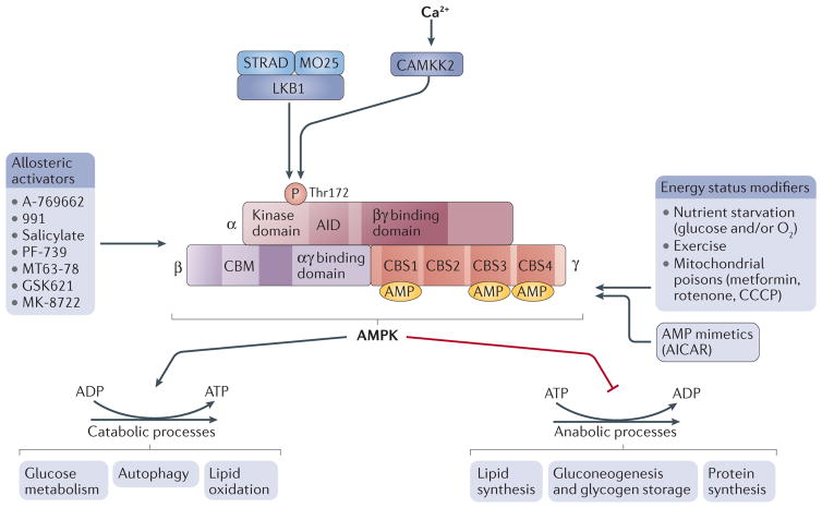
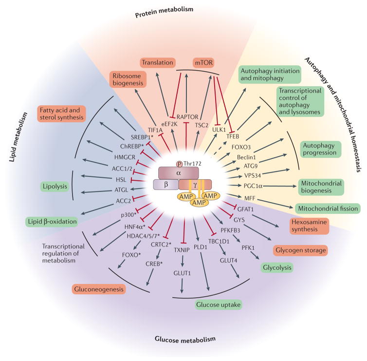
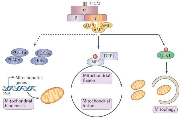
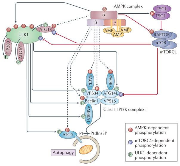
Cells constantly adapt their metabolism to meet their energy needs and respond to nutrient availability. Eukaryotes have evolved a very sophisticated system to sense low cellular ATP levels via the serine/threonine kinase AMP-activated protein kinase (AMPK) complex. Under conditions of low energy, AMPK phosphorylates specific enzymes and growth control nodes to increase ATP generation and decrease ATP consumption. In the past decade, the discovery of numerous new AMPK substrates has led to a more complete understanding of the minimal number of steps required to reprogramme cellular metabolism from anabolism to catabolism. This energy switch controls cell growth and several other cellular processes, including lipid and glucose metabolism and autophagy. Recent studies have revealed that one ancestral function of AMPK is to promote mitochondrial health, and multiple newly discovered targets of AMPK are involved in various aspects of mitochondrial homeostasis, including mitophagy. This Review discusses how AMPK functions as a central mediator of the cellular response to energetic stress and mitochondrial insults and coordinates multiple features of autophagy and mitochondrial biology.


Conflict of interest statement

Competing interests statement

The authors declare no competing interests.

Figures

Figure 1
Figure 1. AMPK structure and activation
Domain structure of the AMP-activated protein kinase (AMPK) trimer, showing the α-, β- and γ-subunits with their respective domains. The upstream kinases CAMKK2 and liver kinase B1 (LKB1) are shown above the AMPK complex. LKB1 is a heterotrimer composed of LKB1, STRAD and MO25; CAMKK2 is activated by intracellular calcium. Several factors lead to AMPK activation, such as mitochondrial poisons and oxygen or glucose starvation, as well as exercise. Drugs that activate AMPK include the AMP mimetic AICAR and several small-molecule allosteric activators (listed on the left-hand side). The effect of AMPK activation is to rewire metabolism to decrease anabolic processes (that is, ATP consumption) and increase catabolism (that is, ATP production) to restore a more favourable energy balance. Downstream substrates are grouped by biological functions. ATP-producing processes are activated, while ATP-consuming processes are inhibited. AID, auto-inhibitory domain; CBM, carbohydrate-binding module; CBS, cystathionine-β-synthase.
Figure 2
Figure 2. AMPK regulates a variety of metabolic processes
Once activated, the AMP-activated protein kinase (AMPK) complex phosphorylates key targets to rewire metabolism. The direct targets of AMPK are shown in the first concentric circle. The arrow indicates whether the phosphorylation is activating or inhibitory for the function of the target protein. The general process in which each target is involved is indicated in the next circle, and the box colour indicates whether that general process is activated (green) or inhibited (red). For certain targets, an intermediate mediator of the effect is indicated between the two circles. The pathways modulated by AMPK are grouped into four general categories — protein metabolism, lipid metabolism, glucose metabolism, and autophagy and mitochondrial homeostasis — denoting the wide range of processes that are controlled by AMPK. mTOR is modulated by AMPK while also modulating several direct or indirect targets of AMPK. This is illustrated by arrows from mTOR to its targets and serves to emphasize the complex relationship between these two signalling pathways. Transcriptional regulators are denoted by an asterisk. It is important to note that only a subset of AMPK substrates is included in the figure. ChREBP, carbohydrate-responsive element-binding protein; CREB, cAMP response element-binding protein; FOXO, forkhead box protein O; HDAC, histone deacetylase; HMGCR, HMG-CoA reductase; HNF4α, hepatocyte nuclear factor 4α; MFF, mitochondrial fission factor; PGC1α, peroxisome proliferator-activated receptor-γ co-activator 1α; PLD1, phospholipase D1; SREBP1, sterol regulatory element-binding protein 1; TFEB, transcription factor EB.
Figure 3
Figure 3. Regulation of mitochondrial homeostasis by AMPK
AMP-activated protein kinase (AMPK) directly phosphorylates mitochondrial fission factor (MFF) to regulate mitochondrial fission through dynamin-like protein DRP1 and activates ULK1, the upstream kinase in autophagy and mitophagy. Mitochondrial fission is required for mitophagy and allows damaged mitochondria to be degraded by mitophagy. During energy stress, AMPK also activates peroxisome proliferator-activated receptor-γ (PPARγ) co-activator 1α (PGC1α), which activates mitochondrial biogenesis genes through interaction with PPARγ or oestrogen-related receptors (ERRs). The dashed arrow indicates potentially indirect regulation.
Figure 4
Figure 4. Details of the regulation of autophagy by mTOR, AMPK and ULK1
ULK1-mediated phosphorylation events are shown in green, mTOR-mediated phosphorylation events in purple and AMP-activated protein kinase (AMPK)-mediated phosphorylation events in red. Dashed arrows indicate reported phosphorylation events by AMPK that are insufficiently documented or that involve sites that do not conform to the AMPK motif. Upon activation, AMPK phosphorylates TSC2 and RAPTOR, which leads to the downregulation of mTOR complex 1 (mTORC1) (purple complex, composed of mTOR, RAPTOR, mLST8, DEPTOR and PRAS40 (also known as AKT1S1); not all shown) activity. AMPK phosphorylates ULK1 on at least four serines to promote its activity. ULK1 is part of a complex with ATG101, ATG13 and FIP200. All of these proteins have been shown to be targets of ULK1, while ATG13 has also been reported as a target of both AMPK and mTORC1. PI3K complex I mediates the conversion of phosphatidylinositol (PI) to phosphatidylinositol-3-phosphate (PtdIns3P) at the surface of the forming autophagosome (beige membrane shown engulfing cellular components, including a mitochondrion), a step required for proper recruitment of cargo and adaptor proteins. The PI3K complex I components (light blue) and accessory factors (dark blue) are shown with their reported phosphorylation. ATG9 is an integral membrane protein localized at the autophagosome membrane. The arrows indicate whether the phosphorylation event is activating or inhibitory for the function of the protein.
Figure 5
Figure 5. Modulation of the transcription of autophagy and lysosome genes by AMPK
AMP-activated protein kinase (AMPK) regulates the activity of forkhead box protein O3 (FOXO3) through direct phosphorylation. In parallel, AMPK decreases mTOR complex 1 (mTORC1) (purple complex, composed of mTOR, RAPTOR, mLST8, DEPTOR and PRAS40; not all shown) activity through phosphorylation of RAPTOR and the upstream regulator TSC2. This leads to downregulation of mTORC1 activity. This results in dephosphorylation of mTORC1 targets FOXK1 and FOXK2, as well as transcription factor EB (TFEB). As a result, dephosphorylated FOXK1 and FOXK2 can no longer act as transcriptional repressors of FOXO3 targets, allowing higher transcription of autophagy genes downstream of FOXO3 binding. However, dephosphorylation of TFEB allows its nuclear translocation and activation of its target genes, including genes involved in lysosome biogenesis. In addition, an increased CARM1 protein level resulting from FOXO3-dependent gene activation further enhances TFEB-dependent gene expression. Acetyl-CoA carboxylase 2 (ACC2) phosphorylation by AMPK stimulates its nuclear import where it enhances TFEB target gene expression.

References

    1. Celenza JL, Carlson M. A yeast gene that is essential for release from glucose repression encodes a protein kinase. Science. 1986;233:1175–1180. - PubMed
    1. Gancedo JM. Carbon catabolite repression in yeast. Eur J Biochem. 1992;206:297–313. - PubMed
    1. Crozet P, et al. Mechanisms of regulation of SNF1/AMPK/SnRK1 protein kinases. Front Plant Sci. 2014;5:190. - PMC - PubMed
    1. Inoki K, Zhu T, Guan KL. TSC2 mediates cellular energy response to control cell growth and survival. Cell. 2003;115:577–590. - PubMed
    1. Gwinn DM, et al. AMPK phosphorylation of raptor mediates a metabolic checkpoint. Mol Cell. 2008;30:214–226. - PMC - PubMed

Publication types

Substances