

Transcranial Brain Sonography in Parkinson's Disease and Other Parkinsonian Disorders: a Hospital Study from Tuzla, Bosnia and Herzegovina
- PMID: 28974846
- PMCID: PMC5585815
- DOI: 10.5455/medarh.2017.71.261-264
Transcranial Brain Sonography in Parkinson's Disease and Other Parkinsonian Disorders: a Hospital Study from Tuzla, Bosnia and Herzegovina
Abstract
Introduction: Transcranial sonography (TCS) is a relatively new ultrasound modality which could display echogenicity of human brain tissue through the intact skull. TCS may be useful in differentiation of idiopathic Parkinson's disease (PD) from other parkinsonian disorders. Therefore, we studied different ultrasound markers by TCS in individuals with Parkinson's disease.
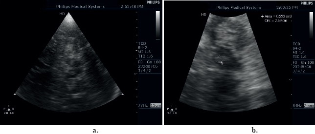
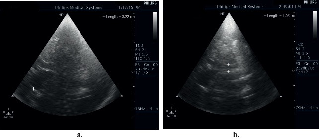
Patients and methods: We performed TCS in 44 patients with PD and 22 patients with other parkinsonian disorders. Echogenic sizes of the substantia nigra (SN) and the lentiform nuclei (LN), as well as the width of the third ventricle and the frontal horns of the lateral ventricle, were measured. We also analyzed the echogenicity of the brainstem raphe (BR).
Results: An unilateral hyperechogenic SN was observed in 31 (70%) patients with PD and only in 2 patients (9%) with other parkinsonian disorders (P<0.0001). Hyperechogenicity of the LN was no observed in patients with PD; however, it was present in 7 (32%) patients with other parkinsonian disorders (P=0.0002). Diameter of third ventricle (8.6+/-2.2 mm vs. 6.9+/-1.7mm, P=0.001), right (18.5+/-2.6 mm vs. 16.5+/-2.3 mm, P=0.003) and left frontal horn of lateral ventricle (19.0+/-3.7 mm vs. 16.2+/-2.6 mm, P=0.0006) was significantly wider in patients with other parkinsonian disorders compared with patients with PD. There was no difference in presence of hypoechogenic or interrupted BR in patients with PD and patients with other parkinsonian disorders (39% vs. 27%, P=0.4).
Conclusion: TCS is a promising diagnostic technique and can be very helpful in differentiating between idiopathic Parkinson's disease and other parkinsonian disorders.
Keywords: Basal ganglia echogenicity; Differential diagnosis; Parkinsonian syndromes; Parkinson’s disease; Transcranial Brain Sonography.
Conflict of interest statement
• Conflict of interest: none declared.
Figures
Similar articles
-
Transcranial sonography in movement disorders.Biomed Pap Med Fac Univ Palacky Olomouc Czech Repub. 2008 Dec;152(2):251-8. doi: 10.5507/bp.2008.039. Biomed Pap Med Fac Univ Palacky Olomouc Czech Repub. 2008. PMID: 19219216 Review.
-
Substantia nigra hyperechogenicity and brain ventricular size as biomarkers of early dementia with Lewy bodies.Alzheimers Res Ther. 2024 Oct 15;16(1):227. doi: 10.1186/s13195-024-01590-w. Alzheimers Res Ther. 2024. PMID: 39407323 Free PMC article.
-
Transcranial brain sonography findings in discriminating between parkinsonism and idiopathic Parkinson disease.Arch Neurol. 2007 Nov;64(11):1635-40. doi: 10.1001/archneur.64.11.1635. Arch Neurol. 2007. PMID: 17998447
-
Hypoechogenicity of brainstem raphe correlates with depression in migraine patients.J Headache Pain. 2019 May 15;20(1):53. doi: 10.1186/s10194-019-1011-2. J Headache Pain. 2019. PMID: 31092190 Free PMC article.
-
[Sonography of the parenchyma in Parkinson's disease].Nervenarzt. 2010 Oct;81(10):1189-95. doi: 10.1007/s00115-010-3025-5. Nervenarzt. 2010. PMID: 20802993 Review. German.
Cited by
-
Transcranial Sonography of the Substantia Nigra for the Differential Diagnosis of Parkinson's Disease and Other Movement Disorders: A Meta-Analysis.Parkinsons Dis. 2021 Apr 30;2021:8891874. doi: 10.1155/2021/8891874. eCollection 2021. Parkinsons Dis. 2021. PMID: 34007439 Free PMC article. Review.
-
Simple biomarkers to distinguish Parkinson's disease from its mimics in clinical practice: a comprehensive review and future directions.Front Neurol. 2024 Sep 19;15:1460576. doi: 10.3389/fneur.2024.1460576. eCollection 2024. Front Neurol. 2024. PMID: 39364423 Free PMC article. Review.
-
Lentiform Nucleus Hyperechogenicity in Parkinsonian Syndromes: A Systematic Review and Meta-Analysis with Consideration of Molecular Pathology.Cells. 2019 Dec 18;9(1):2. doi: 10.3390/cells9010002. Cells. 2019. PMID: 31861253 Free PMC article.
-
Progressive Supranuclear Palsy-Parkinsonism Predominant (PSP-P)-A Clinical Challenge at the Boundaries of PSP and Parkinson's Disease (PD).Front Neurol. 2020 Mar 10;11:180. doi: 10.3389/fneur.2020.00180. eCollection 2020. Front Neurol. 2020. PMID: 32218768 Free PMC article. Review.
References
-
- Becker G, Seufert J, Bogdahn U, Reichmann H, Reiners K. Degeneration of substantia nigra in chronic Parkinson's disease visualized by transcranial color-coded real-time sonography. Neurology. 1995;45:182–4. - PubMed
-
- Mijajlovic MD, Tsivgoulis G, Sternic N. Transcranial Brain Parenchymal Sonography in Neurodegenerative and Psychiatric Diseases. J Ultrasound Med. 2014;33:2061–8. - PubMed
-
- Litvan I, Agid Y, Calne D, et al. Clinical research criteria for the diagnosis of progressive supranuclear palsy (Steele-Richardson-Olszewski syndrome): report of the NINDS-SPSP international workshop. Neurology. 1996;47:1–9. - PubMed
-
- Gilman S, Low PA, Quinn N, et al. Consensus statement on the diagnosis of multiple system atrophy. J Neurol Sci. 1999;163:4–5. - PubMed
MeSH terms
LinkOut - more resources
Full Text Sources
Other Literature Sources