Evidences supporting the vascular etiology of post-double balloon enteroscopy pancreatitis: Study in porcine model
- PMID: 28974886
- PMCID: PMC5603486
- DOI: 10.3748/wjg.v23.i34.6201
Evidences supporting the vascular etiology of post-double balloon enteroscopy pancreatitis: Study in porcine model
Abstract
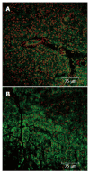
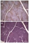
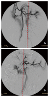
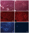
Double balloon enteroscopy (DBE) is an endoscopic technique broadly used to diagnose and treat small bowel diseases. Among the associated complications of the oral DBE, post-procedure pancreatitis has taken the most attention due to its gravity and the thought that it might be associated to the technique itself and anatomical features of the pancreas. However, as the etiology has not been clarified yet, this paper aims to review the published literature and adds new results from a porcine animal model. Biochemical markers, histological sections and the vascular perfusion of the pancreas were monitored in the pig during DBE practice. A reduced perfusion of the pancreas and bowel, the presence of defined hypoxic areas and disseminated necrotic zones were found in the pancreatic tissue of pigs. All these evidences contribute to support a vascular distress as the most likely etiology of the post-DBE pancreatitis.
Keywords: Animal model; Double balloon enteroscopy; Pancreas; Pancreatitis; Pig.
Conflict of interest statement
Conflict-of-interest statement: To the best of all authors’ knowledge, no conflict of interest exists.
Figures


References
-
- Yamamoto H, Sekine Y, Sato Y, Higashizawa T, Miyata T, Iino S, Ido K, Sugano K. Total enteroscopy with a nonsurgical steerable double-balloon method. Gastrointest Endosc. 2001;53:216–220. - PubMed
-
- May A, Nachbar L, Schneider M, Neumann M, Ell C. Push-and-pull enteroscopy using the double-balloon technique: method of assessing depth of insertion and training of the enteroscopy technique using the Erlangen Endo-Trainer. Endoscopy. 2005;37:66–70. - PubMed
-
- Pérez-Cuadrado E, Pons V, Bordas JM, González B, Llach J, Menchén P, Pellicer F, Rodríguez S. Small bowel endoscopic exploration Spanish GI endoscopy society recommendations. Endoscopy. 2012;44:979–987. - PubMed
-
- Pohl J, Blancas JM, Cave D, Choi KY, Delvaux M, Ell C, Gay G, Jacobs MA, Marcon N, Matsui T, et al. Consensus report of the 2nd International Conference on double balloon endoscopy. Endoscopy. 2008;40:156–160. - PubMed
-
- Pennazio M, Spada C, Eliakim R, Keuchel M, May A, Mulder CJ, Rondonotti E, Adler SN, Albert J, Baltes P, et al. Small-bowel capsule endoscopy and device-assisted enteroscopy for diagnosis and treatment of small-bowel disorders: European Society of Gastrointestinal Endoscopy (ESGE) Clinical Guideline. Endoscopy. 2015;47:352–376. - PubMed
Publication types
MeSH terms
Substances
LinkOut - more resources
Full Text Sources
Other Literature Sources
Medical

