. 2017 Aug 20;4(3):140-150.
doi: 10.1002/reg2.81. eCollection 2017 Jun.

The periosteal requirement and temporal dynamics of BMP2-induced middle phalanx regeneration in the adult mouse

Affiliations

The periosteal requirement and temporal dynamics of BMP2-induced middle phalanx regeneration in the adult mouse

Lindsay A Dawson et al. Regeneration (Oxf). .

Abstract

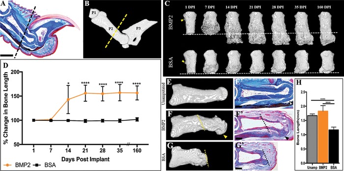
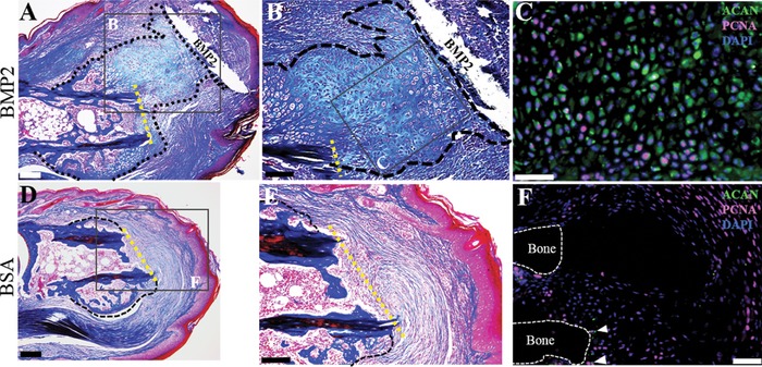
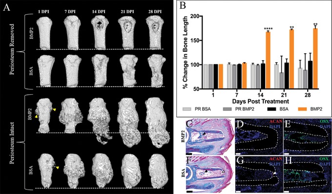
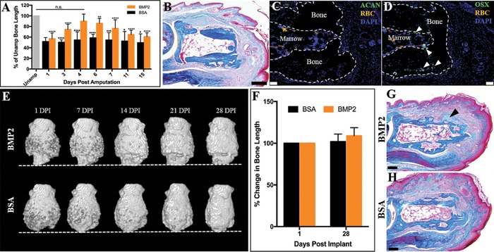
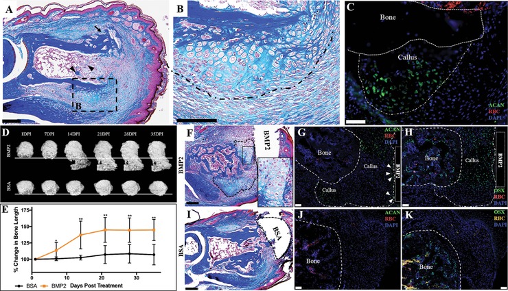
Regeneration of mammalian limbs is restricted to amputation of the distal digit tip, the terminal phalanx (P3). The adjacent skeletal element, the middle phalanx (P2), has emerged as a model system to investigate regenerative failure and as a site to test approaches aimed at enhancing regeneration. We report that exogenous application of bone morphogenetic protein 2 (BMP2) stimulates the formation of a transient cartilaginous callus distal to the amputation plane that mediates the regeneration of the amputated P2 bone. BMP2 initiates a significant regeneration response during the periosteal-derived cartilaginous healing phase of P2 bone repair, yet fails to induce regeneration in the absence of periosteal tissue, or after boney callus formation. We provide evidence that a temporal component exists in the induced regeneration of P2 that we define as the "regeneration window." In this window, cells are transiently responsive to BMP2 after the amputation injury. Simple re-injury of the healed P2 stump acts to reinitiate endogenous bone repair, complete with periosteal chondrogenesis, thus reopening the "regeneration window" and thereby recreating a regeneration-permissive environment that is responsive to exogenous BMP2 treatment.

Keywords: BMP2; digit; endochondral ossification; periosteum; regeneration.


Figures

Figure 1
Figure 1
Exogenous BMP2 induces P2 regeneration. (A) Mallory trichrome stained section illustrating the anatomy of the adult mouse hindlimb digit. (B) μCT 3D rendering of the adult mouse hindlimb digit, showing the first (P1), middle (P2), and terminal phalanx (P3). (A), (B) The dashed line indicates the amputation plane. (C) Representative μCT renderings show that BMP2 induces segment‐specific regeneration of P2 via formation of the distal callus, evident by 14 DPI, while BSA‐treated digits do not show distal bone elongation. The amputation plane is shown as the dashed line. (D) Bone length measurements over 160 days, normalized to 1 DPI, illustrate a significant increase in bone length after BMP2 treatment, resulting in an average 56% enhancement of length by 160 DPI, while BSA treatment does not induce distal bone regeneration (t test, ±SEM, *P ≤ 0.05, ****P ≤ 0.0001). (E), (E′) Representative μCT 3D rendering and Mallory trichrome stained histological section of the unamputated P2 digit. Open arrowhead denotes ventral tendon and associated fibrocartilage. (F), (F′) Representative μCT 3D rendering and corresponding Mallory trichrome stained histological section of a BMP2‐treated regenerated P2 bone at 160 DPI. Arrowheads indicate the region of distal bone end curvature and associated ventral tendon attachment sites. The amputation plane is shown as the dashed line. (G), (G′) Representative μCT 3D rendering and corresponding Mallory trichrome stained histological section of the BSA‐treated digit at 160 DPI. The amputation plane is shown as the dashed line. (H) BMP2 treatment restores the bone length of amputated P2 digits (ANOVA, ±SEM, P ≤ 0.0001). (A), (B), (E)–(G′) Distal is to the right, dorsal is to the top. (C) Distal is to the bottom. Scale bars: (A), (E), (F′) 500 μm; (G′) 200 μm
Figure 2
Figure 2
BMP2‐induced regeneration is mediated by a cartilaginous distal callus. (A)−(C) Serial sections of a representative BMP2‐treated sample at 8 DPI. (A) Mallory trichrome staining illustrating the cartilaginous distal callus in close association with the BMP2 XeroGel. The amputation plane is shown as the yellow dashed line. (B) Inset region of (A). Higher magnification view of the cartilaginous distal callus. (C) Inset region of (B). Double immunostaining for ACAN and PCNA show proliferating chondrocytes within the distal callus. Sample counterstained with DAPI. (D)−(F) Serial sections of a representative BSA‐treated sample at 8 DPI. (D) Mallory staining showing BSA control digits exhibit the characteristic peripheral callus but does not show chondrogenesis distal to the amputation plane, shown as the yellow dashed line. (E) Inset region of (D). Fibrotic tissue caps the distal end of the amputated P2 bone. (F) Double immunostaining for ACAN and PCNA shows few immunopositive chondrocytes (arrowheads), with proliferation localized to the fibrotic region. Sample counterstained with DAPI. (A)–(F) Distal is to the right, dorsal is to the top. Scale bars: (A), (D) 200 μm; (B), (E) 100 μm; (C), (F) 50 μm
Figure 3
Figure 3
BMP2‐induced regeneration is inhibited in P2 digits lacking periosteum. (A) Representative μCT 3D renderings of periosteum removed or periosteum intact (sham) P2 digits treated with BMP2 or BSA XeroGel. Peripheral callus formation is not observed in BMP2‐ or BSA‐treated digits lacking the periosteum, and BMP2 treatment does not induce P2 regeneration in the absence of the periosteum. Peripheral callus formation is evident at 1 DPI (arrowheads) in BMP2‐ or BSA‐treated samples with intact periosteum, and distal callus formation is evident by 14 DPI in BMP2‐treated digits with intact periosteum. (B) Bone length changes over 4 weeks, normalized to 1 DPI, show no BMP2‐induced increase in bone length in digits lacking periosteum (ANOVA, ±SEM, **P ≤ 0.01, ****P ≤ 0.0001). (C)–(E) Serial sections of a representative BMP2‐treated digit lacking periosteum at 8 DPI. (C) Mallory trichrome staining showing no peripheral callus formation or distal callus formation. Bone formation is evident within the endosteal/marrow space (arrowhead). (D) ACAN immunostaining confirms no cartilage is present in digits lacking the periosteum. (E) OSX immunopositive cells are localized to the endosteal/marrow region. Samples counterstained with DAPI. (F)–(H) Serial sections of a representative BSA‐treated digit lacking periosteum at 8 DPI. (F) Mallory trichrome staining illustrating no peripheral callus formation or distal callus formation. Endosteal/marrow bone formation is evident (closed arrowhead), and residual scab is present within the marrow space of the distal bone stump (open arrowhead). (G) Immunostaining for ACAN shows no immunopositive cells distal to the amputation plane or associated with the periosteum. Arrowhead denotes background fluorescence associated with the residual scab. (H) OSX immunostaining is localized to the endosteal/marrow region. Samples counterstained with DAPI. (A) Distal is to the bottom. (C)–(H) Distal is to the right, dorsal is to the top. Scale bars: (C), (F) 200 μm; (D), (E), (G), (H) 50 μm
Figure 4
Figure 4
P2 wound maturation attenuates BMP2‐induced regeneration. (A) Neonate P2 skeletal length is restored after BMP2 treatment at 4 DPA, the approximate time of wound closure. BMP2 treatment prior to or after wound closure attenuates the regeneration response (ANOVA, ±SEM, *P or ♦P ≤ 0.05, **P ≤ 0.01, ***P or ♦♦♦P ≤ 0.001, ****P or ♦♦♦♦P ≤ 0.0001; asterisks denote significance in BMP2‐treated digits compared to BMP2 treatment at 4 DPA; diamonds denote significance between BMP2 and BSA treatment). (B)–(D) Serial sections of a representative 24 DPA P2 digit. (B) Mallory trichrome staining showing the cartilaginous peripheral callus has been completely remodeled to boney tissue. (C) Immunohistochemistry for ACAN shows no immunopositive cells localized to the peripheral callus at 24 DPA. (D) OSX immunostaining shows osteoblasts localized to the endosteal/marrow region and within the boney peripheral callus (arrowheads). Samples counterstained with DAPI. (E), (F) Representative μCT 3D renderings of amputated P2 digits treated with BMP2 at 24 DPA. The amputation plane is shown as the dashed line. Peripheral callus formation is observed by 24 DPA. BMP2 does not function to induce regeneration when implanted at 24 DPA, resulting in no enhancement of bone length from 1 to 28 DPI (t test, ±SEM, P > 0.05, no significance). (G) Mallory trichrome staining of a representative 24 DPA BMP2‐treated digit harvested at 8 DPI showing cartilage associated with the peripheral callus (arrowhead), not distal to the amputation plane. (H) Mallory trichrome staining of a representative 24 DPA BSA‐treated digit harvested at 8 DPI illustrating that the peripheral callus has been completely replaced with boney tissue and no evidence of distal elongation. RBC, red blood cells. (B)–(D), (G), (H) Distal is to the right, dorsal is to the top. (E) Distal is to the bottom. Scale bars: (B), (G), (H) 200 μm; (C), (D) 50 μm
Figure 5
Figure 5
BMP2 induces regeneration of the re‐injured mature P2 bone stump. (A)–(C) Serial sections of a representative 24 DPA reamputated P2 digit at 9 DPA. (A) Mallory trichrome staining showing peripheral callus formation is reinitiated after stump re‐injury. (B) Inset illustrating that peripheral callus chondrogenesis develops external to the pre‐existing peripheral callus. (C) The peripheral callus associated with P2 re‐injury tests immunopositive for ACAN. Sample counterstained with DAPI. (D) Representative μCT renderings illustrate that BMP2 induces regeneration of mature P2 wounds after re‐injury. The regeneration response, evident by 14 DPI, is mediated by the formation of a distal callus, whereas BSA‐treated digits show no distal callus formation or associated bone elongation. The line indicates the amputation plane. (E) Statistical analysis of bone length measurements over 35 days, normalized to 1 DPI, show that BMP2 induces a significant increase in bone length compared to re‐injured digits treated with BSA (t test, ±SEM, *P ≤ 0.05, **P ≤ 0.01). (F)–(H) Serial sections of a representative BMP2‐treated digit at 10 DPI. (F) Mallory trichrome staining shows chondrogenesis associated with the BMP2 XeroGel. Inset displaying high magnification of chondrocytes. Distal callus is outlined. (G) ACAN immunostaining is localized to the boundaries of the distal callus, while (H) OSX immunostaining is localized to the central region of the distal callus, in areas not immunopositive for ACAN staining. Samples counterstained with DAPI. (I)–(K) Serial sections of a representative BSA‐treated digit at 10 DPI. (I) Mallory trichrome staining of BSA‐treated digits show peripheral callus with no evidence of distal callus formation. (J) BSA‐treated digits test negative for ACAN immunostaining. (K) OSX immunostaining is localized to the bone stump (outlined). Samples counterstained with DAPI. (A)–(C), (F)–(K) Distal is to the right, dorsal is to the top. (D) Distal is to the bottom. Scale bars: (A), (F), (I) 200 μm; (B) 100 μm; (C), (G), (H), (J), (K) 50 μm

Similar articles

Cited by

References

    1. Agrawal, V. , Johnson, S. A. , Reing, J. , Zhang, L. , Tottey, S. , Wang, G. , … Badylak, S. F. (2010). Epimorphic regeneration approach to tissue replacement in adult mammals. Proceedings of the National Academy of Sciences of the United States of America, 107(8), 3351–3355. - PMC - PubMed
    1. Agrawal, V. , Kelly, J. , Tottey, S. , Daly, K. A. , Johnson, S. A. , Siu, B. F. , … Badylak, S. F. (2011). An isolated cryptic peptide influences osteogenesis and bone remodeling in an adult mammalian model of digit amputation. Tissue Engineering Part A, 17(23‐24), 3033–3044. - PMC - PubMed
    1. Agrawal, V. , & Sinha, M. (2016). A review on carrier systems for bone morphogenetic protein‐2. Journal of Biomedical Materials, 105(4), 904–925. - PubMed
    1. Agrawal, V. , Siu, B. F. , Chao, H. , Hirschi, K. K. , Raborn, E. , Johnson, S. A. , … Badylak, S. F. (2012). Partial characterization of the Sox2+ cell population in an adult murine model of digit amputation. Tissue Engineering Part A, 18(13‐14), 1454–1463. - PMC - PubMed
    1. Agrawal, V. , Tottey, S. , Johnson, S. A. , Freund, J. M. , Siu, B. F. , & Badylak, S. F. (2011). Recruitment of progenitor cells by an extracellular matrix cryptic peptide in a mouse model of digit amputation. Tissue Engineering Part A, 17(19‐20), 2435–2443. - PMC - PubMed

LinkOut - more resources