. 2017 Dec 1;595(23):7123-7134.
doi: 10.1113/JP275184. Epub 2017 Oct 25.

Rapid decline in MyHC I(β) mRNA expression in rat soleus during hindlimb unloading is associated with AMPK dephosphorylation

Affiliations

Rapid decline in MyHC I(β) mRNA expression in rat soleus during hindlimb unloading is associated with AMPK dephosphorylation

Natalia A Vilchinskaya et al. J Physiol. .

Abstract

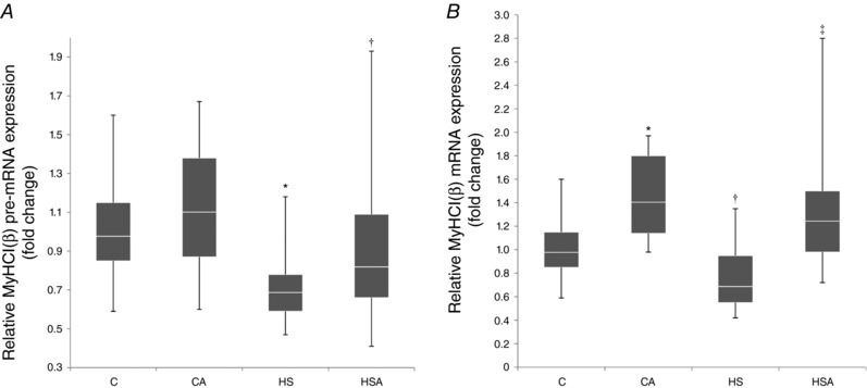
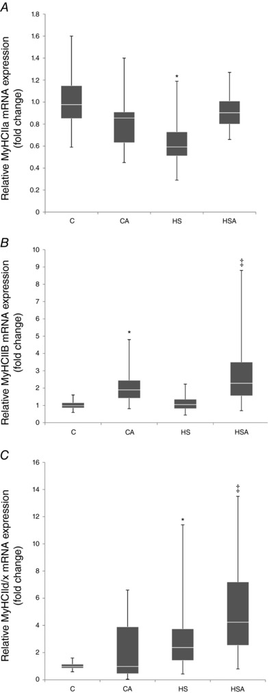
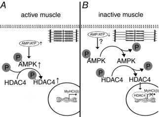
Key points: Inactivation of a skeletal muscle results in slow to fast myosin heavy chain (MyHC) shift. AMP-activated protein kinase (AMPK) can be implicated in the regulation of genes encoding the slow MyHC isoform. Here we report that AMPK dephosphorylation after 24 h of mechanical unloading can contribute to histone deacetylase (HDAC) nuclear translocation; activation of AMPK prevents HDAC4 nuclear accumulation after 24 h of unloading and AMPK dephosphorylation inhibits slow MyHC expression following 24 h of unloading. Our data indicate that AMPK dephosphorylation during the first 24 h of mechanical unloading has a significant impact on the expression of MyHC isoforms in rat soleus causing a decrease in MyHC I(β) pre-mRNA and mRNA expression as well as MyHC IIa mRNA expression.

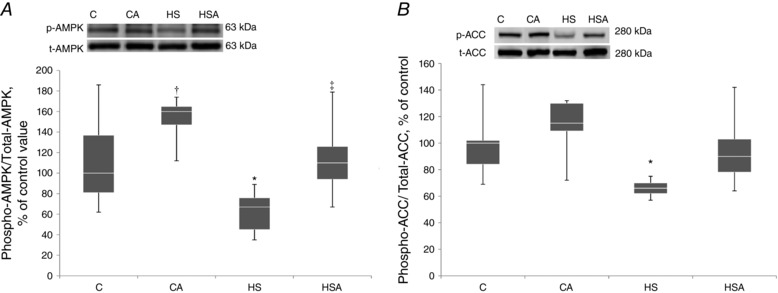
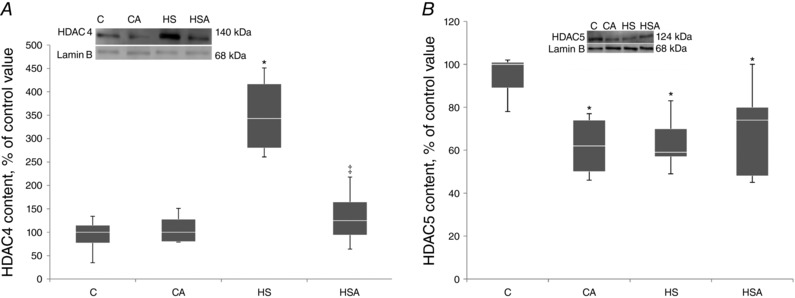
Abstract: One of the key events that occurs during skeletal muscle inactivation is a change in myosin phenotype, i.e. increased expression of fast isoforms and decreased expression of the slow isoform of myosin heavy chain (MyHC). It is known that calcineurin/nuclear factor of activated T-cells and AMP-activated protein kinase (AMPK) can regulate the expression of genes encoding MyHC slow isoform. Earlier, we found a significant decrease in phosphorylated AMPK in rat soleus after 24 h of hindlimb unloading (HU). We hypothesized that a decrease in AMPK phosphorylation and subsequent histone deacetylase (HDAC) nuclear translocation can be one of the triggering events leading to a reduced expression of slow MyHC. To test this hypothesis, Wistar rats were treated with AMPK activator (AICAR) for 6 days before HU as well as during 24 h of HU. We discovered that AICAR treatment prevented a decrease in pre-mRNA and mRNA expression of MyHC I as well as MyHC IIa mRNA expression. Twenty-four hours of hindlimb suspension resulted in HDAC4 accumulation in the nuclei of rat soleus but AICAR pretreatment prevented this accumulation. The results of the study indicate that AMPK dephosphorylation after 24 h of HU had a significant impact on the MyHC I and MyHC IIa mRNA expression in rat soleus. AMPK dephosphorylation also contributed to HDAC4 translocation to the nuclei of soleus muscle fibres, suggesting an important role of HDAC4 as an epigenetic regulator in the process of myosin phenotype transformation.

Keywords: AICAR; AMPK; HDAC; MyHC; hindlimb unloading; soleus muscle.


Figures

Figure 1
Figure 1. Quantification of p‐AMPK/t‐AMPK (A) and p‐ACC/t‐ACC (B) ratios expressed relative (%) to control; representative immunoblots are shown above the graphs
C, control group; CA, control with AICAR pretreatment; HS, hindlimb suspension for 24 h; HSA, hindlimb suspension for 24 h with AICAR pretreatment. *Significant difference from control (P < 0.05); trend towards a significant difference from control (P = 0.09); significant difference from HS (P < 0.05). Box plots show 25–75 percentiles and median values and the whiskers represent the minimum and the maximum; n = 8/group.
Figure 2
Figure 2. Quantification of HDAC4 (A) and HDAC5 (B) in the nuclear fraction of the rat soleus expressed relative (%) to control and representative immunoblots for HDAC4 and lamin B (used for normalization)
C, control group; CA, control with AICAR pretreatment; HS, hindlimb suspension for 24 h; HSA, hindlimb suspension for 24 h with AICAR pretreatment. *Significant difference from control (P < 0.05); significant difference from HS (P < 0.05). Box plots show 25–75 percentiles and median values and the whiskers represent the minimum and the maximum; n = 8/group.
Figure 3
Figure 3. Quantification of p‐AMPK (Ser 485/491) (A) and p‐PKD (B) in the rat soleus expressed relative (%) to control and representative immunoblots for p‐AMPK (Ser 485/491) (A) and p‐PKD (B) and GAPDH (used for normalization)
C, control group; CA, control with AICAR pretreatment; HS, hindlimb suspension for 24 h; HSA, hindlimb suspension for 24 h with AICAR pretreatment. *Significant difference from HS (P < 0.05). Box plots show 25–75 percentiles and median values and the whiskers represent the minimum and the maximum; n = 8/group.
Figure 4
Figure 4. PGC‐1α protein expression (A) and PGC‐1α mRNA expression (B) in the rat soleus muscle expressed relative to control
C, control group; CA, control with AICAR pretreatment; HS, hindlimb suspension for 24 h; HSA, hindlimb suspension for 24 h with AICAR pretreatment. Box plots show 25–75 percentiles and median values and the whiskers represent the minimum and the maximum; n = 8/group.
Figure 5
Figure 5. Relative MyHC I(β) pre‐mRNA (A) and mRNA (B) expression in the rat soleus muscle
C, control group; CA, control with AICAR pretreatment; HS, hindlimb suspension for 24 h; HSA, hindlimb suspension for 24 h with AICAR pretreatment. *Significant difference from control (P < 0.05); a trend towards a significant difference from HS (P = 0.09); significant difference from HS (P < 0.05). Box plots show 25–75 percentiles and median values and the whiskers represent the minimum and the maximum; n = 8/group.
Figure 6
Figure 6. Relative mRNA expression of MyHC IIa (A), MyHC IIb (B) and MyHC IId/x (C) in the rat soleus muscle
C, control group; CA, control with AICAR pretreatment; HS, hindlimb suspension for 24 h; HSA, hindlimb suspension for 24 h with AICAR pretreatment. *Significant difference from control (P < 0.05); significant difference from HS (P < 0.05). Box plots show 25–75 percentiles and median values and the whiskers represent the minimum and the maximum; n = 8/group.
Figure 7
Figure 7. A schematic representation depicting a hypothetical mechanism of the AMPK influence on MyHC I(β) mRNA expression via HDAC4 in an active and unloaded rat soleus muscle
A, rat soleus muscle under the conditions of normal activity. Normal contractile activity determines an equilibrium in the AMP:ATP ratio when the level of AMPK phosphorylation maintains an equilibrium in the level of HDAC4 phosphorylation, the main pool of which is localized in the cytoplasm (Cohen et al. 2015). No blocking of the promoter of the myh7 gene occurs and MyHC I(β) mRNA is expressed normally. B, rat soleus muscle under the unloading conditions. Sharply reduced contractile activity may lead to an accumulation of ATP and reduction in AMP. As a result, the level of AMPK phosphorylation decreases and induces partial HDAC4 dephosphorylation and its nuclear translocation. The myh7 gene promoter gets blocked and MyHC I (β) mRNA expression decreases.

References

    1. Cannavino J, Brocca L, Sandri M, Bottinelli R & Pellegrino MA (2014). PGC1‐α over‐expression prevents metabolic alterations and soleus muscle atrophy in hindlimb unloaded mice. J Physiol 592, 4575–4589. - PMC - PubMed
    1. Cohen TJ, Choi MC, Kapur M, Lira VA, Yan Z & Yao TP (2015). HDAC4 regulates muscle fibre type‐specific gene expression programs. Mol Cells 38, 343–348. - PMC - PubMed
    1. Coughlan KA, Valentine RJ, Sudit BS, Allen K, Dagon Y, Kahn BB, Ruderman NB & Saha AK (2016). PKD1 inhibits AMPKα2 through phosphorylation of serine 491 and impairs insulin signalling in skeletal muscle cells. J Biol Chem 291, 5664–5675. - PMC - PubMed
    1. De‐Doncker L, Kasri M, Picquet F & Falempin M (2005). Physiologically adaptive changes of the L5 afferent neurogram and of the rat soleus EMG activity during 14 days of hindlimb unloading and recovery. J Exp Biol 208, 4585–4592. - PubMed
    1. Dupré‐Aucouturier S, Castells J, Freyssenet D & Desplanches D (2015). Trichostatin A, a histone deacetylase inhibitor, modulates unloaded induced skeletal muscle atrophy. J Appl Physiol 119, 342–351. - PubMed

LinkOut - more resources