. 2017 Oct 25;17(21):3692-3703.
doi: 10.1039/c7lc00740j.

Cardiac microphysiological devices with flexible thin-film sensors for higher-throughput drug screening

Affiliations

Cardiac microphysiological devices with flexible thin-film sensors for higher-throughput drug screening

Johan U Lind et al. Lab Chip. .

Abstract

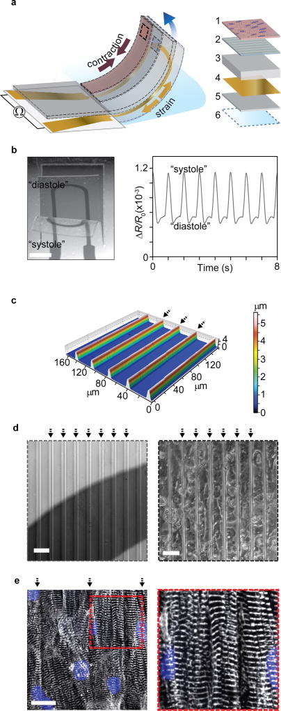
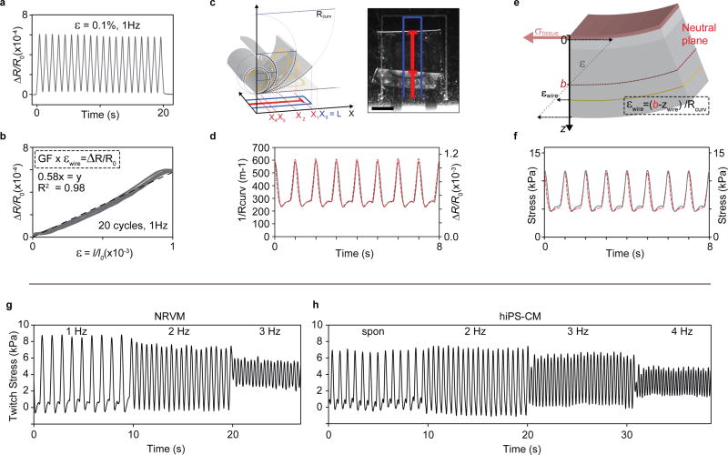
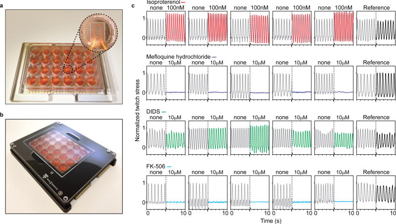
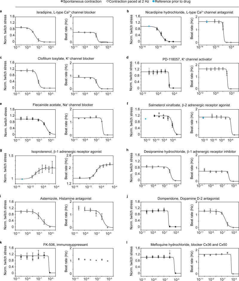
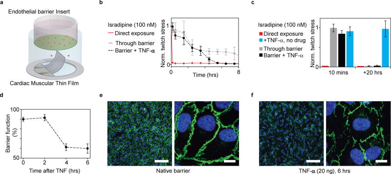
Microphysiological systems and organs-on-chips promise to accelerate biomedical and pharmaceutical research by providing accurate in vitro replicas of human tissue. Aside from addressing the physiological accuracy of the model tissues, there is a pressing need for improving the throughput of these platforms. To do so, scalable data acquisition strategies must be introduced. To this end, we here present an instrumented 24-well plate platform for higher-throughput studies of engineered human stem cell-derived cardiac muscle tissues that recapitulate the laminar structure of the native ventricle. In each well of the platform, an embedded flexible strain gauge provides continuous and non-invasive readout of the contractile stress and beat rate of an engineered cardiac tissue. The sensors are based on micro-cracked titanium-gold thin films, which ensure that the sensors are highly compliant and robust. We demonstrate the value of the platform for toxicology and drug-testing purposes by performing 12 complete dose-response studies of cardiac and cardiotoxic drugs. Additionally, we showcase the ability to couple the cardiac tissues with endothelial barriers. In these studies, which mimic the passage of drugs through the blood vessels to the musculature of the heart, we regulate the temporal onset of cardiac drug responses by modulating endothelial barrier permeability in vitro.


Figures

Figure 1
Figure 1. Engineered cardiac muscle tissue on cantilever with embedded flexible thin-film strain gauge
(a) Principle sketch of device cantilever and constituent layers 1: Engineered cardiac muscle tissue 2: tissue-aligning micro-molded or micro-patterned layer 3: PDMS layer, 4: Ti-Au thin-film sensor layer, 5: bottom PDMS layer 6: PNIPAAM release layer. (b) Example microscope image of the deflecting cantilever and corresponding electrical readout, scale bar 2 mm. (c) Optical profilometer characterization of molded 500 kPa PDMS surface. Arrows indicate barrier peaks. (d) Left: Bright field optical micrograph of grooves on cantilevers applied for structural guidance of hiPS-CM tissues, strain gauge wire seen a dark area. Right: Bright field optical micrograph of hiPS-CM tissue on molded PDMS top layer, scale bars 40 µm. (e) Confocal microscopy of immunostained hiPS-CM tissue, blue: DAPI nuclei stain, white α-actinin. Scale bar 20 µm.
Figure 2
Figure 2. Titanium-Gold thin-film micro-structure, appearance and mechanical robustness
(a) Optical micrographs (left, scale bars 2 mm) and SEM images (right, scale bars 5 µm) of 20 nm Au thin films deposited using 3, 5 or 7 nm Ti adhesion layers, on 5 µm thick PDMS substrates. For 5 nm and 7 nm Ti adhesion layers, buckled micro-structures were observed. These micro-buckled films frequently displayed macroscopic failure cracks in device formulations, deposited through shadow masks. For all thin films applied in devices, 1 nm Ti was added onto Au surface to ensure adhesion to subsequent PDMS layers. Thus, in final devices 3 nm Ti, 20 nm Au, and 1 nm Ti was deposited sequentially. These films yielded robust and reproducible devices. (b-c) Uniaxial strain tests of 3-20-1 nm Ti-Au-Ti thin films deposited on 5 µm thick PDMS substrates. (b) Cyclic 10 % strain did not lead to failure in thin-film conductivity. (c) Cyclic strain in the 0.1 % regime relevant to the final devices, displayed negligible dependency on strain rate, in the frequency range of tissue contractions.
Figure 3
Figure 3. Thin-film sensor calibration and device mechanical model
(a) Relative resistance change of 3-20-1 nm Ti-Au-Ti thin films deposited on 5 µm PDMS substrates, upon cyclic 0.1 % uniaxial strain at 1 Hz. (b) Relative resistance change vs. strain for cyclic 0.1 % uniaxial strain at 1 Hz. Linear fit (dashed line) indicate a gauge factor (GF) of app. 0.58. (c) optical tracking of cantilever deflection, scale bar 1 mm. (d) Concurrent optical tracking of cantilever curvature (left axis, dashed red line) and electrical readout of relative resistance change (right axis, solid grey line) from cantilever with NRVM tissue. (e) Principle sketch of mechanical model based on Stoney’s equation, applied to convert electrical and optical readout to stress generated by the engineered muscle tissues. By taking advantage of the concurrent optical and electrical readout, the placement of cantilever neutral axis b could be determined, see supplementary information (f) Stress values obtained applying mechanical model to convert optical (left axis, dashed red line) and electrical (right axis, solid grey line) readouts displayed in (d) to tissue stress values. (g) Force-frequency of twitch stress of NRVM-based tissues, paced at 1, 2, 3 Hz at day 4 after seeding on devices (h) Force-frequency of twitch stress of hiPS-CM tissues, spontaneously contracting and paced at 2, 3, 4 Hz at day 6 after seeding.
Figure 4
Figure 4. Instrumented 24-well platform enables multiple parallel experiments
(a) Example 24-well device, with polycarbonate multi-well housing. Insert: Example cantilever in well. (b) 24-well device in recording holder applied for data acquisition inside incubator environments. (c) 24 example readouts of hiPS-CM tissues applied in drug dose-response experiments, with parallel replicates and references. From top: 100 nM Isoproterenol increased beat rate and spontaneous contractile stress, 10 µM Mefloquine hydrochloride completely disrupted contraction, 10 µM Disodium 4,4'-diisothiocyanatostilbene-2,2'-disulfonate (DIDS) did not cause any notably effects, 10 µM FK-506 markedly decreased contractile stress, but not beat rate.
Figure 5
Figure 5. Sequential drug dose-response studies on hiPS-CM-derived tissues
Increasing drug doses were sequentially added, and tissue spontaneous beat rate and spontaneous twitch stress normalized to initial twitch stress, were recorded (grey circles). For a subset of the studies, the twitch stress while pacing electrically at 2 Hz (hollow circles) was also acquired. Error bars are S.E.M. Lines indicated 4-parameter logistic fits. EC/ IC50 values obtained from fits presented in Table 1. (a) Isradipine, N=5 (b) Nicardipine, N=4, (c) Clofilium tosylate, N=5 (d) PD-118057, N=5, (e) Flecanide acetate, N=5, (f) Salmeterol xinafoate, N=5, (g) Isoproterenol, N=6, (h) Desipramine hydrochloride N=4, (i) Astemizole N=5, (j) Domperidone N=5, (k) FK-506, N=5, (l) Mefloquine hydrochloride N=5.
Figure 6
Figure 6. Coupling endothelial barriers with cardiac MTFs to study drug transport
(a) Schematic illustration of experiments. Endothelial barrier tissues in Transwell® inserts are introduced into the wells of the instrumented cardiac 24-well MTF platform. A cardiac drug (Isradipine) is introduced in the volume shielded by the endothelial barrier. Drug transport across barrier and resultant cardiac effects are recorded in real-time through embedded sensors. (b) Decrease in normalized contractile twitch stress over time. Direct exposure of Isradipine (100 nM) immediately halts contraction (red). Exposure of Isradipine (100 nM) through endothelial barrier inserts (grey line), leads to delayed drug effect, where contraction is still observed after > 7 h exposure. (N=3). Compromising endothelial barrier function by co exposure with TNF-α significantly accelerates drug diffusion, leading to a full effect of the drug after ~5 h. (c) After 20 h full effect of Isradipine was observed for all samples. TNF-α did not directly introduce changes in twitch stress of cardiac tissues. (d) Endothelial tissue barrier function decreases upon exposure to TNF-α (20 ng/ml). Barrier function was evaluated as percent of fluorescent marker (400 Da) contained inside barrier during 20 mins diffusion into reservoir without tracer (e) Confocal microscopy of immunostained intact endothelial barrier. Blue: DAPI nuclei stain, Green: VE-Cadherin, Scale bars left: 100 µm, right: 10 µm (f) Confocal microscopy of immunostained endothelial barrier after exposure to 20 ng/ml TNF-α for 6 h. Blue: DAPI nuclei stain, Green: VE-Cadherin, Scale bars left: 100 µm, right: 10 µm.

References

    1. Schachter AD, Ramoni MF. Nature reviews Drug discovery. 2007;6:107–108. - PubMed
    1. DiMasi JA, Grabowski HG, Hansen RW. Journal of health economics. 2016;47:20–33. - PubMed
    1. Olson H, Betton G, Robinson D, Thomas K, Monro A, Kolaja G, Lilly P, Sanders J, Sipes G, Bracken W. Regulatory Toxicology and Pharmacology. 2000;32:56–67. - PubMed
    1. Go AS, Mozaffarian D, Roger VL, Benjamin EJ, Berry JD, Blaha MJ, Dai S, Ford ES, Fox CS, Franco S. Circulation. 2013;127:e62–e245. - PMC - PubMed
    1. Buckberg GD, Hoffman JI, Coghlan HC, Nanda NC. European Journal of Cardio-Thoracic Surgery. 2015;47:587–601. - PubMed

Publication types

Substances