. 2017;13(12):2018-2027.
doi: 10.1080/15548627.2017.1382782. Epub 2017 Nov 23.

Conserved and unique features of the fission yeast core Atg1 complex

Affiliations

Conserved and unique features of the fission yeast core Atg1 complex

Tamiza Nanji et al. Autophagy. 2017.

Abstract

Although the human ULK complex mediates phagophore initiation similar to the budding yeast Saccharomyces cerevisiae Atg1 complex, this complex contains ATG101 but not Atg29 and Atg31. Here, we analyzed the fission yeast Schizosaccharomyces pombe Atg1 complex, which has a subunit composition that resembles the human ULK complex. Our pairwise coprecipitation experiments showed that while the interactions between Atg1, Atg13, and Atg17 are conserved, Atg101 does not bind Atg17. Instead, Atg101 interacts with the HORMA domain of Atg13 and this enhances the stability of both proteins. We also found that S. pombe Atg17, the putative scaffold subunit, adopts a rod-shaped structure with no discernible curvature. Interestingly, S. pombe Atg17 binds S. cerevisiae Atg13, Atg29, and Atg31 in vitro, but it cannot complement the function of S. cerevisiae Atg17 in vivo. Furthermore, S. pombe Atg101 cannot substitute for the function of S. cerevisiae Atg29 and Atg31 in vivo. Collectively, our work generates new insights into the subunit organization and structural properties of an Atg101-containing Atg1/ULK complex.

Keywords: Atg1; Atg101; Atg17; Schizosaccharomyces pombe; ULK1; cross-linking coupled to mass spectrometry; electron microscopy (EM); protein-protein interaction.


Figures
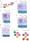
Figure 1.
Figure 1.
Intersubunit interactions of the S. pombe Atg1 core complex. (A) A schematic of the S. pombe Atg1 complex subunits: the CTD of Atg1 (gray), the HORMA domain (pink), and CTD of Atg13 (pink), full-length Atg17 (yellow) and Atg101 (green). Panels (B to E) show results of coprecipitation experiments performed by co-expressing one of the proteins of interest with a His-tag and another protein with a GST-tag or the GST tag alone. The Commassie Brilliant Blue-stained gel (top panel) shows the GST construct inputs (cell lysate) as well as the samples precipitated by glutathione beads (affinity isolate [pull down]). The GST-tagged baits are indicated by an asterisk. We loaded equal amounts of GST-tagged protein except in the case of GST-Atg13CTD co-expressing with His-Atg17, in which we could only detect a faint band of GST-Atg13CTD even after 10 times the amount of cells were used. For our western blot analyses, we loaded equal amounts of cell lysate and twice as much of the affinity isolate, with the exception of GST-Atg1CTD, His-Atg13CTD (bottom panel C) and GST-Atg13CTD, His-Atg1CTD (bottom panel D) where half the volume of the cell lysate was loaded due to saturation of western blot signals. (B) His-Atg17 was co-expressed either with GST or GST-tagged Atg1CTD, Atg13CTD, or Atg101. (C) His-Atg13CTD was co-expressed either with GST, GST-tagged Atg1CTD, or Atg101. (D) His-Atg1CTD was co-expressed either with GST or GST-tagged Atg13(476-545), Atg13CTD, or Atg101. (E) His-Atg101 was co-expressed either with GST, GST-tagged Atg13HORMA (Atg13H), or GST-tagged Atg1CTD. (F) Schematic comparing the subunit interactions between the fission (left panel) and budding (right panel) yeasts Atg1 complex.
Figure 2.
Figure 2.
Negative stain EM analysis of S. pombe His-MBP-Atg17. (A) Representative class averages obtained from classification of 5,922 negatively-stained His-MBP-tagged S. cerevisiae Atg17 particles showed that S. cerevisiae Atg17 can adopt 3 major conformations: S-shaped, asymmetric, and extended. Side length of each panel is 64 nm. (B) Representative class averages obtained from the classification of 5,382 negatively-stained His-MBP-tagged S. pombe Atg17 particles showed that this protein adopts a rigid rod-shape conformation. Side length of each panel is 64 nm. (C) Distribution of junction-to-junction lengths of S. pombe Atg17 showing that most particles have lengths consistent with S. cerevisiae Atg17-Atg31-Atg29 with 2 minor populations having shorter lengths.
Figure 3.
Figure 3.
S. pombe Atg17 binds S. cerevisiae Atg29 and Atg31 but cannot complement S. cerevisiae Atg17. (A) His-tagged S. pombe Atg17 was co-expressed either with GST or GST-tagged S. pombe Atg13CTD, S. cerevisiae Atg29, or S. cerevisiae Atg31. Commassie Brilliant Blue-stained gel (top panel) shows the GST construct inputs. Glutathione resin was used for precipitation; 25% of each affinity isolate was loaded. Western blot (bottom panel) was probed using anti-His antibody; 10% of the affinity isolate (pull down) was loaded for both the GST construct alone and with S. cerevisiae Atg29, while 17% was loaded for the S. pombe Atg13CTD construct and only 0.6% loaded for the S. cerevisiae Atg31 construct. (B) S. pombe Atg17 complementation assay using the Pho8Δ60 activity to measure autophagy in S. cerevisiae. The wild-type strain was transformed with an empty vector, while the atg17Δ strain was transformed with either an empty vector or vectors harboring S. cerevisiae (Sc) Atg17, S. pombe (Sp) Atg17, HA-SpAtg17 or GFP-SpAtg17. Cells were grown in nutrient-rich medium (SMD-Ura) and then shifted to nitrogen starvation medium (SD-N) for 4 h. Phosphatase activity was measured and plotted. Experiments were performed in triplicate. (C) His-tagged S. pombe Atg17 was co-expressed either with GST or GST-tagged S. pombe Atg13CTD or full-length S. cerevisiae Atg13 (ScAtg13FL). Commassie Brilliant Blue-stained gel (top panel) shows the GST construct inputs. Glutathione resin was used for precipitation; 25% of each affinity isolate was loaded. Western blot (bottom panel) was probed using anti-His antibody where 10% of the affinity isolate was loaded. (D) S. cerevisiae Atg17 failed to complement the autophagy defect of S. pombe atg17Δ. Autophagy in S. pombe was assayed by monitoring CFP-Atg8 cleavage. Coomassie Brilliant Blue (CBB) staining of the PVDF membrane was used as a loading control. (E) The wild-type (WLY176) S. cerevisiae strain was transformed with an empty vector, while the S. cerevisiae atg29Δ atg31Δ (XLY160) strain was transformed with either an empty vector, or vector harboring a gene encoding HA-SpAtg101. These cells were cultured to mid-log phase in SMD-Trp before they were shifted to nitrogen starvation medium (SD-N) for 3 h. Samples were collected and the Pho8Δ60 assay was performed. The value of starved wild-type cells was set to 100% and other values were normalized. The error bars indicate the standard deviation (SD) of 3 independent experiments. (F) The S. cerevisiae atg17Δ (XLY134) or atg17Δ ATG31-PA (XLY159) strains were transformed with either pCu(416)-GFP-ScAtg17 or pCu(416)-GFP-SpAtg17. These cells were cultured to mid-log phase in SMD-Ura before they were shifted to nitrogen starvation medium (SD-N) for 3 h. Cell lysates were prepared and incubated with IgG-Sepharose for affinity isolation. The total lysates and eluted proteins were analyzed using SDS-PAGE and detected with monoclonal anti-YFP antibody and an antibody that binds to PA. S.E., short exposure; L.E., long exposure.

References

    1. Mizushima N, Yoshimori T, Ohsumi Y. The role of Atg proteins in autophagosome formation. Annu Rev Cell Dev Biol. 2011;27:107-32. https://doi.org/10.1146/annurev-cellbio-092910-154005. PMID:21801009 - DOI - PubMed
    1. Reggiori F, Klionsky DJ. Autophagic processes in yeast: Mechanism, machinery and regulation. Genetics. 2013;194:341–61. https://doi.org/10.1534/genetics.112.149013. PMID:23733851 - DOI - PMC - PubMed
    1. Ragusa MJ, Stanley RE, Hurley JH. Architecture of the Atg17 complex as a scaffold for autophagosome biogenesis. Cell. 2012;151:1501–12. https://doi.org/10.1016/j.cell.2012.11.028. PMID:23219485 - DOI - PMC - PubMed
    1. Shintani T, Klionsky DJ. Autophagy in health and disease: A double-edged sword. Science. 2004;306:990–5. https://doi.org/10.1126/science.1099993. PMID:15528435 - DOI - PMC - PubMed
    1. Xie Z, Klionsky DJ. Autophagosome formation: Core machinery and adaptations. Nat Cell Biol. 2007;9:1102–9. https://doi.org/10.1038/ncb1007-1102. PMID:17909521 - DOI - PubMed

Publication types

MeSH terms

Substances