. 2018 Feb;43(3):665-672.
doi: 10.1038/npp.2017.235. Epub 2017 Oct 4.

Activation of Nigrostriatal Dopamine Neurons during Fear Extinction Prevents the Renewal of Fear

Affiliations

Activation of Nigrostriatal Dopamine Neurons during Fear Extinction Prevents the Renewal of Fear

Courtney A Bouchet et al. Neuropsychopharmacology. 2018 Feb.

Abstract

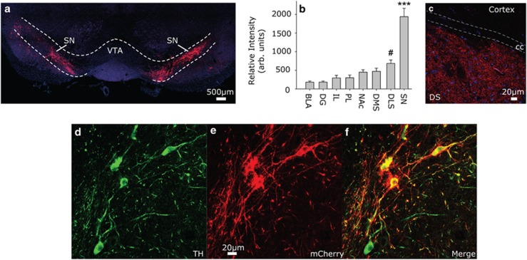
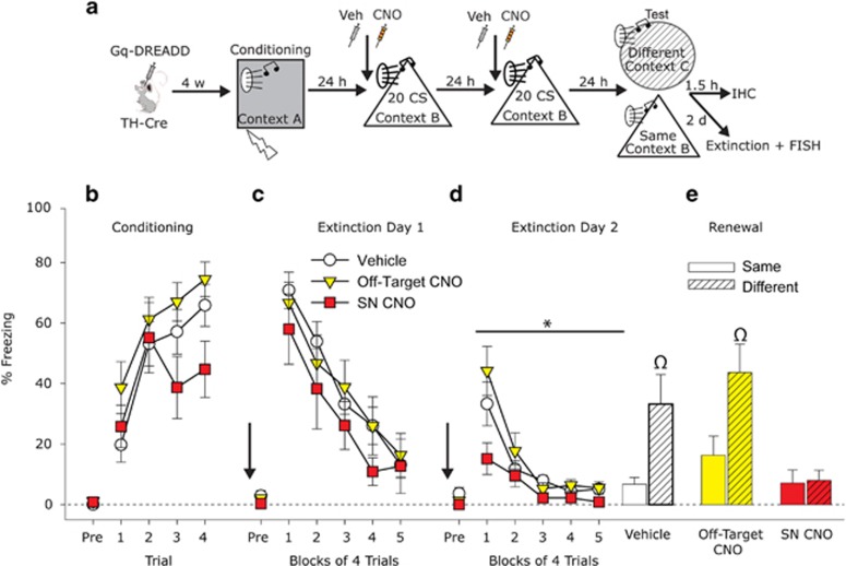
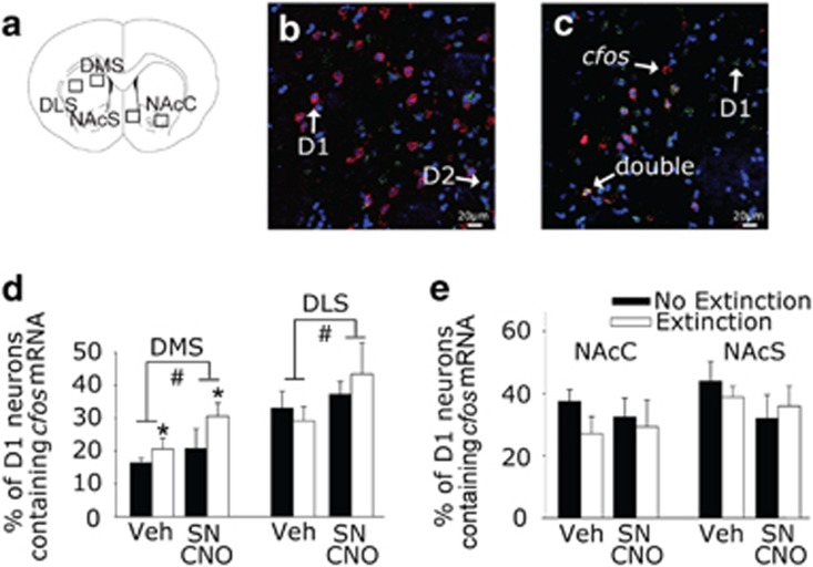
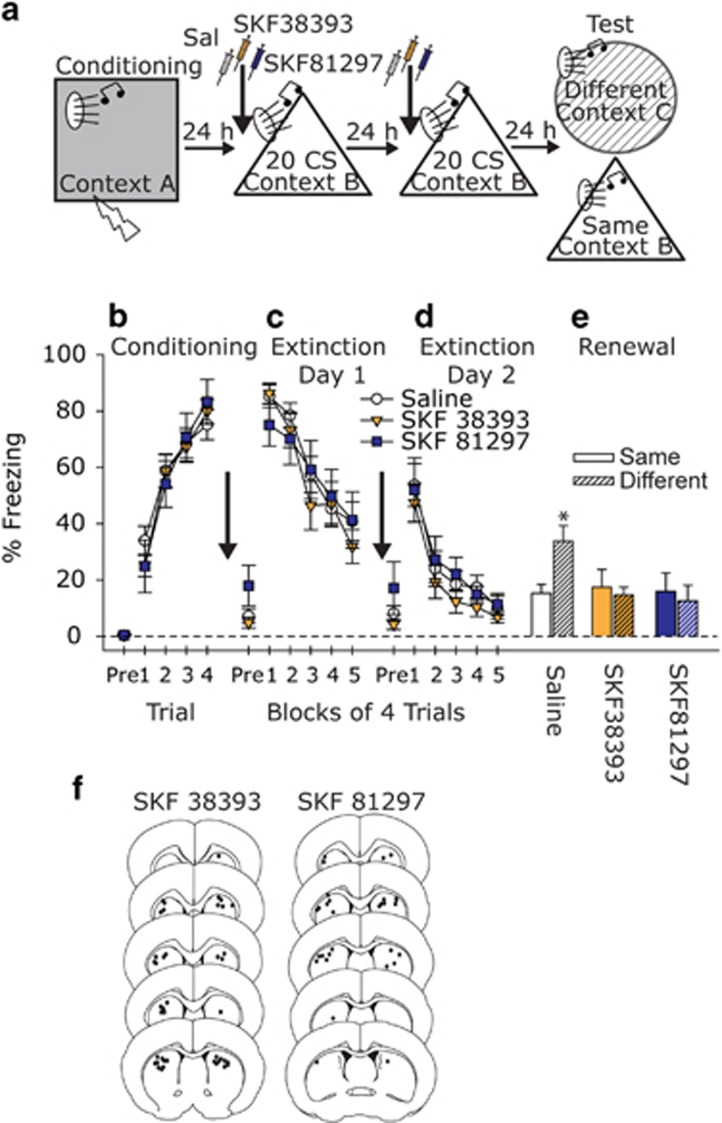
Manipulations that increase dopamine (DA) signaling can enhance fear extinction, but the circuits involved remain unknown. DA neurons originating in the substantia nigra (SN) projecting to the dorsal striatum (DS) are traditionally viewed in the context of motor behavior, but growing data implicate this nigrostriatal circuit in emotion. Here we investigated the role of nigrostriatal DA in fear extinction. Activation of SN DA neurons with designer Gq-coupled receptors exclusively activated by designer drugs (Gq-DREADD) during fear extinction had no effect on fear extinction acquisition, but enhanced fear extinction memory and blocked the renewal of fear in a novel context; a pattern of data paralleled by cFos expression in the central amygdala. D1 receptors in the DS are a likely target mediating the effects of SN DA activation. D1-expressing neurons in the medial DS (DMS) were recruited during fear extinction, and Gq-DREADD-induced DA potentiated activity of D1-expressing neurons in both the DMS and the lateral DS (DLS). Pharmacological activation of D1 receptors in the DS did not impact fear extinction acquisition or memory, but blocked fear renewal in a novel context. These data suggest that activation of SN DA neurons and DS D1 receptors during fear extinction render fear extinction memory resistant to the disrupting effects of changes in contextual contingencies, perhaps by recruiting habitual learning strategies involving the DLS. Nigrostriatal DA thus represents a novel target to enhance long-term efficacy of extinction-based therapies for anxiety and trauma-related disorders.


Figures

Figure 1
Figure 1
Expression of viral Gq-coupled designer receptors exclusively activated by designer drugs (Gq-DREADD). (a) Representative photomicrograph depicting bilateral mCherry (red) in the substantia nigra (SN). Blue is 4',6-diamidino-2-phenylindole (DAPI). (b) mCherry intensity in SN and terminal regions including basolateral amygdala (BLA), dentate gyrus (DG), infralimbic cortex (IL), prelimbic cortex (PL), nucleus accumbens (NAc), dorsal medial striatum (DMS), and dorsal lateral striatum (DLS). (c) Representative photomicrograph of terminal mCherry expression in the dorsal striatum (DS). (d) Tyrosine hydroxylase (TH) immunoreactivity in the SN, (e) mCherry autofluorescence in the SN, (f) merge of TH and mCherry. Data presented represent mean±SEM. #P<0.01 relative to BLA, DG, IL, and PL; ***p<0.0001 relative to all other groups.
Figure 2
Figure 2
Effect of activation of substantia nigra (SN) dopamine neurons on fear extinction and fear renewal. (a) Experimental design. TH-Cre rats received injections of a viral vector containing a construct coding for a Cre-dependent Gq-coupled designer receptor exclusively activated by a designer drug (Gq-DREADD). Rats were exposed to auditory fear conditioning, followed by 2 consecutive days of auditory fear extinction training preceded by administration of Vehicle (Veh) or Clozapine-N-Oxide (CNO; 1 mg/kg). (b) Levels of freezing during fear conditioning. (c,d) Levels of freezing during fear extinction training days. Arrows indicate Veh or CNO administration 30 min before fear extinction. (e) Levels of freezing during the renewal test. Rats in the Vehicle and Off-Target CNO groups exhibited an increase in fear in response to the tone when placed into a novel context (Different) as compared with those placed into the same context as extinction was learned (Same). This fear renewal was absent in the SN CNO group. Data presented represent mean±SEM. *P=0.05 SN CNO compared with Vehicle and Off-Target CNO groups; Ωp<0.05 relative to all other groups except each other.
Figure 3
Figure 3
Double fluorescent in situ hybridization (FISH) labeling of cfos and D1 mRNAs after fear extinction. TH-Cre rats that received intra-SN injections of a viral vector containing a construct coding for a Gq-coupled designer receptor exclusively activated by a designer drug were injected with either Vehicle (Veh) or Clozapine-N-Oxide (SN CNO) and then were either left in their home cages (No Extinction) or were exposed to auditory fear extinction (Extinction) 30 min later. (a) Coronal section modified from the Paxinos and Watson rat brain atlas (Paxinos and Watson, 1998) showing the regions counted. (b) Representative photomicrograph from dorsal medial striatum (DMS) showing D1 (red) and D2 (green) mRNA expression. Blue is 4',6-diamidino-2-phenylindole (DAPI). (c) Representative photomicrograph from the DMS showing D1 (green) and cfos (red) mRNA expression. (d,e) Percent of D1 neurons containing cfos mRNA within the DMS, dorsal lateral striatum (DLS), nucleus accumbens core (NAcC), and nucleus accumbens shell (NAcS). Data presented represent mean±SEM. *P<0.05 main effect of fear extinction; #p<0.05 main effect of drug.
Figure 4
Figure 4
The effects of D1 agonists in the DS during fear extinction on fear extinction memory and fear renewal. (a) Experimental design. Rats received injections of Saline or D1 agonists (SKF 38393 or SKF 81297; 1 μl/hemisphere) into the DS before fear extinction learning, and then were placed drug free into either the familiar fear extinction context (Same) or a novel context (Different) to test for fear renewal. (b) Levels of freezing during auditory fear conditioning. (c,d) Levels of freezing during two sequential days of auditory fear extinction training in the presence of either Saline or D1 agonist. (e) Levels of freezing during the fear renewal test. (f) Black dots represent location of cannulae tips in the dorsal striatum. Data presented represent mean±SEM. *P<0.05 compared with all other groups.

Similar articles

Cited by

References

    1. Abraham AD, Neve KA, Lattal KM (2014). Dopamine and extinction: a convergence of theory with fear and reward circuitry. Neurobiol Learn Mem 108: 65–77. - PMC - PubMed
    1. Abraham AD, Neve KA, Lattal KM (2016). Activation of D1/5 dopamine receptors: a common mechanism for enhancing extinction of fear and reward-seeking behaviors. Neuropsychopharmacology 41: 2072–2081. - PMC - PubMed
    1. Agnoli L, Mainolfi P, Invernizzi RW, Carli M (2013). Dopamine D1-like and D2-like receptors in the dorsal striatum control different aspects of attentional performance in the five-choice serial reaction time task under a condition of increased activity of corticostriatal inputs. Neuropsychopharmacology 38: 701–714. - PMC - PubMed
    1. Alexander GM, Rogan SC, Abbas AI, Armbruster BN, Pei Y, Allen JA et al (2009). Remote control of neuronal activity in transgenic mice expressing evolved G protein-coupled receptors. Neuron 63: 27–39. - PMC - PubMed
    1. Badrinarayan A, Wescott SA, Vander Weele CM, Saunders BT, Couturier BE, Maren S et al (2012). Aversive stimuli differentially modulate real-time dopamine transmission dynamics within the nucleus accumbens core and shell. J Neurosci 32: 15779–15790. - PMC - PubMed

Publication types

MeSH terms

LinkOut - more resources