. 2017 Oct 13;45(18):10481-10491.
doi: 10.1093/nar/gkx676.

A sequence-specific core promoter-binding transcription factor recruits TRF2 to coordinately transcribe ribosomal protein genes

Affiliations

A sequence-specific core promoter-binding transcription factor recruits TRF2 to coordinately transcribe ribosomal protein genes

Douglas G Baumann et al. Nucleic Acids Res. .

Abstract

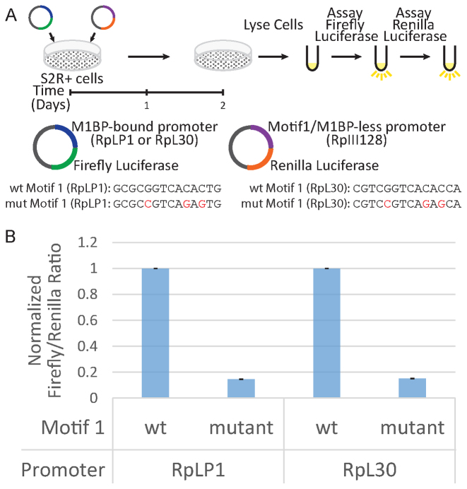
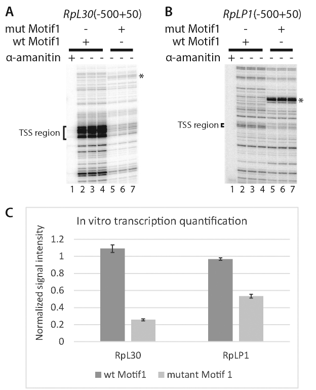
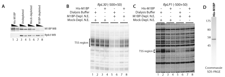
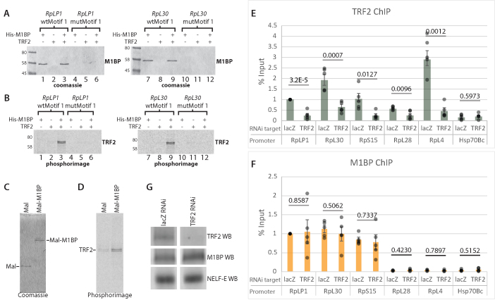
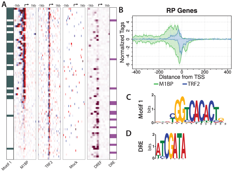
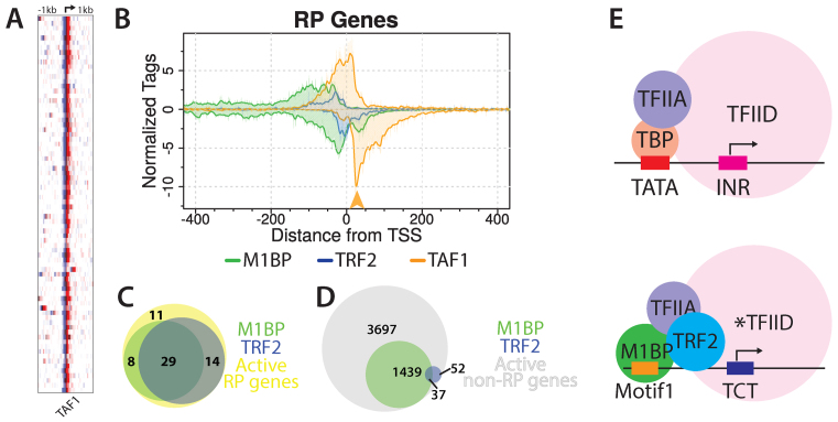
Ribosomal protein (RP) genes must be coordinately expressed for proper assembly of the ribosome yet the mechanisms that control expression of RP genes in metazoans are poorly understood. Recently, TATA-binding protein-related factor 2 (TRF2) rather than the TATA-binding protein (TBP) was found to function in transcription of RP genes in Drosophila. Unlike TBP, TRF2 lacks sequence-specific DNA binding activity, so the mechanism by which TRF2 is recruited to promoters is unclear. We show that the transcription factor M1BP, which associates with the core promoter region, activates transcription of RP genes. Moreover, M1BP directly interacts with TRF2 to recruit it to the RP gene promoter. High resolution ChIP-exo was used to analyze in vivo the association of M1BP, TRF2 and TFIID subunit, TAF1. Despite recent work suggesting that TFIID does not associate with RP genes in Drosophila, we find that TAF1 is present at RP gene promoters and that its interaction might also be directed by M1BP. Although M1BP associates with thousands of genes, its colocalization with TRF2 is largely restricted to RP genes, suggesting that this combination is key to coordinately regulating transcription of the majority of RP genes in Drosophila.


Figures

Figure 1.
Figure 1.
Motif 1 is required for RP gene transcription in cells. (A) Schematic of the RP gene luciferase reporter assay. Mutant RpLP1 and RpL30 have three highly conserved nucleotides in Motif 1 mutated. The wt and mutant Motif 1 sequences for both promoters are shown below the illustration. The RpIII128 promoter lacks Motif 1 and serves an internal control. (B) Firefly/Renilla luciferase ratio of relative light unit measurements. Ratios are normalized to the wt Motif 1 sample for each promoter. Error bars represent standard deviation (n = 3).
Figure 2.
Figure 2.
Motif 1 is required for transcription of RpL30 and RpLP1 in vitro. (A and B) Primer extension analysis of transcripts produced from the (A) RpL30 and (B) RpLP1 promoters (−500 to ∼+50) during transcription in Drosophila embryo nuclear extracts. Transcription reactions lacking α-amanitin were performed in triplicate. The bracketed region encompasses the M1BP-dependent TSS region and a portion of the TCT motif (Parry et al. (11). The M1BP-dependent transcription start sites (TSSs) observed in vitro correspond to the TSSs detected in vivo using PRO-cap (Kwak et al.(30)). (C) Quantification of bracketed TSS region transcripts from (A and B). Error bars represent standard deviation (n = 3). Samples have been normalized to the first wt replicate for each promoter. ‘*’ Denotes an artifact band arising in the Motif 1 region following mutation.
Figure 3.
Figure 3.
M1BP is required for RP gene transcription in vitro. (A) M1BP-probed (top) and Rpb3-probed (bottom) western blot of purified His-M1BP and undepleted, mock-depleted or M1BP-depleted nuclear extracts from 0–12 h embryos. A total of 10 or 30 ng purified His-M1BP and 20 or 60 µg of each extract type was loaded for SDS-PAGE western blot analysis. (B and C) Primer extension analysis of transcripts produced from the (B) RpL30 or (C) RpLP1 promoter in embryo nuclear extracts. The bracketed region denotes the same TSS region described in Figure 2A. Each transcription reaction was performed in duplicate. Lanes 1 and 2: mock-depleted extract supplemented with dialysis buffer. Lanes 3 and 4: mock depleted extract supplemented with recombinant M1BP. Lanes 5 and 6: M1BP-depleted extract supplemented with dialysis buffer. Lanes 7 and 8: M1BP-depleted extract supplemented with enough recombinant M1BP to replace the amount that was immunodepleted. (D) Coomassie-stained SDS-PAGE analysis of purified, N-terminally His-tagged M1BP expressed in and purified from Escherichia coli.
Figure 4.
Figure 4.
M1BP recruits TRF2 to RP gene promoters. (A and B) Immobilized template pulldown assay. 35S-labeled TRF2 was synthesized in vitro using Promega’s TnT T7 Quick Coupled Transcription/Translation System. His-M1BP was expressed in and purified from Escherichia coli. His-M1BP, TRF2 or TRF2 and His-M1BP were added to either RpLP1 or RpL30 template-bound streptavidin dynabeads containing either a wild-type (wt) or mutant Motif 1 sequence (wtMotif 1 or mutMotif 1, respectively). Panel A shows coomassie-stained images from SDS-PAGE analysis of bound protein recovered from RpLP1 and RpL30 immobilized templates. Panel B shows phosphorimager scans of the same gels in panel A. M1BP binds only to the wt Motif 1 template regardless of whether TRF2 is present in the reaction. TRF2 only binds to the wt Motif 1 promoter template when M1BP is present. (C and D) Maltose-binding protein (Mal) and Mal-M1BP fusion pulldown assay. 35S-labeled TRF2 was synthesized as described above and added to either purified and amylose resin-bound Mal or Mal-M1BP. Panel C shows coomassie-stained images from SDS-PAGE analysis of bound protein recovered after binding and washing. Panel D shows the phosphorimage scans of the same gels in panel C. Recovery of TRF2 is increased with the Mal-M1BP fusion compared to the Mal alone. (E and F) RNAi-mediated depletion of TRF2. Following 3 days RNAi knockdown using dsRNA targeting either lacZ (negative control) or TRF2, cells were lysed and ChIP experiments were performed for TRF2 or M1BP. qPCR quantifications were normalized to lacZ RNAi signal at RpLP1. Hsp70Bc lacks both factors and thus serves as a negative control. RpL28 and RpL4 lack M1BP. Individual data points are displayed as gray dots. Each experiment was performed at least four-times. Error bars indicate standard deviation. P-values from two-tailed t-tests are provided for each promoter. (G) Western blots from S2R+ chromatin lysates used for ChIP following 3 days RNAi. The RNAi targets are indicated above the blot images. lacZ RNAi served as a negative control.
Figure 5.
Figure 5.
M1BP and TRF2 co-occupy the majority of RP gene promoters. (A) M1BP, TRF2 and Mock ChIP-exo reads and DNA replication-related element-binding factor (DREF) ChIP-seq reads mapped relative to the TSS of 78 RP genes and sorted by M1BP ChIP-exo reads summed in a 2 kb window centered on the TSS. RP genes having Motif 1 or a DRE within 200 bp of the TSS are indicated in green in the far left panel or purple in the far right panel, respectively. The arrow at the top of each heatmap marks the TSS. Eight paralogs lacking a TCT motif or TRF2 peak have been removed. (B) ChIP-exo analysis of M1BP (green trace) and TRF2 (blue trace) for RP genes. Composite plots in single nucleotide bins were generated from the same RP gene list used for the heatmap in panel A. (C and D) Logo representation of the Motif 1 or DRE position weight matrices (PWM) used to identify genomic motif locations.
Figure 6.
Figure 6.
TAF1 is present at RP gene promoters. (A) Heatmaps display TAF1 ChIP-exo reads from S2R+ cells piled from −1 to +1 kb relative to RP gene TSS. Rows represent individual genes and are sorted by M1BP reads summed in a 2kb window as in Figure 5A. The TSS position is indicated by the arrow at the top of the panel. Duplicate genes were refined to a single isoform by removing eight paralogs lacking TRF2 or a TCT motif in the promoter. (B) Composite plots for M1BP, TRF2 and TAF1 were generated from the same RP gene list used for the heatmap. The orange arrow highlights a TAF1 peak that aligns with an M1BP peak. (C and D) Venn diagrams showing the overlap between M1BP and TRF2 peaks present at all active RP gene promoters (n = 62) or all other active gene promoters (n = 5225). (E) Model depicting M1BP’s recruitment of TRF2 at RP gene promoters. At TATA-containing promoters, TBP-bound TFIID engages with promoter sequences both up- and downstream of the TSS. At the majority of RP gene promoters, which lack both a TATA box and initiator sequence, M1BP and TRF2 bind the core promoter upstream of the TSS. The asterisk(*) denotes a noncanonical TFIID complex proposed to have TRF2 substituting for TBP.

Similar articles

Cited by

References

    1. Perry R.P. Balanced production of ribosomal proteins. Gene. 2007; 401:1–3. - PMC - PubMed
    1. Reja R., Vinayachandran V., Ghosh S., Pugh B.F.. Molecular mechanisms of ribosomal protein gene coregulation. Genes Dev. 2015; 29:1942–1954. - PMC - PubMed
    1. Hu H., Li X.. Transcriptional regulation in eukaryotic ribosomal protein genes. Genomics. 2007; 90:421–423. - PubMed
    1. Tanay A., Regev A., Shamir R.. Conservation and evolvability in regulatory networks: the evolution of ribosomal regulation in yeast. Proc. Natl. Acad. Sci. U.S.A. 2005; 102:7203–7208. - PMC - PubMed
    1. Li X., Zhong S., Wong W.H.. Reliable prediction of transcription factor binding sites by phylogenetic verification. Proc. Natl. Acad. Sci. U.S.A. 2005; 102:16945–16950. - PMC - PubMed

MeSH terms