. 2017 Nov 2;45(19):11193-11212.
doi: 10.1093/nar/gkx723.

Abasic and oxidized ribonucleotides embedded in DNA are processed by human APE1 and not by RNase H2

Affiliations

Abasic and oxidized ribonucleotides embedded in DNA are processed by human APE1 and not by RNase H2

Matilde Clarissa Malfatti et al. Nucleic Acids Res. .

Abstract

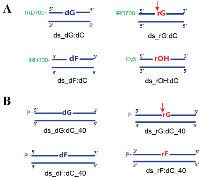
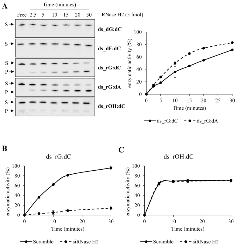
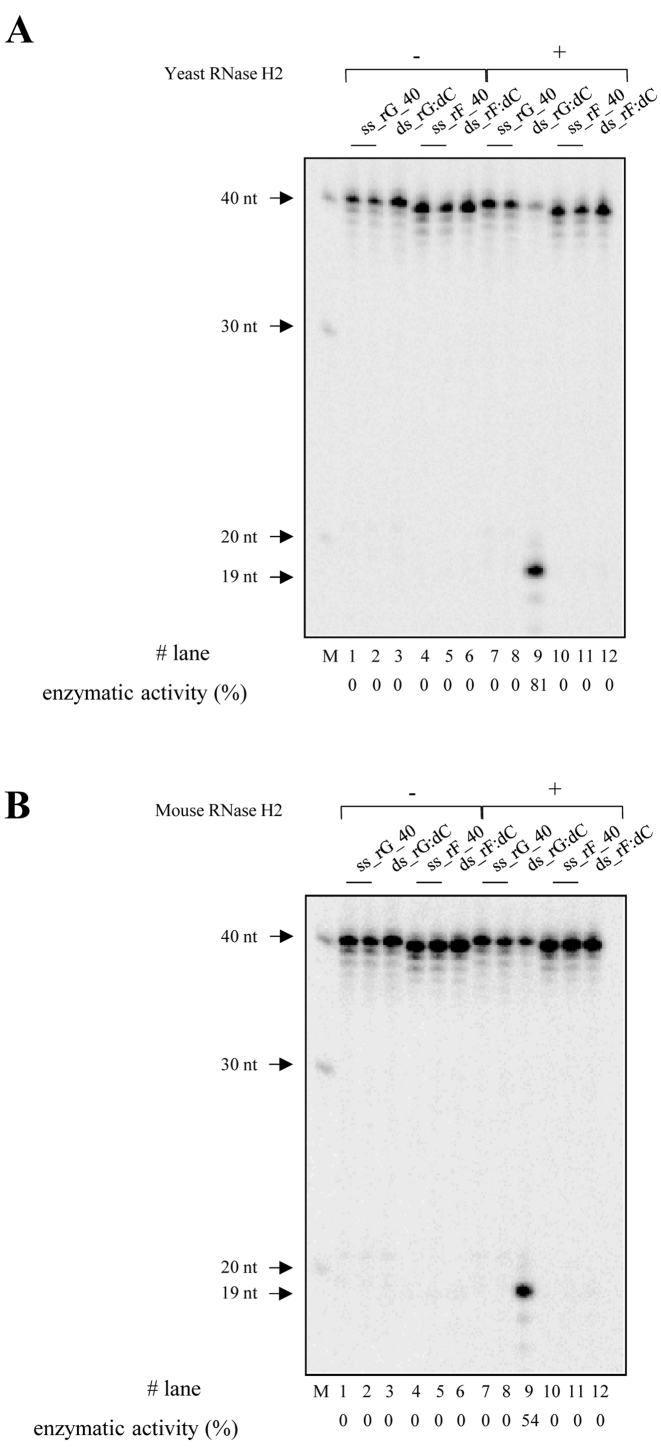
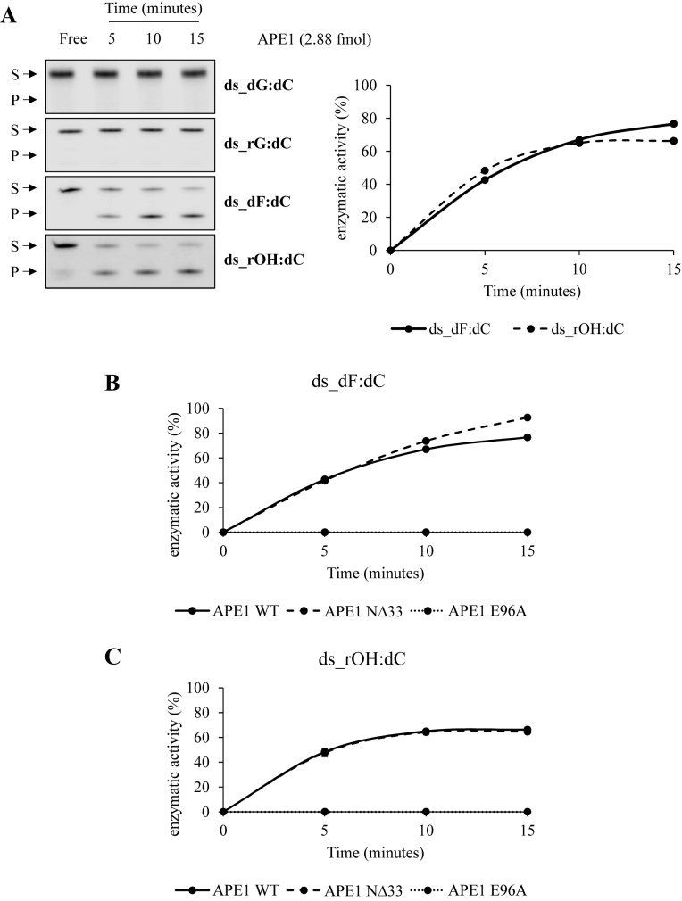
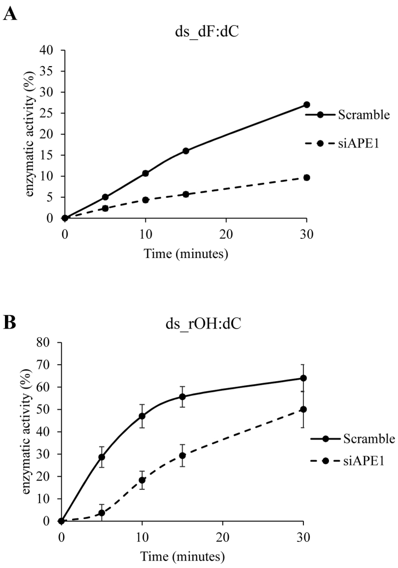
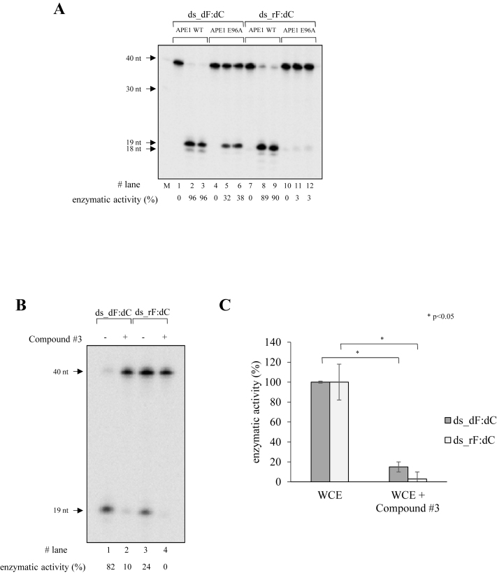
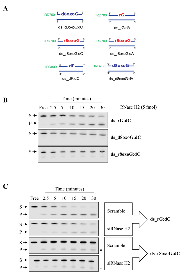
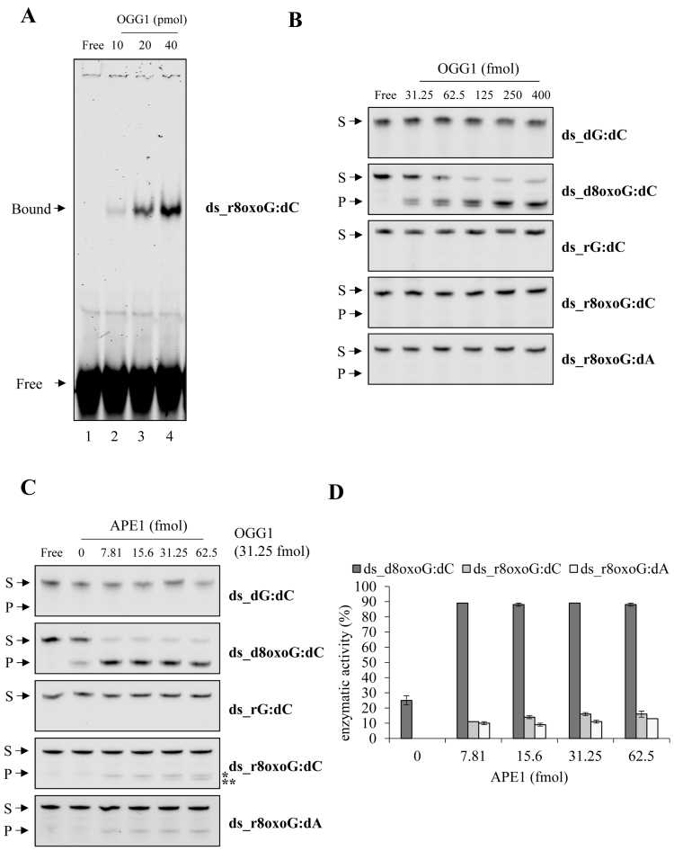
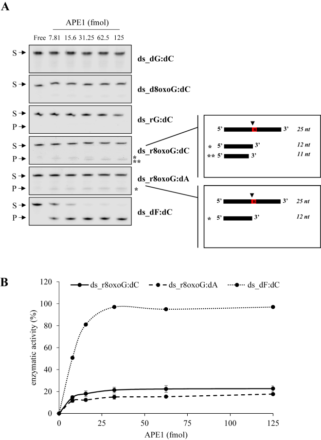
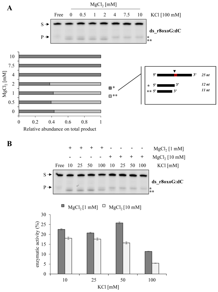
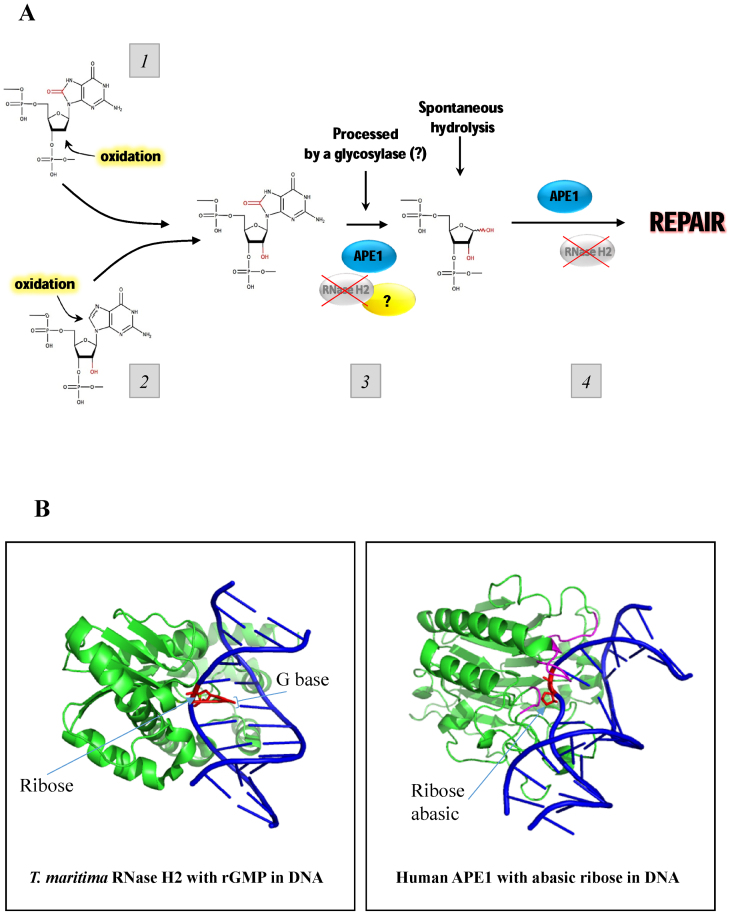
Ribonucleoside 5'-monophosphates (rNMPs) are the most common non-standard nucleotides found in DNA of eukaryotic cells, with over 100 million rNMPs transiently incorporated in the mammalian genome per cell cycle. Human ribonuclease (RNase) H2 is the principal enzyme able to cleave rNMPs in DNA. Whether RNase H2 may process abasic or oxidized rNMPs incorporated in DNA is unknown. The base excision repair (BER) pathway is mainly responsible for repairing oxidized and abasic sites into DNA. Here we show that human RNase H2 is unable to process an abasic rNMP (rAP site) or a ribose 8oxoG (r8oxoG) site embedded in DNA. On the contrary, we found that recombinant purified human apurinic/apyrimidinic endonuclease-1 (APE1) and APE1 from human cell extracts efficiently process an rAP site in DNA and have weak endoribonuclease and 3'-exonuclease activities on r8oxoG substrate. Using biochemical assays, our results provide evidence of a human enzyme able to recognize and process abasic and oxidized ribonucleotides embedded in DNA.


Figures

Figure 1.
Figure 1.
Scheme of substrates used to test cleavage of an rAP site in DNA. DNA nucleotides are in blue, RNA in red. The 5′ and 3′ ends of each DNA strand are indicated. (A) Scheme of double strand (ds) DNA 25-mer substrates (single strand (ss) oligonucleotide annealed to ss_dC containing-complementary oligonucleotide) containing a dGMP, rGMP, dF and 1′ OH abasic rNMP (rOH) site in the 13th position. The ‘IRD700’ in green indicate IRDye 700 phosphoramidite dye tagged at the 5′ end. The ‘IRD800’ in green indicate IRDye 800 phosphoramidite dye tagged at the 5′ end. The ‘Cy5’ in green indicates cyanine dye tagged at the 5′-end of the top strand of the duplex. (B) Scheme of the ds_DNA 40-mer substrate (ss oligonucleotide annealed to its ss complementary oligonucleotide) containing a dGMP, rGMP, dF and rF site in the 20th position. The P in purple indicates radiolabelled 32P at the 5′-end of the top strand of the duplex. The red arrow indicates the cleavage position by RNase H2 5′ to the rGMP site.
Figure 2.
Figure 2.
Human RNase H2 is not able to process an rAP site embedded in a duplex DNA substrate. (A) Representative denaturing polyacrylamide gel of oligonucleotides (25 nM) incision by recombinant human RNase H2 (0.5 nM). The reaction was performed in RNase H2-buffer (20 mM Tris–HCl, 25 mM KCl, 0.1% BSA, 0.01% Tween20, 4 mM MgCl2, pH 7.4) for different time points, expressed in minutes and shown on the top of the figure, at 37°C. ds_dG:dC and ds_dF:dC oligonucleotides were used as negative controls whereas paired and mismatched ds_rG oligonucleotides as positive controls. S indicates the substrate position while P indicates the product position (left). Relative graph illustrating the time-course kinetics activity of the recombinant protein on ds_rG:dC and ds_rG:dA oligonucleotides. Data are expressed as mean ± SD of three independent technical replicas (right). (B) Graph illustrating the time-course kinetics activity of NCE on ds_rG:dC in control and RNase H2-knocked down conditions. Enzymatic reaction was performed at 37°C in RNase H2-buffer with 500 ng of NCE. Data are expressed as mean ± SD of three independent technical replicas. Standard deviation values were always less than 10% of the mean of the experimental points. (C) Graph illustrating the time-course kinetics activity of NCE on ds_rOH:dC oligonucleotide in control and RNase H2-knocked down conditions. Enzymatic reaction was performed at 37°C in RNase H2-buffer with 500 ng of NCE. Data are expressed as mean ± SD of three independent technical replicas. Standard deviation values were always less than 10% of the mean of the experimental points.
Figure 3.
Figure 3.
Mouse and yeast RNase H2 are not able to process an rAP site embedded in a duplex DNA substrate. (A and B) Denaturing PAGE gels showing cleavage result using 10 nM of 40-mer radioactive substrate containing an rG or an rF site, without (lanes 1–6) or with (lanes 7–12) 10 nM of yeast RNase H2 protein (A) or 10 nM of mouse RNase H2 protein (B). All reactions were carried out at 37°C for 1 h in yeast/mouse reaction 1× buffer (15 mM Tris–HCl pH 8.0, 50 mM NaCl, 10 mM MgCl2, 5% glycerol, 1 mM DTT and 0.1 mg/mL BSA). M indicates the DNA ladder and the black arrows on the left of each panel show specific band sizes. Lanes 1,7 have ss-substrate containing rG (ss_rG_40), lanes 2,8 have ss-substrate containing ss_rG_40 that is cooled slowly at room temperature to demonstrate the absence of any self-annealing structures, and lanes 3,9 show the ds-substrate containing rG (ds_rG_dC). Lanes 4,10 have ss-substrate containing abasic ribo site (ss_rF_40), lanes 5,11 have ss_rF_40 that is cooled slowly in room temperature to observe any self-annealing; and lanes 6,12 have ds substrate containing abasic ribo site (ds_rF:dC). The percentages of cleavage of each reaction are displayed below the images as enzymatic activity (%).
Figure 4.
Figure 4.
Human APE1 efficiently processes an rAP site embedded in a duplex DNA substrate. (A) Representative denaturing polyacrylamide gel of oligonucleotides (0.25 μM) incision by recombinant human APE1 (0.288 nM). The reaction was performed in APE1-buffer (20 mM Tris–HCl, 100 mM KCl, 0.1% BSA, 0.01% Tween20, pH 7.4) for different time points, expressed in minutes and shown on the top of the figure, at 37°C. ds_dG:dC and ds_rG:dC oligonucleotides were used as negative controls, whereas ds_dF:dC oligonucleotide as positive control. S indicates the substrate position, while P indicates the product position (left). Relative graph illustrating the time-course kinetics activity of the recombinant protein on ds_dF:dC and ds_rOH:dC oligonucleotides. Data are expressed as mean ± SD of three independent technical replicas. Standard deviation values were always <10% of the mean of the experimental points (right). (B) Graph illustrating the time-course kinetics activity of APE1 mutants on ds_dF:dC oligonucleotide. Data are expressed as mean ± SD of three independent technical replicas. Standard deviation values were always <10% of the mean of the experimental points. (C) Graph illustrating the time-course kinetics activity of APE1 mutants on ds_rOH:dC oligonucleotide. Data are expressed as mean ± SD of three independent technical replicas. Standard deviation values were always <10% of the mean of the experimental points.
Figure 5.
Figure 5.
APE1 knock down in human cells impairs the processing of an rAP site embedded in a duplex DNA substrate. (A) Graph illustrating the time-course kinetics activity of NCE on ds_dF:dC in control and APE1-knocked down conditions. Data are expressed as mean ± SD of three independent technical replicas. Standard deviation values were always less than 10% of the mean of the experimental points. (B) Graph illustrating the time-course kinetics activity of NCE on ds_rOH:dC oligonucleotide in control and APE1-knocked down conditions. Enzymatic reaction was performed at 37°C in APE1-buffer with 10 ng of NCE. Data are expressed as mean ± SD of three independent technical replicas.
Figure 6.
Figure 6.
Recombinant human APE1 efficiently processes a tetrahydrofuran ribonucleotide mimicking an abasic residue site (rF) embedded in a duplex DNA substrate. (A) Cleavage result of 10 nM of 32P double-stranded oligonucleotides ds_dF:dC or ds_rF:dC substrates with different concentrations of APE1 WT protein (lanes 1–3 and 7–9) and mutant APE1 E96A protein (lanes 4–6 and 10–12) at 37°C for 1 h in APE1-reaction 1× buffer containing 50 mM Tris–HCl pH 7.5, 50 mM KCl, 10 mM MgCl2, 0.001 mg/mL BSA and 0.05% Triton X-100. First lane on the left, M is a ssDNA ladder (barely visible) and the black arrows on the left show specific band sizes. Both oligonucleotides were incubated with 0 nM (lanes 1,4,7,10), 5 nM (lanes 2,5,8,11) or 10 nM (lanes 3,6,9,12) of APE1 WT protein (lanes 1–3 and 7–9) or mutant E96A protein (lanes 4–6 and 10–12), respectively. The percentages of cleavage of each reaction are displayed below the image as enzymatic activity (%). (B) Cleavage result of 10 nM ds_dF:dC or ds_rF:dC radioactive substrates using 12.5 ng of whole HeLa cell extracts untreated (lanes 1, 3) and treated (lanes 2, 4) with 0.20 nM of Compound #3, a specific APE1-endonuclease inhibitor, at 37°C for 10 min. The black arrows on the left of the gel image indicate the size of uncut and cut substrates following denaturation. The cleavage percentage of this experiment is displayed below the image as enzymatic activity (%). (C) Histograms showing data from four independent experimental replicas shown in panel (B) with ranges as bars. P values of <0.05 are marked by asterisk. WCE, whole cell extracts.
Figure 7.
Figure 7.
Human RNase H2 is not able to process an r8oxoG site embedded in a duplex DNA substrate. (A) Scheme of ds_DNA 25-mer substrates (single strand (ss) oligonucleotide annealed to ss_dC containing- or ss_dA containing-complementary oligonucleotide) containing a d8oxoG, r8oxoG and dF site in the 13th position. The ‘IRD700’ and ‘IRD800’ in green indicates IRDye 800 phosphoramidite and IRDye700 phosphoramidite dye tagged at the 5′ end. (B) Representative denaturing polyacrylamide gel of oligonucleotides (25 nM) incision by human recombinant RNase H2 (5 fmol). Reaction was performed in RNase H2-buffer (20 mM Tris–HCl, 25 mM KCl, 0.1% BSA, 0.01% Tween20, 4 mM MgCl2, pH 7.4) at 37°C. ds_rG:dC and ds_d8oxoG:dC oligonucleotides were used as positive and negative controls, respectively. Time points are shown on the top of the figure. S indicates the substrate position, while P indicates the product position. (C) Representative denaturing polyacrylamide gel of oligonucleotides (0.25 μM) incision by nuclear HeLa cell extracts (NCE). In order to discriminate the activity of RNase H2, 500 ng of NCE in which RNase H2 expression was previously knocked down through specific siRNA (indicated as siRNase H2) were tested in comparison to control cells (Scramble) at different time points (minutes), shown on top of the figure. The endoribonuclease activity detected for r8oxoG oligonucleotide was indicated with an asterisk on the right side of each panel. S indicates the substrate position while P indicates the product position.
Figure 8.
Figure 8.
Human OGG1 has neither lyase nor glycosylase activities on r8oxoG-containing oligonucleotide. (A) Representative native EMSA polyacrylamide gel of OGG1 binding on ds_r8oxoG:dC oligonucleotide (25 nM) is shown. The ‘Bound’ arrow indicates the retarded complex between OGG1 and the probe whereas the ‘Free’ arrow the unbound substrate. Amounts of OGG1 protein, expressed in pico moles, are shown on the top of the figure. Reactions were performed as explained in ‘Materials and Methods’ section. (B) Representative denaturing polyacrylamide gel of lyase activity of human OGG1 on different duplex DNA oligonucleotides (25 nM). Doses of OGG1 protein expressed in femto moles are shown on the top of the figure. Reactions were performed in OGG1-buffer (20 mM Tris–HCl, 100 mM KCl, 0.1% BSA, 0.01% Tween20, pH 7.4) at 37°C for 30 min. S indicates the substrate position, while P indicates the product position. (C) Representative denaturing polyacrylamide gel of incision by different doses of APE1 co-incubated with a fixed amount of OGG1 (3.125 nM) on different duplex DNA oligonucleotides (25 nM) in order to investigate glycosylase activity of OGG1. Different doses of APE1 protein, expressed in femto moles, are shown on top of the figure. Reactions were performed in a buffer containing 20 mM Tris–HCl, 100 mM KCl, 0.1% BSA, 0.01% Tween20, pH 7.4 at 37°C for 30 min. S indicates the substrate position while P indicates the product position. Moreover, at the right of the panel, a longer product of about 12 nucleotides is indicated by an asterisk whereas a smaller one of 11 nucleotides is indicated by a double asterisk. (D) Histograms represent the dose response of OGG1 glycosylase activity on ds_d8oxoG:dC and paired and mismatched ds_r8oxoG oligonucleotides. ds_d8oxoG oligonucleotide was used as a positive control whereas ds_dG:dC and ds_rG:dC oligonucleotides were used as negative controls. The activity is reported as percentage of substrate converted to product. Data are expressed as mean ± SD of three independent technical replicas.
Figure 9.
Figure 9.
Human APE1 shows a weak endo- and a 3′-exonuclease activities on r8oxoG substrate. (A) Representative denaturing polyacrylamide gel of APE1 incision on different duplex DNA oligonucleotides (25 nM), in which ds_dF:dC oligonucleotide was used as a positive control, whereas ds_dG:dC and ds_rG:dC oligonucleotides were used as negative controls. The doses of APE1 protein used, expressed in femto moles, are shown on the top of the figure. On the right side, a schematic representation of the cleavage products, showing the position of the ribonucleotide (red box with R) embedded in the DNA oligonucleotide and the APE1 cleavage on it, producing a longer product of about 12 nucleotides (*) and a smaller one of 11 nucleotides (**). Reactions were performed in APE1-buffer containing 20 mM Tris–HCl, 100 mM KCl, 0.1% BSA, 0.01% Tween20, pH 7.4 for 30 min at 37°C. S indicates the substrate position while P indicates the product position. (B) Relative graph indicates a dose–response APE1 activity on paired and mismatched ds_r8oxoG:dC oligonucleotide in comparison to ds_dF:dC positive control. Data are expressed as mean ± SD of three independent technical replicas.
Figure 10.
Figure 10.
Human APE1 activities on r8oxoG substrate depend on mono- and di-valent cations. (A) Representative denaturing polyacrylamide gel of APE1 (5 nM) incision on ds_r8oxoG:dC oligonucleotide under different MgCl2 concentrations that is expressed in mM (top) performing a reaction long 30 minutes in APE1 buffer. S indicates the substrate position while P indicates the product position. Moreover, at the right of the panel, a longer product of about 12 nucleotides is indicated by an asterisk whereas a smaller one of 11 nucleotides is indicated by a double asterisk Also shown on the right is a schematic representation of the cleavage products, showing the position of the ribonucleotide (red box with R) embedded in the DNA oligonucleotide and the APE1 cleavage on it, producing a longer product of about 12 nucleotides (*) and a smaller one of 11 nucleotides (**). Relative graph shows the ratio between two products obtained as a function of MgCl2 concentration (bottom). (B) Representative denaturing polyacrylamide gel of APE1 (5 nM) incision on ds_r8oxoG:dC oligonucleotide under different KCl concentrations in combination with two different MgCl2 concentrations, 1 mM and 10 mM (top) performing a reaction long 30 min in a buffer containing 20 mM Tris–HCl, 0.1% BSA, 0.01% Tween20, pH 7.4. S indicates the substrate position, while P indicates the product position. Moreover, at the right of the panel, a longer product of about 12 nucleotides is indicated by an asterisk whereas a smaller one of 11 nucleotides is indicated by a double asterisk. Relative graph shows product levels in association with different salts concentrations (bottom).
Figure 11.
Figure 11.
Model for repair of oxidized rNMPs and rAP sites embedded in DNA by APE1. (A) 8oxo-ribonucleotides could be generated in the cellular nucleotide pool or even when they are already incorporated in DNA as a result of an oxidation of the sugar (1) and/or the base (2). After the evidence that human RNase H2 is not able to process an oxidized rNMP embedded in DNA, we found that APE1 shows a weak but significant activity on it (3). Similarly, RNase H2 does not process an rAP embedded in DNA, which could be generated spontaneously or by the r8oxoG processing, and again APE1 possesses a strong activity on this type of damage (4). Because APE1 activity on 8oxo-ribonucleotides in DNA is low, we hypothesize that other proteins (some glycosylases?) may participate in their repair. (B) Structural models with the active site of RNase H2 and APE1 with an rNMP or an rAP. T. maritima (left) and human APE1 (right) is in a complex with DNA having a single rNMP or single abasic residue, respectively. DNA is indicated in blue, while the single rNMP and the abasic residues are shown in red as sticks. The arrows points towards ribose or G base or abasic sites in the panels. Proteins are shown in green except for three of the several regions on APE1 that engulf the abasic sugar which are in magenta. PDB for RNase H2 is 303-F; for APE1 as 1DEW (92).

Similar articles

Cited by

References

    1. Williams J.S., Kunkel T.A.. Ribonucleotides in DNA: origins, repair and consequences. DNA Repair. 2014; 19:27–37. - PMC - PubMed
    1. Koh K.D., Balachander S., Hesselberth J.R., Storici F.. Ribose-seq: global mapping of ribonucleotides embedded in genomic DNA. Nat. Methods. 2015; doi:10.1038/nmeth.3259. - PMC - PubMed
    1. Williams J.S., Lujan S.A., Kunkel T.A.. Processing ribonucleotides incorporated during eukaryotic DNA replication. Nat. Rev. Mol. Cell Biol. 2016; 17:350–363. - PMC - PubMed
    1. Clausen A.R., Zhang S., Burgers P.M., Lee M.Y., Kunkel T.A.. Ribonucleotide incorporation, proofreading and bypass by human DNA polymerase δ. DNA Repair. 2013; 12:121–127. - PMC - PubMed
    1. Randerath K., Reddy R., Danna T.F., Watson W.P., Crane A.E., Randerath E.. Formation of ribonucleotides in DNA modified by oxidative damage in vitro and in vivo. Characterization by 32P-postlabeling. Mutat. Res. 1992; 275:355–366. - PubMed