. 2017 Nov 2;45(19):11401-11412.
doi: 10.1093/nar/gkx766.

Involvement of G-triplex and G-hairpin in the multi-pathway folding of human telomeric G-quadruplex

Affiliations

Involvement of G-triplex and G-hairpin in the multi-pathway folding of human telomeric G-quadruplex

Xi-Miao Hou et al. Nucleic Acids Res. .

Abstract

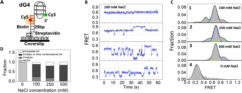
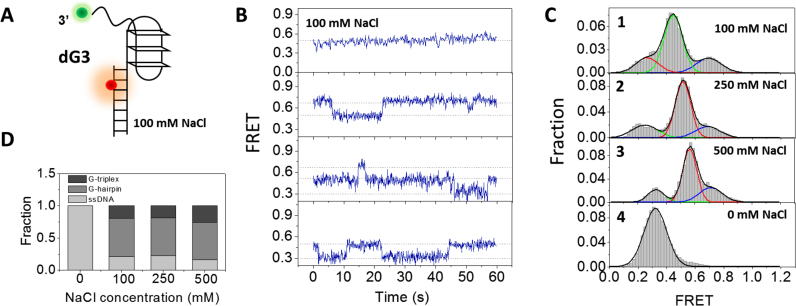
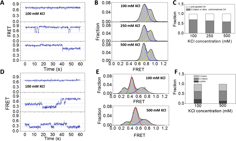
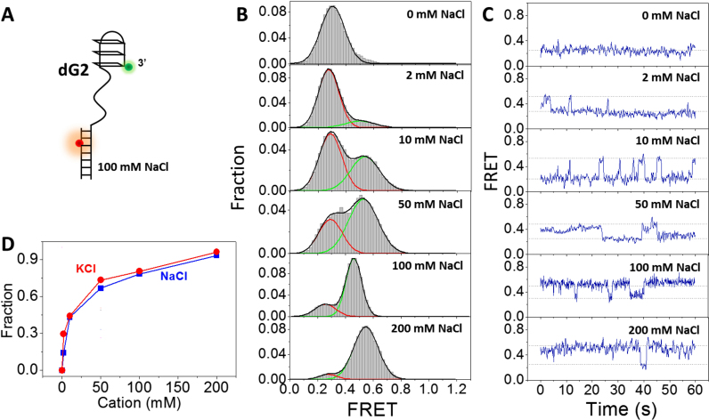
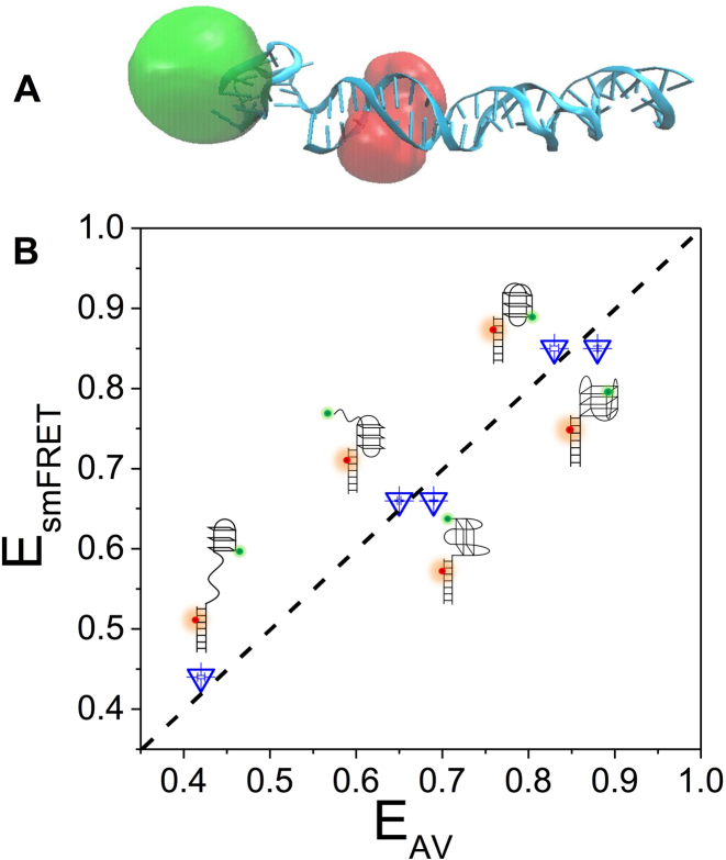
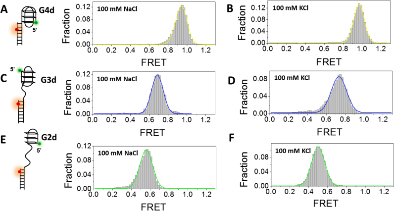
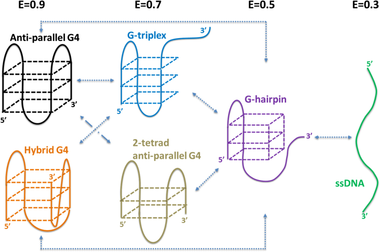
G-quadruplex (G4) can be formed by G-rich DNA sequences that are widely distributed throughout the human genome. Although G-triplex and G-hairpin have been proposed as G4 folding intermediates, their formation still requires further investigation by experiments. Here, we employed single-molecule FRET to characterize the folding dynamics of G4 from human telomeric sequence. First, we observed four states during G4 folding initially assigned to be anti-parallel G4, G-triplex, G-hairpin and unfolded ssDNA. Then we constructed putative intra-strand G-triplex, G-hairpin structures and confirmed their existences in both NaCl and KCl. Further studies revealed those structures are going through dynamic transitions between different states and show relatively weak dependence on cations, unlike G4. Based on those results and molecular dynamics simulations, we proposed a multi-pathway folding mechanism for human telomeric G4. The present work may shed new light on our current understanding about the existence and stability of G4 intermediate states.


Figures

Figure 1.
Figure 1.
Conformational dynamics of G4 at the 3′ end of duplex DNA in NaCl. (A) Schematic representation of experimental set-up. (B) Representative smFRET traces of dG4 in 100 mM NaCl. (C) FRET histograms of dG4 in 20 mM Tris–HCl, pH 8.0 buffer containing different concentrations of NaCl. Multi-peak and single-peak Gaussian distributions were used to fit those histograms. (D) Fractions of different folding structures at increasing concentrations of NaCl, obtained from the area of individual Gaussian peaks in (C).
Figure 2.
Figure 2.
Conformational dynamics of G-triplex at the 3′ end of duplex DNA in NaCl. (A) Design of dG3 substrate. (B) Representative traces of dG3 in 100 mM NaCl. (C) FRET histograms of dG3 in 20 mM Tris–HCl, pH 8.0 buffer containing different concentrations of NaCl. Multi-peak and single-peak Gaussian distributions were used to fit those histograms. (D) The fractions of different folding structures at increasing concentrations of NaCl.
Figure 3.
Figure 3.
Conformational dynamics of G4 and G-triplex at 3′ end of duplex DNA in KCl. (A) Representative traces of dG4 in 100 mM KCl. (B) In 100–500 mM KCl, the FRET histograms of dG4 display two peaks at E0.9 and E0.75. (C) The fractions of different folding structures at increasing concentrations of KCl. (D) Representative traces of dG3 in 100 mM KCl. (E) FRET histograms of dG3 in 20 mM Tris–HCl, pH 8.0 buffer containing different concentrations of KCl. Multi-peak Gaussian distributions were used to fit those histograms. (F) The fractions of different folding structures at increasing concentrations of KCl.
Figure 4.
Figure 4.
Formation of G-hairpin at the 3′ end of ss/dsDNA. (A) Design of dG2 substrate. (B) FRET distributions of dG2 in 0–200 mM NaCl. Single-peak and two-peak Gaussian distributions were used to fit those histograms. (C) Selected FRET traces of dG2 in 0–200 mM NaCl. (D) Fractions of G-hairpin in different concentrations of NaCl and KCl.
Figure 5.
Figure 5.
Molecular dynamics simulations. (A) Selective structure of anti-parallel G4 adjacent to duplex DNA. Donor and acceptor positions were obtained using AV approach and shown as clouds. (B) FRET efficiencies obtained from smFRET experiments (EsmFRET) and AV calculations based on the MD simulations (EAV). Standard errors from Gaussian fitting to obtain the peak values of EsmFRET and EAV are <0.01. The diagonal dashed line indicates the identity line.
Figure 6.
Figure 6.
Formation of G4, G-triplex and G-hairpin at the 5′ end of a duplex DNA in 100 mM NaCl or KCl. (A) smFRET histograms of G4d in 100 mM NaCl has 1 peak at E0.94. (B) smFRET histograms of G4d structures in 100 mM KCl has 1 peak at E0.96. (C) smFRET histograms of G3d structures in 100 mM NaCl has 1 peak at E0.69. (D) smFRET histograms of G3d structures in 100 mM KCl has 1 peak at E0.73. (E) smFRET histograms of G2d structures in 100 mM NaCl has 1 peak at E0.56. (F) smFRET histograms of G-hairpin structures in 100 mM KCl has 1 peak at E0.50.
Figure 7.
Figure 7.
Proposed folding pathway of human telomeric G4 at 3′ end of duplex DNA.

References

    1. Lane A.N., Chaires J.B., Gray R.D., Trent J.O.. Stability and kinetics of G-quadruplex structures. Nucleic Acids Res. 2008; 36:5482–5515. - PMC - PubMed
    1. Bochman M.L., Paeschke K., Zakian V.A.. DNA secondary structures: stability and function of G-quadruplex structures. Nat. Rev. Genet. 2012; 13:770–780. - PMC - PubMed
    1. Maizels N., Gray L.T.. The G4 genome. PLoS Genet. 2013; 9:e1003468. - PMC - PubMed
    1. Todd A.K., Johnston M., Neidle S.. Highly prevalent putative quadruplex sequence motifs in human DNA. Nucleic Acids Res. 2005; 33:2901–2907. - PMC - PubMed
    1. Huppert J.L., Balasubramanian S.. Prevalence of quadruplexes in the human genome. Nucleic Acids Res. 2005; 33:2908–2916. - PMC - PubMed

LinkOut - more resources