. 2017 Nov 2;45(19):11019-11032.
doi: 10.1093/nar/gkx757.

Occurrence and stability of lone pair-π stacking interactions between ribose and nucleobases in functional RNAs

Affiliations

Occurrence and stability of lone pair-π stacking interactions between ribose and nucleobases in functional RNAs

Mohit Chawla et al. Nucleic Acids Res. .

Abstract

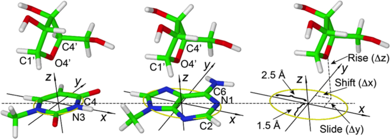
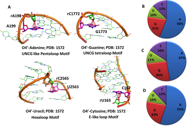
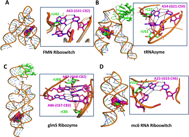
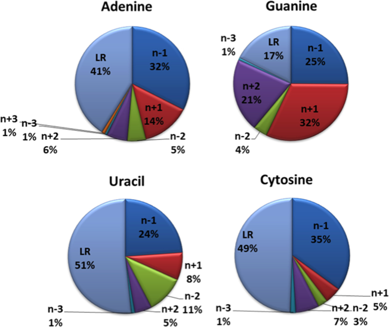
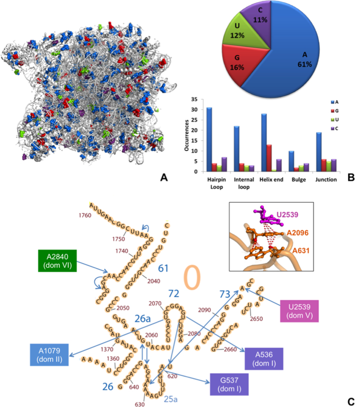
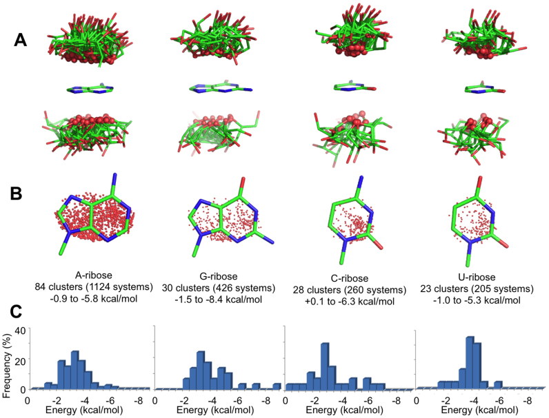
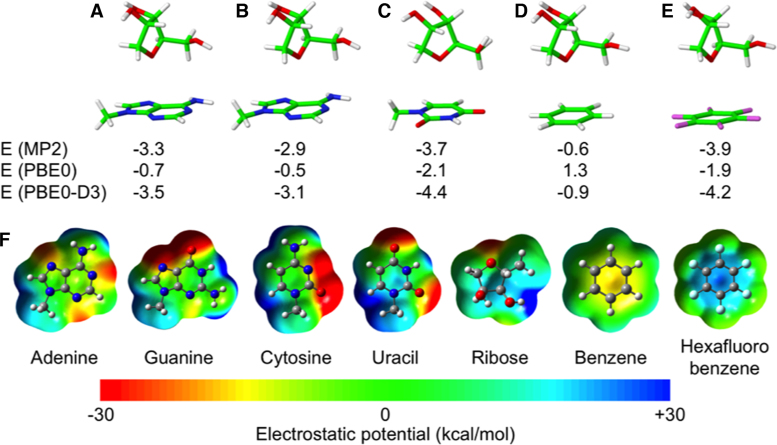
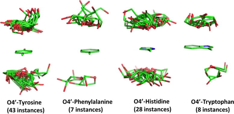
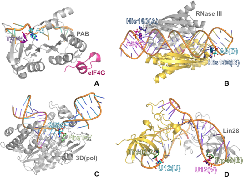
The specific folding pattern and function of RNA molecules lies in various weak interactions, in addition to the strong base-base pairing and stacking. One of these relatively weak interactions, characterized by the stacking of the O4' atom of a ribose on top of the heterocycle ring of a nucleobase, has been known to occur but has largely been ignored in the description of RNA structures. We identified 2015 ribose-base stacking interactions in a high-resolution set of non-redundant RNA crystal structures. They are widespread in structured RNA molecules and are located in structural motifs other than regular stems. Over 50% of them involve an adenine, as we found ribose-adenine contacts to be recurring elements in A-minor motifs. Fewer than 50% of the interactions involve a ribose and a base of neighboring residues, while approximately 30% of them involve a ribose and a nucleobase at least four residues apart. Some of them establish inter-domain or inter-molecular contacts and often implicate functionally relevant nucleotides. In vacuo ribose-nucleobase stacking interaction energies were calculated by quantum mechanics methods. Finally, we found that lone pair-π stacking interactions also occur between ribose and aromatic amino acids in RNA-protein complexes.


Figures

Figure 1.
Figure 1.
Definition of the reference Cartesian frame on the nucleobases and of the shift, slide and rise parameters used to define the position of the O4′ atom of ribose relative to the base. The origin is at the geometrical center of the heterocycle skeleton, the x-axis passing through the N3 atom for pyrimidines and through the middle point of the N1–C2 bond for purines; the y-axis forms a 90° angle with the x-axis, with the C6 atom of purines and the C4 atom of pyrimidines lying in the xy-plane at positive y values; the z-axis is the cross product of the versors (unit vectors indicating the directions) along the x- and y-axes, thus forming a right-handed frame. The yellow circle defines a circle of radius 1.5 Å in the xy-plane of pyrimidines, while the yellow ellipse defines an ellipse in the xy-plane of purines, with minor and major axes equal to 1.5 and 2.5 Å. A ribose and a base are considered to be interacting if the projection of the O4′ atom of the ribose on the xy-plane is within the yellow circle for pyrimidines or the yellow ellipse for purines, with the rise parameter in the –4.0 to +4.0 Å range.
Figure 2.
Figure 2.
(A) Examples of ribose–base stacking contacts involving different nucleobases in different structural motifs. (B–D) Percentage of nucleobases involved in ribose–base stacking interactions in different RNA molecules: (B) nrRNA3.0 (non-redundant RNA structure dataset with a resolution ≤ 3.0 Å); (C) as in B) but not including ribosomal structures; (D) subset of nrRNA3.0 with a resolution ≤ 2.0 Å. Number of occurrences corresponding to A, G, C, U are 1124, 426, 260 and 205 respectively in (B), 374, 207, 126 and 88 in (C) and 87, 36, 19 and 16 in (D).
Figure 3.
Figure 3.
Examples of A-minor motifs, including a ribose-adenine stacking interaction in different RNA molecules. (A) FMN riboswitch (PDB ID: 3F2Q); (B) tRNAzyme (PDB ID: 3CUN), with an adenine stacked between two riboses; (C) glmS Ribozyme (PDB ID: 2NZ4), featuring an A-patch interaction motif (75). (D) mc6 RNA riboswitch (PDB ID: 3LA5), with an adenine giving a tertiary interaction with the G53:C46 base pair.
Figure 4.
Figure 4.
Sequence distance between the ribose(n) and the nucleobase(n±i) moieties involved in stacking interactions, reported for each of the four bases. The residue numbering is in the 5′→3′ direction of the RNA chain. LR refers to the long-range contacts, with a sequence spacing of at least 4 residues (i ≥ 4 or i ≤ 4).
Figure 5.
Figure 5.
ribose–base stacking contacts in 23S rRNA from H. marismortui. (A) 3D representation (PDB ID: 1S72), with blue, red, green and purple spheres representing adenine, guanine, uracil, cytosine bases involved in ribose–base interactions, respectively. (B) Fraction of different nucleobases involved in the interactions (top) and number of occurrences of bases involved in the interactions in different structural motifs (bottom). (C) ribose–base stacking contacts of Domain 0. In the 2D representation (adapted from http://apollo.chemistry.gatech.edu/RibosomeGallery/), the arrows are directed from the riboses to the nucleobases. A 3D representation of the interactions involving nucleotides A631, A2096 and U2539 (on domain V) is also provided.
Figure 6.
Figure 6.
(A) Stick representations of the ribose orientation relative to the base in the 165 structures representative of different interaction clusters, organized by nucleobase identity. The ribose O4′ atoms are shown as small spheres. (B) The projection of the O4′ atom of riboses on the base plane is shown as a red dot. (C) Distribution of the interaction energies calculated for all 165 cluster representatives (kcal/mol).
Figure 7.
Figure 7.
Geometries and energies of model T-shaped geometries corresponding to ribose stacked on top of the baricenter of adenine (A) on top of the center of the 6 membered aromatic ring of adenine (B), uracil (C), benzene (D), hexafluorobenzene (E). (F) electrostatic potential of nucleobases and of bezene and hexafluorobenzene. Electrostatic potentials were mapped on electron density isosurfaces corresponding to a value of 0.0004 atomic units, and are scaled between –30 and 30 kcal/mol. Interaction energies, in kcal/mol, were calculated at the MP2 level and at the DFT level using the PBE0 functional, and at the PBE0-D3 level, which means adding an empirical dispersion energy term to the PBE0 energy.
Figure 8.
Figure 8.
Stick representation of the ribose orientation relative to the side chains of the four aromatic amino acids in the lp–π ribose-amino acid stacking interactions.
Figure 9.
Figure 9.
An example of lp–π ribose-amino acid stacking interactions for each aromatic amino acid: (A) ribose-Tyr stacking contact in a ternary complex between the human translation factors polyadenylate-binding protein-1 (PABP) and eIF4G and a poly(A)(11) RNA (PDB ID: 4F02); (B) ribose-His stacking contact in a complex between A. aeolicus dimeric ribonuclease III (RNase III) and a product double-stranded RNA (dsRNA) (PDB ID: 2EZ6); (C) ribose-Phe stacking contact in the complex between rhinovirus RNA-dependent RNA polymerase (3D(pol)) and synthetic RNA (PDB ID: 4K50); (D) ribose-Trp stacking contact in a complex between mouse dimeric Lin28 and the microRNA (miRNA) let-7 (PDB ID: 3TS0).

References

    1. Hendrix D.K., Brenner S.E., Holbrook S.R.. RNA structural motifs: building blocks of a modular biomolecule. Q. Rev. Biophys. 2005; 38:221–243. - PubMed
    1. Lescoute A., Leontis N.B., Massire C., Westhof E.. Recurrent structural RNA motifs, Isostericity Matrices and sequence alignments. Nucleic Acids Res. 2005; 33:2395–2409. - PMC - PubMed
    1. Djebali S., Davis C.A., Merkel A., Dobin A., Lassmann T., Mortazavi A., Tanzer A., Lagarde J., Lin W., Schlesinger F. et al. . Landscape of transcription in human cells. Nature. 2012; 489:101–108. - PMC - PubMed
    1. Wilusz J.E. Long noncoding RNAs: Re-writing dogmas of RNA processing and stability. Bba-Gene Regul. Mech. 2016; 1859:128–138. - PMC - PubMed
    1. Sponer J., Sponer J.E., Mladek A., Jurecka P., Banas P., Otyepka M.. Nature and magnitude of aromatic base stacking in DNA and RNA: Quantum chemistry, molecular mechanics, and experiment. Biopolymers. 2013; 99:978–988. - PubMed