. 2017 Oct 1;158(10):3235-3248.
doi: 10.1210/en.2017-00469.

Subdomain 2, Not the Transmembrane Domain, Determines the Dimerization Partner of Growth Hormone Receptor and Prolactin Receptor

Affiliations

Subdomain 2, Not the Transmembrane Domain, Determines the Dimerization Partner of Growth Hormone Receptor and Prolactin Receptor

Ying Liu et al. Endocrinology. .

Abstract

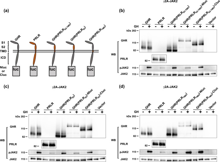
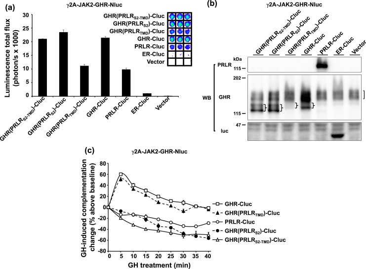
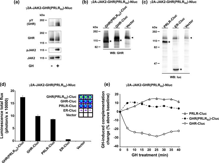
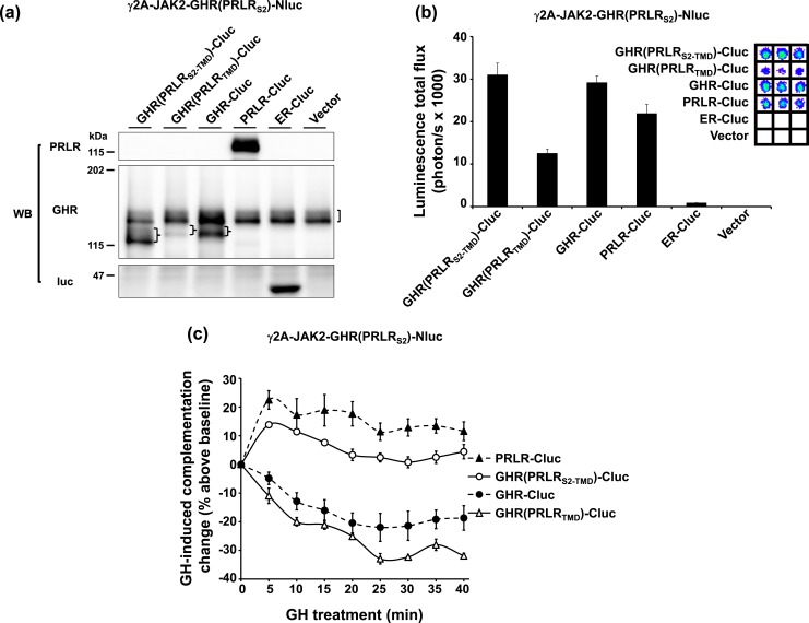
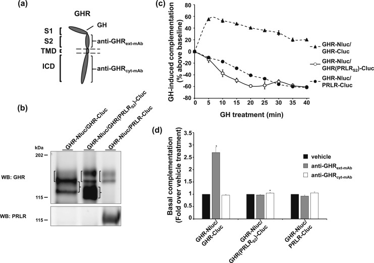
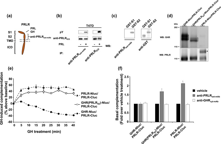
Growth hormone receptor (GHR) and prolactin (PRL) receptor (PRLR) are homologous transmembrane class I cytokine receptors. In humans, GH interacts with GHR homodimers or PRLR homodimers and PRL interacts with only PRLR homodimers to promote signaling. In human breast cancer cells endogenously expressing both receptors, GHR and PRLR specifically coimmunoprecipitate. We previously devised a split luciferase complementation assay to study GHR and PRLR assemblages. In this technique, firefly luciferase is split into two fragments (N- and C-terminal fragments of the luciferase), each without enzyme activity and tethered to the tails of two receptors. The fragments restore luciferase activity when brought close to each other by the receptors. Real-time ligand-induced complementation changes reflect the arrangement of receptors and indicate that GHR/PRLR is arranged as a heteromultimer comprised of GHR-GHR homodimers and PRLR-PRLR homodimers. We now dissect determinants for GHR and PRLR homodimerization versus heteroassociation. GHR and PRLR have extracellular domains comprised of the ligand-binding N-terminal subdomain 1 and a membrane-proximal subdomain 2 (S2), which fosters receptor-receptor contact. Based on previous studies of S2 versus the transmembrane domain (TMD) in GHR dimerization, we constructed GHR(PRLRS2), GHR(PRLRS2-TMD), and GHR(PRLRTMD), replacing GHR's S2 alone, S2 plus TMD, and TMD alone with PRLR's counterpart. We tested by complementation the ability of these chimeras and GHR or PRLR to homodimerize or heteroassociate. Comparing various combinations, we found GHR(PRLRS2) and GHR(PRLRS2-TMD) behaved as PRLR, whereas GHR(PRLRTMD) behaved as GHR regarding their dimerization partners. We conclude that S2 of GHR and PRLR, rather than their TMDs, determines their dimerization partner.


Figures

Figure 1.
Figure 1.
Replacement of GHR S2 or TMD with S2 or TMD of PRLR does not impair GH signaling. (a) Schematic diagram of domain-swapped human GHR/PRLR chimeras tethered with a Nluc/Cluc fragment at their cytoplasmic tails. GHR is in gray. PRLR is in orange. GHR(PRLRS2-TMD) is GHR with the S2 and TMD replaced with those of PRLR. GHR(PRLRS2) is GHR with its S2 replaced with the S2 of PRLR. GHR(PRLRTMD) is GHR with its TMD replaced with the TMD of PRLR. (b) GHR(PRLRS2-TMD), GHR(PRLRS2-TMD)-Nluc, and GHR(PRLRS2-TMD)-Cluc allow acute GH signaling. γ2A-JAK2 cells were transiently transfected with 3 μg GHR, 4.5 μg PRLR, 3 μg GHR(PRLRS2-TMD), 4.5 μg GHR(PRLRS2-TMD)-Nluc, 4 μg GHR(PRLRS2-TMD)-Cluc, or 3 μg empty vector per 6-cm dish, respectively, treated with ±GH, and analyzed by immunoblotting with anti-GHR (anti-GHRcyt-AL47, polyclonal antibody against the rabbit GHR intracellular domain), anti-PRLR (anti-PRLRcyt-AL84, polyclonal antibody against the human PRLR intracellular domain), anti-pJAK2, and anti-JAK2 (anti-JAK2AL33). (c) GHR(PRLRS2), GHR(PRLRS2)-Nluc, and GHR(PRLRS2)-Cluc allow acute GH signaling. γ2A-JAK2 cells were transiently transfected with 3 μg GHR, 4.5 μg PRLR, 3 μg GHR(PRLRS2), 4.5 μg GHR(PRLRS2)-Nluc, 4 μg GHR(PRLRS2)-Cluc, or 3 μg empty vector per 6-cm dish, respectively, treated with ±GH, and analyzed by immunoblotting with anti-GHR, anti-PRLR, anti-pJAK2, and anti-JAK2. (d) GHR(PRLRTMD), GHR(PRLRTMD)-Nluc, and GHR(PRLRTMD)-Cluc allow acute GH signaling. γ2A-JAK2 cells were transiently transfected with 3 μg GHR, 4.5 μg PRLR, 3 μg GHR(PRLRTMD), 4.5 μg GHR(PRLRTMD)-Nluc, 3 μg GHR(PRLRTMD)-Cluc, or 3 μg empty vector per 6-cm dish, respectively, split into 6-well plates, treated with ±GH, and analyzed by immunoblotting with anti-GHR, anti-PRLR, anti-pJAK2, and anti-JAK2.
Figure 2.
Figure 2.
S2 of GHR is necessary for a chimeric receptor to dimerize with GHR. (a) Specific luciferase complementation of GHR-Nluc, a GHR-Nluc stably expressing cell line, and chimeric receptor-Clucs. γ2A-JAK2-GHR-Nluc cells were transiently transfected with GHR(PRLRS2-TMD)-Cluc, GHR(PRLRS2)-Cluc, GHR(PRLRTMD)-Cluc, GHR-Cluc, PRLR-Cluc, ER-Cluc, or empty vector, respectively, and measured for bioluminescence in triplicate in a 96-well plate format (inset shows actual signals). The quantifications are displayed graphically as mean ± SE of total flux (photons/s × 1000). (b) Transient expression of chimeric receptor-Clucs in γ2A-JAK2-GHR-Nluc. The same pools of the transfected cells in (a) were analyzed by immunoblotting with anti-PRLR, anti-GHR, and anti-luc. Stably expressed GHR-Nluc is indicated by a bracket. Transfected chimeric receptor-Clucs are indicated by braces. (c) GH-induced complementation change. After basal bioluminescence was measured (a), GH was added and bioluminescence was again serially detected at 5-minute intervals over 40 minutes. Data are displayed as mean ± SE of GH-induced signal as a percentage above baseline signal (n = 3 per condition).
Figure 3.
Figure 3.
S2 of PRLR is sufficient for a chimeric receptor to dimerize with PRLR. (a) GH signaling in γ2A-JAK2-GHR(PRLRS2)-Nluc, a GHR(PRLRS2)-Nluc stably expressing cell line. γ2A-JAK2-GHR(PRLRS2)-Nluc cells were treated with ±GH (10 minutes) and analyzed by immunoblotting with anti-pY, anti-GHR, anti-pJAK2, and anti-JAK2. (b and c) Transient expression of GHR(PRLRS2)-Cluc, GHR-Cluc, PRLR-Cluc, ER-Cluc, or empty vector in γ2A-JAK2-GHR(PRLRS2)-Nluc cells. γ2A-JAK2-GHR(PRLRS2)-Nluc cells were transiently transfected with GHR(PRLRS2)-Cluc, GHR-Cluc, PRLR-Cluc, ER-Cluc, or empty vector, respectively, and analyzed by immunoblotting with (b) anti-GHR or (c) anti-luc. Stably expressed GHR(PRLRS2)-Nluc is indicated by black arrowheads. Transfected receptor-Clucs are indicated by asterisks. (d) Specific luciferase complementation of GHR(PRLRS2)-Nluc and GHR(PRLRS2)-Cluc, GHR-Cluc, or PRLR-Cluc. The same pool of the transfected cells in (b) and (c) was measured for bioluminescence in triplicate in a 96-well plate format (inset shows actual signals). The quantifications are displayed graphically as mean ± SE of total flux (photons/s × 10,000). (e) GH-induced complementation change. After basal bioluminescence was measured (d), GH was added and bioluminescence was again serially detected at 5-minute intervals over 40 minutes. Data are displayed as mean ± SE of GH-induced signal as a percentage above baseline signal (n = 3 per condition).
Figure 4.
Figure 4.
TMD does not alter the S2-dictated ligand-induced profile of complementation. (a) Transient expression of GHR(PRLRS2-TMD)-Cluc and GHR(PRLRTMD)-Cluc in γ2A-JAK2-GHR(PRLRS2)-Nluc cells. γ2A-JAK2-GHR(PRLRS2)-Nluc cells were transiently transfected with GHR(PRLRS2-TMD)-Cluc, GHR(PRLRTMD)-Cluc, GHR-Cluc, PRLR-Cluc, ER-Cluc, or empty vector, respectively, and analyzed by immunoblotting with anti-PRLR, anti-GHR, or anti-luc. Stably expressed GHR(PRLRS2)-Nluc is indicated by a bracket. Transfected receptor-Clucs are indicated by braces. (b) Specific luciferase complementation of GHR(PRLRS2)-Nluc and GHR(PRLRS2-TMD)-Cluc, or GHR(PRLRTMD)-Cluc. The same pool of the transfected cells in (a) was measured for bioluminescence in triplicate in a 96-well plate format (inset shows actual signals). The quantifications are displayed graphically as mean ± SE of total flux (photons/s × 1000). (c) GH-induced complementation change. After basal bioluminescence was measured (b), GH was added and bioluminescence was again serially detected at 5-minute intervals over 40 minutes. Data are displayed as mean ± SE of GH-induced signal as a percentage above baseline signal (n = 3 per condition).
Figure 5.
Figure 5.
Effect of anti-GHRext-mAb suggests lack of dimerization between GHR and GHR(PRLRS2). (a) Diagram of the GHR domains that interact with anti-GHRext-mAb, anti-GHRcyt-mAb, and GH. (b) Three double-stable clones with GHR-Nluc but distinct receptor-Clucs. γ2A-JAK2-GHR-Nluc/GHR-Cluc, γ2A-JAK2-GHR-Nluc/GHR(PRLRS2)-Cluc, and γ2A-JAK2-GHR-Nluc/PRLR-Cluc cells were analyzed by immunoblotting with anti-GHR and anti-PRLR. Brackets on the left side of each lane indicate the position of GHR-Nluc, which is comprised of both the upper mature (less distinct) and lower precursor (sharper) bands. Braces on the right of lanes 1 and 2 indicate the positions of GHR-Cluc and GHR(PRLRS2)-Cluc, each comprised of both the mature and precursor bands. Note that the GHR-Nluc precursor and the GHR-Cluc mature form overlap in lane 1 to produce a more intense combined band. (c) GH-induced complementation change in three GHR-Nluc–expressing double-stable clones. γ2A-JAK2-GHR-Nluc/GHR-Cluc, γ2A-JAK2-GHR-Nluc/GHR(PRLRS2)-Cluc, and γ2A-JAK2-GHR-Nluc/PRLR-Cluc cells were measured for basal bioluminescence in triplicate in a 96-well plate format. GH was added, and bioluminescence was again serially detected at 5-minute intervals over 40 minutes. Data are displayed as mean ± SE of GH-induced signal as a percentage above baseline signal (n = 3 per condition). (d) Anti-GHRext-mAb enhances basal complementation of GHR-Nluc/GHR-Cluc, but not of GHR-Nluc/GHR(PRLRS2)-Cluc or GHR-Nluc/PRLR-Cluc. γ2A-JAK2-GHR-Nluc/GHR-Cluc, γ2A-JAK2-GHR-Nluc/GHR(PRLRS2)-Cluc, and γ2A-JAK2-GHR-Nluc/PRLR-Cluc cells were incubated with vehicle buffer, anti-GHRext-mAb, or anti-GHRcyt-mAb for 30 minutes. Bioluminescence was detected in triplicate in a 96-well plate format and normalized to the mean value from the vehicle buffer-incubated cells. Each bar represents data from three independent experiments and is displayed as mean of the fold change ± SE. *P < 0.05 when compared with respective vehicle treatment in each cell line.
Figure 6.
Figure 6.
Effect of anti-PRLRext-mAb suggests the dimerization between GHR(PRLRS2) and PRLR. (a) Diagram of the PRLR domains that interact with anti-PRLRext-mAb, GH, and PRL. (b) Anti-PRLRext-mAb was compared with commercial anti-ICD serum for immunoprecipitation (IP) of T47D cells treated ± PRL. Human breast cancer T47D cells were treated with ±PRL (500 ng/mL, 10 min), immunoprecipitated with anti-PRLRext-mAb or anti-PRLRICD, and analyzed by immunoblotting with anti-pY and anti-PRLRICD. (c) Anti-PRLRext-mAb reacts with PRLR ECD S2, but not S1. Bacterial extracts harboring GST fused to S1 (GST-S1) or S2 (GST-S2) of PRLR ECD were resolved by sodium dodecyl sulfate–polyacrylamide gel electrophoresis and immunoblotted with anti-PRLRext-mAb or anti-GST. (d) Three double-stable clones with PRLR-Cluc, but distinct receptor-Nlucs. γ2A-JAK2-GHR-Nluc/PRLR-Cluc, γ2A-JAK2-GHR(PRLRS2)-Nluc/PRLR-Cluc, and γ2A-JAK2-PRLR-Nluc/PRLR-Cluc cells were analyzed by immunoblotting with anti-GHR and anti-PRLR. GHR-Nluc and GHR(PRLRS2)-Nluc are indicated by brackets. PRLR-Nluc is indicated by an asterisk. (e) GH-induced complementation change in three PRLR-Cluc–expressing double-stable clones. γ2A-JAK2-GHR-Nluc/PRLR-Cluc, γ2A-JAK2-GHR(PRLRS2)-Nluc/PRLR-Cluc, and γ2A-JAK2-PRLR-Nluc/PRLR-Cluc cells were measured for basal bioluminescence in triplicate in a 96-well plate format. GH was added, and bioluminescence was again serially detected at 5-minute intervals over 40 minutes. Data are displayed as mean ± SE of GH-induced signal as a percentage above baseline signal (n = 3 per condition). (f) Anti-PRLRext-mAb enhances basal complementation of GHR(PRLRS2)-Nluc/PRLR-Cluc or PRLR-Nluc/PRLR-Cluc, but not of GHR-Nluc/PRLR-Cluc. γ2A-JAK2-GHR-Nluc/PRLR-Cluc, γ2A-JAK2-GHR(PRLRS2)-Nluc/PRLR-Cluc, and γ2A-JAK2-PRLR-Nluc/PRLR-Cluc cells were incubated with vehicle buffer, anti-PRLRext-mAb, or anti-GHRcyt-mAb for 30 minutes. Bioluminescence was detected in triplicate in a 96-well plate format and normalized to the mean value from the vehicle buffer-incubated cells. Each bar represents data from three independent experiments and is displayed as mean of the fold change ± SE. *P < 0.05 when compared with respective vehicle treatment in each cell line.

Similar articles

Cited by

References

    1. Gent J, van Kerkhof P, Roza M, Bu G, Strous GJ. Ligand-independent growth hormone receptor dimerization occurs in the endoplasmic reticulum and is required for ubiquitin system-dependent endocytosis. Proc Natl Acad Sci USA. 2002;99(15):9858–9863. - PMC - PubMed
    1. Yang N, Wang X, Jiang J, Frank SJ. Role of the growth hormone (GH) receptor transmembrane domain in receptor predimerization and GH-induced activation. Mol Endocrinol. 2007;21(7):1642–1655. - PubMed
    1. Brown RJ, Adams JJ, Pelekanos RA, Wan Y, McKinstry WJ, Palethorpe K, Seeber RM, Monks TA, Eidne KA, Parker MW, Waters MJ. Model for growth hormone receptor activation based on subunit rotation within a receptor dimer. Nat Struct Mol Biol. 2005;12(9):814–821. - PubMed
    1. Biener-Ramanujan E, Ramanujan VK, Herman B, Gertler A. Spatio-temporal kinetics of growth hormone receptor signaling in single cells using FRET microscopy. Growth Horm IGF Res. 2006;16(4):247–257. - PubMed
    1. Brooks AJ, Dai W, O’Mara ML, Abankwa D, Chhabra Y, Pelekanos RA, Gardon O, Tunny KA, Blucher KM, Morton CJ, Parker MW, Sierecki E, Gambin Y, Gomez GA, Alexandrov K, Wilson IA, Doxastakis M, Mark AE, Waters MJ. Mechanism of activation of protein kinase JAK2 by the growth hormone receptor. Science. 2014;344(6185):1249783. - PubMed

Publication types