

Structural insights into species-specific features of the ribosome from the human pathogen Mycobacterium tuberculosis
- PMID: 28977617
- PMCID: PMC5737476
- DOI: 10.1093/nar/gkx785
Structural insights into species-specific features of the ribosome from the human pathogen Mycobacterium tuberculosis
Abstract
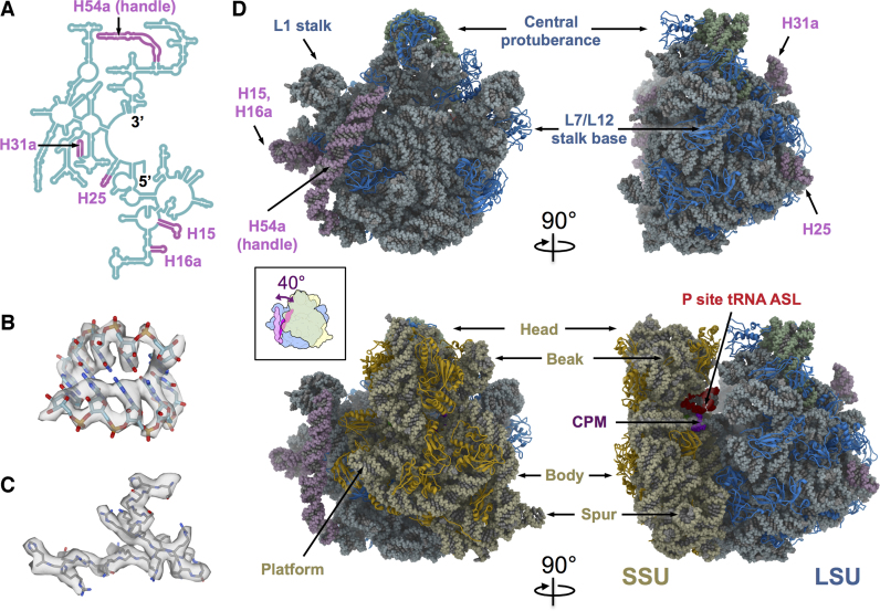
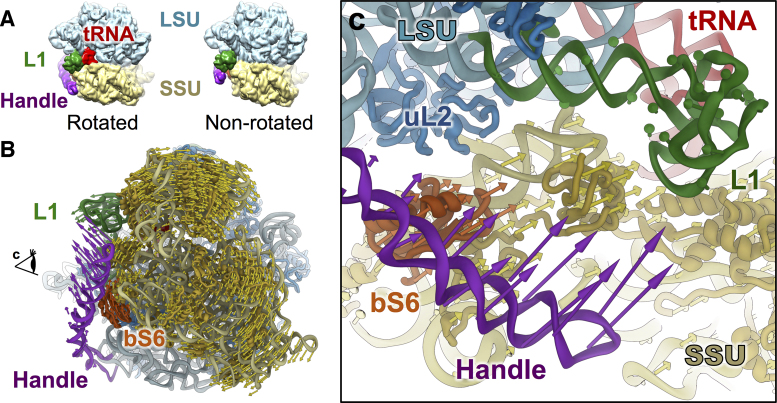
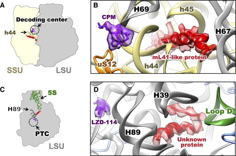
Ribosomes from Mycobacterium tuberculosis (Mtb) possess species-specific ribosomal RNA (rRNA) expansion segments and ribosomal proteins (rProtein). Here, we present the near-atomic structures of the Mtb 50S ribosomal subunit and the complete Mtb 70S ribosome, solved by cryo-electron microscopy. Upon joining of the large and small ribosomal subunits, a 100-nt long expansion segment of the Mtb 23S rRNA, named H54a or the 'handle', switches interactions from with rRNA helix H68 and rProtein uL2 to with rProtein bS6, forming a new intersubunit bridge 'B9'. In Mtb 70S, bridge B9 is mostly maintained, leading to correlated motions among the handle, the L1 stalk and the small subunit in the rotated and non-rotated states. Two new protein densities were discovered near the decoding center and the peptidyl transferase center, respectively. These results provide a structural basis for studying translation in Mtb as well as developing new tuberculosis drugs.
© The Author(s) 2017. Published by Oxford University Press on behalf of Nucleic Acids Research.
Figures

Similar articles
-
Structural diversity in bacterial ribosomes: mycobacterial 70S ribosome structure reveals novel features.PLoS One. 2012;7(2):e31742. doi: 10.1371/journal.pone.0031742. Epub 2012 Feb 24. PLoS One. 2012. PMID: 22384065 Free PMC article.
-
The Complete Structure of the Mycobacterium smegmatis 70S Ribosome.Cell Rep. 2017 Jul 5;20(1):149-160. doi: 10.1016/j.celrep.2017.06.029. Cell Rep. 2017. PMID: 28683309
-
Structure of Ribosomal Silencing Factor Bound to Mycobacterium tuberculosis Ribosome.Structure. 2015 Oct 6;23(10):1858-1865. doi: 10.1016/j.str.2015.07.014. Epub 2015 Aug 20. Structure. 2015. PMID: 26299947 Free PMC article.
-
Unique structural features of the Mycobacterium ribosome.Prog Biophys Mol Biol. 2020 May;152:15-24. doi: 10.1016/j.pbiomolbio.2019.12.001. Epub 2019 Dec 10. Prog Biophys Mol Biol. 2020. PMID: 31858996 Review.
-
The ribosome through the looking glass.Angew Chem Int Ed Engl. 2003 Aug 4;42(30):3464-86. doi: 10.1002/anie.200200544. Angew Chem Int Ed Engl. 2003. PMID: 12900959 Review.
Cited by
-
Structure of the 70S Ribosome from the Human Pathogen Acinetobacter baumannii in Complex with Clinically Relevant Antibiotics.Structure. 2020 Oct 6;28(10):1087-1100.e3. doi: 10.1016/j.str.2020.08.004. Epub 2020 Aug 27. Structure. 2020. PMID: 32857965 Free PMC article.
-
Structural Heterogeneities of the Ribosome: New Frontiers and Opportunities for Cryo-EM.Molecules. 2020 Sep 17;25(18):4262. doi: 10.3390/molecules25184262. Molecules. 2020. PMID: 32957592 Free PMC article. Review.
-
Hierarchical natural move Monte Carlo refines flexible RNA structures into cryo-EM densities.RNA. 2020 Dec;26(12):1755-1766. doi: 10.1261/rna.071100.119. Epub 2020 Aug 21. RNA. 2020. PMID: 32826323 Free PMC article.
-
Cryo-EM structure of Mycobacterium tuberculosis 50S ribosomal subunit bound with clarithromycin reveals dynamic and specific interactions with macrolides.Emerg Microbes Infect. 2022 Dec;11(1):293-305. doi: 10.1080/22221751.2021.2022439. Emerg Microbes Infect. 2022. PMID: 34935599 Free PMC article.
-
Zinc depletion induces ribosome hibernation in mycobacteria.Proc Natl Acad Sci U S A. 2018 Aug 7;115(32):8191-8196. doi: 10.1073/pnas.1804555115. Epub 2018 Jul 23. Proc Natl Acad Sci U S A. 2018. PMID: 30038002 Free PMC article.
References
-
- Wayne L.G., Sohaskey C.D.. Nonreplicating persistence of mycobacterium tuberculosis. Annu. Rev. Microbiol. 2001; 55:139–163. - PubMed
-
- Smith C.V., Sharma V., Sacchettini J.C.. TB drug discovery: addressing issues of persistence and resistance. Tuberculosis (Edinb). 2004; 84:45–55. - PubMed
-
- Chao M.C., Rubin E.J.. Letting sleeping dos lie: does dormancy play a role in tuberculosis. Annu. Rev. Microbiol. 2010; 64:293–311. - PubMed
MeSH terms
Substances
Grants and funding
LinkOut - more resources
Full Text Sources
Other Literature Sources
Research Materials
