Review
. 2017 Oct 13;45(18):10350-10368.
doi: 10.1093/nar/gkx759.

Nuclear speckles: molecular organization, biological function and role in disease

Affiliations
Review

Nuclear speckles: molecular organization, biological function and role in disease

Lukasz Galganski et al. Nucleic Acids Res. .

Abstract

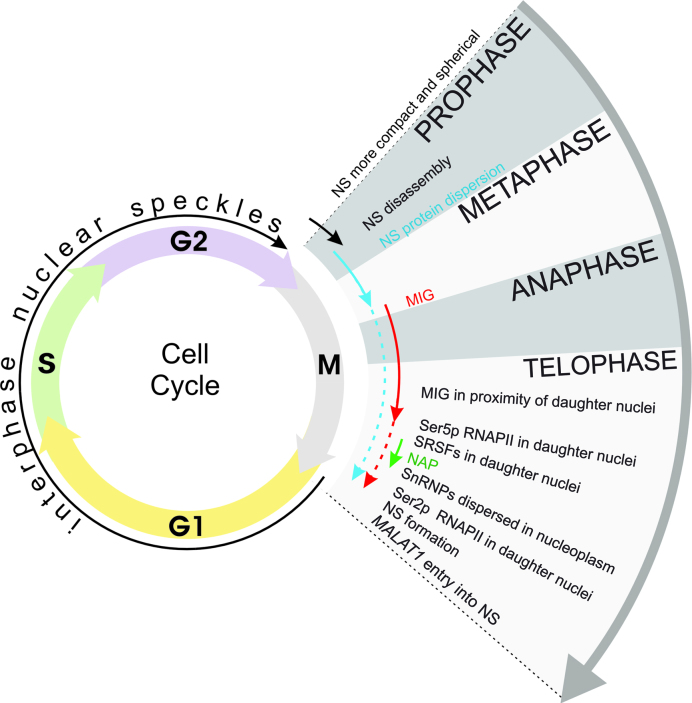
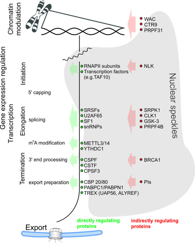
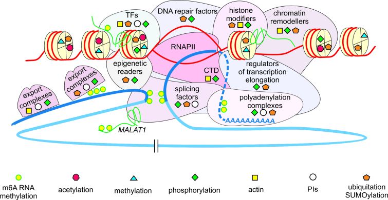
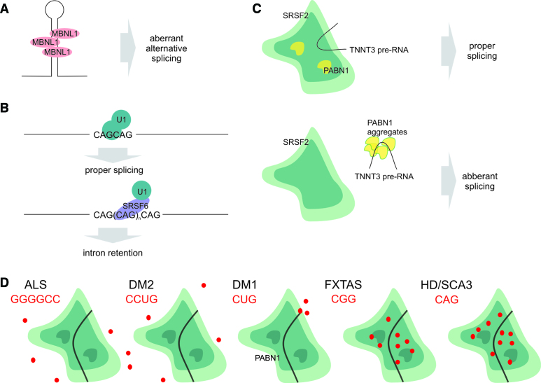
The nucleoplasm is not homogenous; it consists of many types of nuclear bodies, also known as nuclear domains or nuclear subcompartments. These self-organizing structures gather machinery involved in various nuclear activities. Nuclear speckles (NSs) or splicing speckles, also called interchromatin granule clusters, were discovered as sites for splicing factor storage and modification. Further studies on transcription and mRNA maturation and export revealed a more general role for splicing speckles in RNA metabolism. Here, we discuss the functional implications of the localization of numerous proteins crucial for epigenetic regulation, chromatin organization, DNA repair and RNA modification to nuclear speckles. We highlight recent advances suggesting that NSs facilitate integrated regulation of gene expression. In addition, we consider the influence of abundant regulatory and signaling proteins, i.e. protein kinases and proteins involved in protein ubiquitination, phosphoinositide signaling and nucleoskeletal organization, on pre-mRNA synthesis and maturation. While many of these regulatory proteins act within NSs, direct evidence for mRNA metabolism events occurring in NSs is still lacking. NSs contribute to numerous human diseases, including cancers and viral infections. In addition, recent data have demonstrated close relationships between these structures and the development of neurological disorders.


Figures

Figure 1.
Figure 1.
Schematic representation of the dynamics of NS components during the cell cycle. The diverse forms of NS protein assemblies are indicated with arrows: black represents enlarged NSs (possibly caused by transcriptional inhibition), blue represents a diffuse pattern, red represents MIGs and green represents nucleolar organizing region-associated patches (NAP). Importantly, different sets of proteins exhibit diverse patterns of cell cycle-regulated localization and diverse timing of their entry into and their presence in assemblies of NS proteins. Different assemblies of NS proteins can coexist and those in the minority are indicated with dotted lines. Note that NSs and MIG, in contrast to NAP, contain poly(A)+ RNA.
Figure 2.
Figure 2.
RNA processing, from the site of transcription to nuclear export, is regulated by multiple proteins localized within NSs. The RNA pathway in the nucleus starts with transcription initiation and multiple proteins, many of which are found in NSs, are responsible for the processing of primary transcripts, including splicing, m6A modification, 3′ end processing and export. In the figure, examples of proteins with known functions in transcription and RNA maturation are presented (green dots). Additionally, a large group of other NS-associated proteins is indirectly involved in the precise regulation of RNA processing and their examples are shown (red dots).
Figure 3.
Figure 3.
The role of proteins localized to NSs in pre-mRNA synthesis, maturation and DNA template regulation; impact of NS regulatory proteins on depicted protein complexes is indicated with specific symbols explained in the figure. DNA template is shown in red and primary transcript in blue, exon (dark blue) intron (light blue). TFs: transcription factors; RNAPII: RNA Polymerase II; PIs: phosphoinositides; CTD: C-terminal domain of RNAPII.
Figure 4.
Figure 4.
(A) MBNL1 binds to expanded CUG RNA, resulting in aberrant alternative splicing in myotonic dystrophy type 1. (B) HTT mRNA splicing is disturbed by SRSF6 in a CAG length-dependent manner. (C) PABN1 aggregates bind pre-mRNA, which hinders splicing factor binding and results in aberrant splicing. (D) RNA foci harboring transcripts containing expanded simple repeats have different localization patterns in relation to NSs. Schematic representations of RNA foci localization are shown.
Figure 5.
Figure 5.
NSs differ considerably among various human cell lines. Representative microscopy images of cells labeled for SRSF2 (constitutive protein of NSs) are presented. Due to the different expression levels of SRSF2 in different cell lines, the anti-SRSF2 antibody was used at different dilutions: 1:500 in fibroblasts, 1:500 in SK-MCs, 1:200 in lymphoblasts and 1:500 in SK-N-MCs. Bars = 10 μm (for the fibroblast, SK-MC and SK-N-MC) and 5 μm (for the lymphoblast).

Similar articles

Cited by

References

    1. Dundr M., Misteli T.. Biogenesis of nuclear bodies. Cold Spring Harb. Perspect. Biol. 2010; 2:a000711. - PMC - PubMed
    1. Mao Y., Zhang B., Spector D.. Biogenesis and function of nuclear bodies. Trends Genet. 2011; 27:295–306. - PMC - PubMed
    1. Dundr M. Nuclear bodies: multifunctional companions of the genome. Curr. Opin. Cell Biol. 2012; 24:415–422. - PMC - PubMed
    1. Spector D.L., Lamond A.I.. Nuclear speckles. Cold Spring Harb. Perspect. Biol. 2011; 3:a000646. - PMC - PubMed
    1. Sahin U., Lallemand-Breitenbach V., de Thé H.. PML nuclear bodies: regulation, function and therapeutic perspectives. J. Pathol. 2014; 234:289–291. - PubMed

MeSH terms