. 2017 Nov 2;45(19):11222-11235.
doi: 10.1093/nar/gkx763.

Regulation of mitotic recombination between DNA repeats in centromeres

Affiliations

Regulation of mitotic recombination between DNA repeats in centromeres

Faria Zafar et al. Nucleic Acids Res. .

Erratum in

Abstract

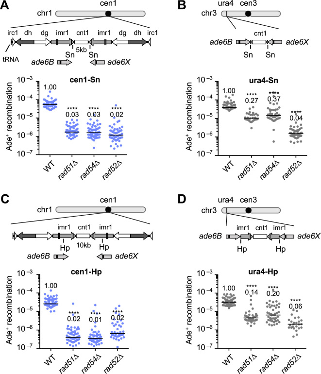
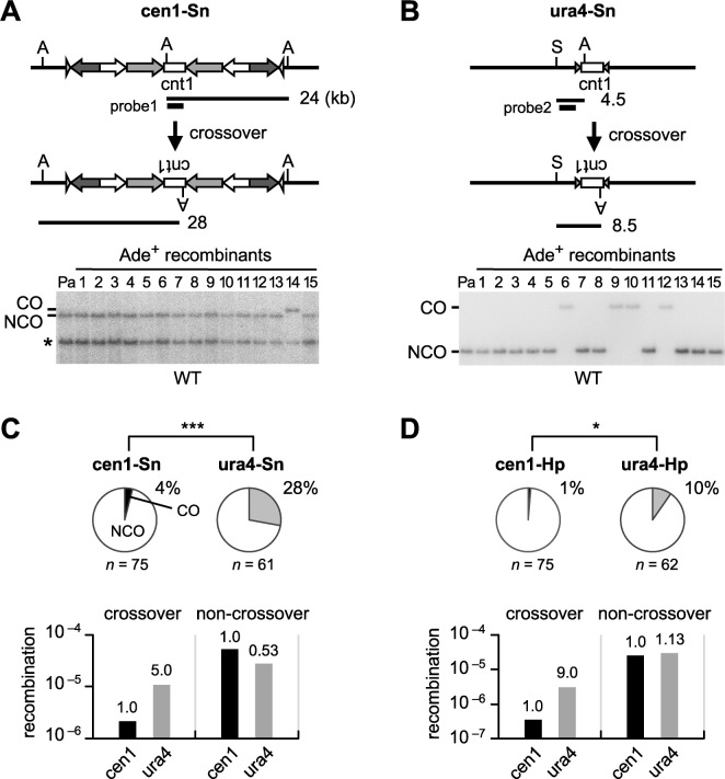
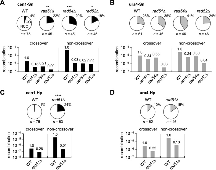
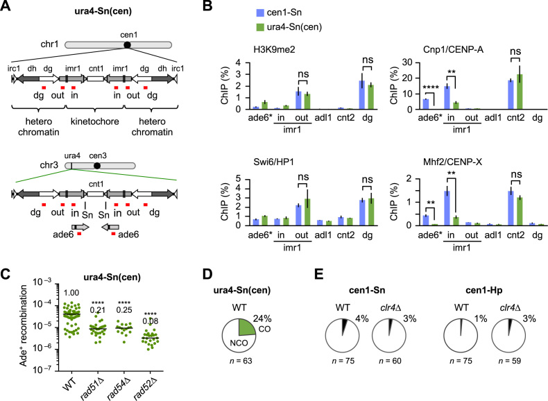
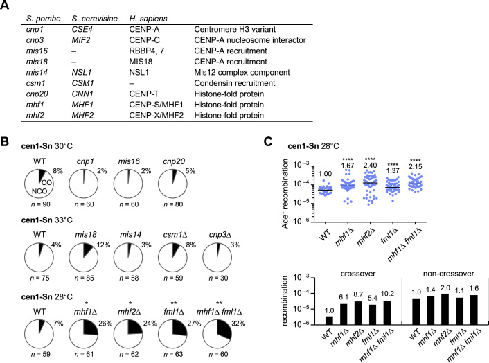
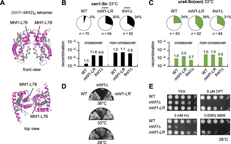
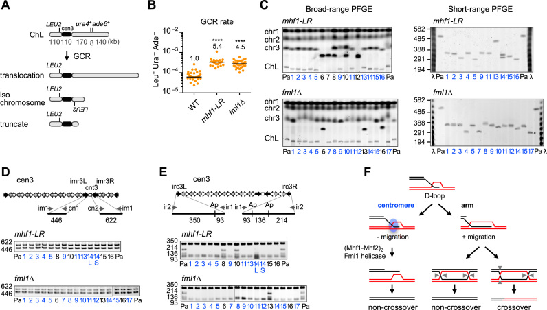
Centromeres that are essential for faithful segregation of chromosomes consist of unique DNA repeats in many eukaryotes. Although recombination is under-represented around centromeres during meiosis, little is known about recombination between centromere repeats in mitotic cells. Here, we compared spontaneous recombination that occurs between ade6B/ade6X inverted repeats integrated at centromere 1 (cen1) or at a non-centromeric ura4 locus in fission yeast. Remarkably, distinct mechanisms of homologous recombination (HR) were observed in centromere and non-centromere regions. Rad51-dependent HR that requires Rad51, Rad54 and Rad52 was predominant in the centromere, whereas Rad51-independent HR that requires Rad52 also occurred in the arm region. Crossovers between inverted repeats (i.e. inversions) were under-represented in the centromere as compared to the arm region. While heterochromatin was dispensable, Mhf1/CENP-S, Mhf2/CENP-X histone-fold proteins and Fml1/FANCM helicase were required to suppress crossovers. Furthermore, Mhf1 and Fml1 were found to prevent gross chromosomal rearrangements mediated by centromere repeats. These data for the first time uncovered the regulation of mitotic recombination between DNA repeats in centromeres and its physiological role in maintaining genome integrity.


Figures

Figure 1.
Figure 1.
Recombination between ade6B/ade6X heteroalleles in centromere and arm regions. (A) Recombination in the cen1-Sn construct. Illustrated are the central sequence cnt1 as well as the imr1, dg, dh and irc1 inverted repeats in the centromere 1 (cen1) of fission yeast. ade6B and ade6X mutant genes were integrated at the Sn sites in imr1. Spontaneous rates of Ade+ prototroph formation were determined in wild-type (WT), rad51Δ, rad54Δ and rad52Δ strains (TNF3347, 3446, 3452 and 3459, respectively). (B) Recombination in the ura4-Sn construct. From the cen1-Sn construct, the ade6B/X heteroalleles flanking the central region of cen1 were amplified and integrated at the ura4 locus. Recombination rates were determined in WT, rad51Δ, rad54Δ and rad52Δ strains (TNF3631, 3635, 3645 and 3643, respectively). (C) Recombination in the cen1-Hp construct. ade6B/X were integrated at the Hp sites in imr1 (35). Recombination rates were determined in WT, rad51Δ, rad54Δ and rad52Δ strains (TNF3144, 3257, 3286 and 3277, respectively). Recombination rates of WT, rad51Δ and rad54Δ in the cen1-Hp construct were previously published (35). (D) Recombination in the ura4-Hp construct. From the cen1-Hp construct, ade6B/X flanking the central region of cen1 were amplified and integrated at the ura4 locus. Recombination rates were determined in WT, rad51Δ, rad54Δ and rad52Δ strains (TNF3650, 3664, 3670 and 3667, respectively). Independent experimental values are shown in scatter plots and lines indicate medians. Rates relative to the WT value are indicated at the top of each column. P-values were determined by the two-tailed Mann–Whitney test. ****P < 0.0001. Sn, SnaBI; Hp, HpaI.
Figure 2.
Figure 2.
Crossovers and non-crossovers between inverted repeats in centromere and arm regions. (A) Crossover and non-crossover recombinants produced in the cen1-Sn construct in WT (TNF3347). DNA was prepared, digested with AfeI, separated by pulse field gel electrophoresis (PFGE) (0.6% agarose, 0.5× TBE, 6 V/cm, 1–6 s switching time, for 11–15 h), transferred to a nylon membrane and subjected to Southern hybridization using probe1. An asterisk indicates the band derived from cen3. (B) Crossover and non-crossover recombinants produced in the ura4-Sn construct in WT (TNF3631). DNA was digested with AfeI and SmaI and separated by PFGE (0.6% agarose, 0.5× TBE, 6 V/cm, 1 to 5 s switching time, for 9 h). Probe2 was used for Southern hybridization. (C) Proportions of crossovers among recombinants in cen1-Sn and ura4-Sn constructs in WT are indicated in Pie charts. Net rates of crossover and non-crossover recombination are shown in bar graphs. (D) Proportions of crossovers and net rates of crossover and non-crossover recombination in cen1-Hp and ura4-Hp constructs in WT (TNF3144 and 3650, respectively). The cen1-Hp data were reported previously (35). Rates relative to the cen1 value are indicated at the top of each bar. P-values were obtained by the two-tailed Fisher’s exact test. *P < 0.05; ***P < 0.001. n, sample number; A, AfeI; S, SmaI; CO, crossover; NCO, non-crossover; Pa, parental.
Figure 3.
Figure 3.
Rad51-dependent recombination preferentially promote non-crossovers in centromeres. (A) Proportions of crossovers among recombinants (pie charts) in the cen1-Sn construct in WT, rad51Δ, rad54Δ and rad52Δ strains (TNF3347, 3446, 3452 and 3459, respectively) and their net rates of crossover and non-crossover recombination (bar graphs). (B) Proportions of crossovers in the ura4-Sn construct in WT, rad51Δ, rad54Δ and rad52Δ strains (TNF3631, 3635, 3645 and 3643, respectively) and their net rates of crossover and non-crossover recombination. (C) Proportions and net rates of recombination in the cen1-Hp construct of WT and rad51Δ (TNF3144 and 3257, respectively). The proportions were published previously (35). (D) The proportions and net rates of recombination in the ura4-Hp construct of WT and rad51Δ (TNF3650 and 3664, respectively). **P < 0.01.
Figure 4.
Figure 4.
Effects of pericentromere repeats on recombination. (A) Illustrated are the cen1 region on chr1 and the ectopic cen1 region introduced at the ura4 locus of chr3 in the ura4-Sn(cen) strain. Kinetochore chromatin and heterochromatin are assembled on the cen1. The positions of PCR amplification in chromatin immunoprecipitation (ChIP) analysis are shown in red. (B) Results of ChIP analysis conducted to examine H3K9me2, Swi6, Cnp1 and Mhf2 levels in the cen1-Sn and the ura4-Sn(cen) strains (TNF3347 and 4684). cnt2 and adl1 are in the centromere and arm regions of chr2, respectively. imr1-in and imr1-out are in the kinetochore and heterochromatin domains, respectively. ade6* is present in the original cen1 in cen1-Sn, while it is present only in the ectopic cen1 in ura4-Sn(cen). Data are represented as mean ± SEM from three biologically independent experiments. P-values were determined by the two-tailed student’s t-test. ns, statistically non-significant. (C) Recombination rates were determined in the ura4-Sn(cen) strain of WT, rad51Δ, rad54Δ and rad52Δ (TNF4684, 5814, 5826 and 5829, respectively). (D) Proportions of crossovers in the ura4-Sn(cen) construct in WT. (E) Proportions of crossovers in the cen1-Sn construct in WT and clr4Δ strains (TNF3347 and 3734, respectively) and in the cen1-Hp construct in WT and clr4Δ strains (TNF3144 and 3550, respectively).
Figure 5.
Figure 5.
Roles of the kinetochore proteins in centromere recombination. (A) List of kinetochore-related proteins examined in this study. (B) Proportions of crossovers in the mutants at their semi-permissive temperatures (30, 33 or 28°C). The cen1-Sn strain of WT, cnp1–76, mis16–53, cnp20-M447T, mis18–262, mis14–271, csm1Δ, cnp3Δ, mhf1Δ, mhf2Δ, fml1Δ and mhf1Δ fml1Δ (TNF3347, 3736, 4656, 5534, 4657, 5376, 4139, 4115, 4779, 5082, 5353 and 5128, respectively) were examined. (C) Recombination rates in the cen1-Sn strain of WT, mhf1Δ, mhf2Δ, fml1Δ and mhf1Δ fml1Δ strains at 28°C are shown in the scatter plot. Net rates of crossover and non-crossover recombination are shown in the bar graph.
Figure 6.
Figure 6.
(Mhf1–Mhf2)2 tetramer formation is important for the suppression of crossovers in centromeres. (A) Ribbon diagram of the crystal structure of chicken (Mhf1–Mhf2)2 tetramers (15). The position of the conserved leucine residue in α3 helix, which corresponds to the Mhf1-L78 of fission yeast is indicated. (B) Proportions of crossovers and net rates of crossover and non-crossover recombination in the cen1-Sn strain of WT, mhf1-LR and fml1Δ (TNF3347, 5444 and 5353, respectively). (C) Proportion of crossovers and net rates of crossover and non-crossover recombination in the ura4-Sn(cen) strain of WT, mhf1-LR and fml1Δ (TNF4684, 5455 and 4806, respectively). (D) Growth of mhf1 mutants. WT, mhf1Δ and mhf1-LR strains (TNF3347, 4779 and 5444, respectively) were streaked on YE+A plates that contain adenine and were incubated at the indicated temperatures for 2 days. (E) Camptothecin (CPT), hydroxyurea (HU) and methyl methanesulphonate (MMS) sensitivities. Exponentially growing cells of WT, mhf1Δ and mhf1-LR strains (TNF3347, 4779 and 5444, respectively) were 5-fold serially diluted with distilled water and spotted on YE+A plates supplemented with the indicated concentrations of CPT, HU and MMS. Plates were incubated for 3–5 days at 28°C.
Figure 7.
Figure 7.
Mhf1 and Fml1 suppress gross chromosomal rearrangements (GCRs) in centromeres. (A) GCR assay using the extra chromosome ChL (36). GCRs associated with loss of the right arm of ChL result in Leu+ Ura Ade. The GCR product can be translocation, isochromosome and truncate of different lengths. (B) Spontaneous GCR rates in WT, mhf1-LR and fml1Δ strains (TNF3896, 5477 and 4813, respectively). Rates relative to WT values are indicated at the top of each column. P-values were determined by the two-tailed Mann–Whitney test. The GCR rate of WT was published previously (35). (C) Chromosomal DNA of mhf1-LR and fml1Δ were separated by broad-range and short-range PFGE and stained with ethidium bromide (EtBr) (see ‘Materials and Methods’ section). Positions of chr1, chr2, chr3 and the parental ChL are indicated on the left of the broad-range gel. Size of λ ladder (ProMega-Markers) bands are indicated on the left of the short-range gel. Pa, parental. Clone #6, 7 and 12 of mhf1-LR may have suffered complex rearrangements, resulting in two chr3 GCR products of similar sizes. Two GCR products of different sizes were detected in clone #14 of mhf1-LR, which may be due to the change in the copy number of centromere repeats. (D) PCR analysis of GCR products. PCR was carried out using ChL DNAs recovered from agarose gel using the indicated primers. cnt3–imr3 junctions were amplified and applied to standard agarose gel electrophoresis and stained with EtBr. (E) irc3L and irc3R regions were amplified and treated with ApoI. Ap, ApoI. WT data were reported previously (35). (F) Model of how (Mhf1–Mhf2)2 tetramers and Fml1 helicase suppress crossovers in centromeres. The 3′ single-stranded DNA (ssDNA) tail invades into homologous double-stranded DNA to form displacement-loops (D-loops). In the arm region, branch migration extends the length of the heteroduplex and stabilizes recombination intermediates, endonucleolytic cleavage of which results in either crossovers or non-crossovers. However, in the centromere, unidentified centromere proteins shown as a blue circle prevents branch migration, thereby stimulating (Mhf1–Mhf2)2 binding to branched DNA that recruits Fml1 helicase to dissociate D-loops, resulting in non-crossovers by synthesis-dependent strand annealing (SDSA) reactions. Arrow heads indicate endonucleotic cleavage sites of Holliday junctions.

Similar articles

Cited by

References

    1. Padeken J., Zeller P., Gasser S.M.. Repeat DNA in genome organization and stability. Curr. Opin. Genet. Dev. 2015; 31:12–19. - PubMed
    1. de Koning A.P., Gu W., Castoe T.A., Batzer M.A., Pollock D.D.. Repetitive elements may comprise over two-thirds of the human genome. PLoS Genet. 2011; 7:e1002384. - PMC - PubMed
    1. Tubbs A., Nussenzweig A.. Endogenous DNA damage as a source of genomic instability in cancer. Cell. 2017; 168:644–656. - PMC - PubMed
    1. Deininger P.L., Batzer M.A.. Alu repeats and human disease. Mol. Genet. Metab. 1999; 67:183–193. - PubMed
    1. Campbell I.M., Gambin T., Dittwald P., Beck C.R., Shuvarikov A., Hixson P., Patel A., Gambin A., Shaw C.A., Rosenfeld J.A. et al. . Human endogenous retroviral elements promote genome instability via non-allelic homologous recombination. BMC Biol. 2014; 12:74. - PMC - PubMed

MeSH terms