. 2017 Dec;284(23):4017-4034.
doi: 10.1111/febs.14285. Epub 2017 Oct 26.

An unconventional family 1 uracil DNA glycosylase in Nitratifractor salsuginis

Affiliations

An unconventional family 1 uracil DNA glycosylase in Nitratifractor salsuginis

Jing Li et al. FEBS J. 2017 Dec.

Abstract

The uracil DNA glycosylase superfamily consists of at least six families with a diverse specificity toward DNA base damage. Family 1 uracil N-glycosylase (UNG) exhibits exclusive specificity on uracil-containing DNA. Here, we report a family 1 UNG homolog from Nitratifractor salsuginis with distinct biochemical features that differentiate it from conventional family 1 UNGs. Globally, the crystal structure of N. salsuginisUNG shows a few additional secondary structural elements. Biochemical and enzyme kinetic analysis, coupled with structural determination, molecular modeling, and molecular dynamics simulations, shows that N. salsuginisUNG contains a salt bridge network that plays an important role in DNA backbone interactions. Disruption of the amino acid residues involved in the salt bridges greatly impedes the enzymatic activity. A tyrosine residue in motif 1 (GQDPY) is one of the distinct sequence features setting family 1 UNG apart from other families. The crystal structure of Y81G mutant indicates that several subtle changes may account for its inactivity. Unlike the conventional family 1 UNG enzymes, N. salsuginisUNG is not inhibited by Ugi, a potent inhibitor specific for family 1 UNG. This study underscores the diversity of paths that a uracil DNA glycosylase may take to acquire its unique structural and biochemical properties during evolution.

Database: Structure data are available in the PDB under accession numbers 5X3G and 5X3H.

Keywords: DNA repair; deamination; protein-DNA interactions; salt bridge; uracil DNA glycosylase inhibitor.


Conflict of interest statement

Conflict of Interest: The authors declare that they have no conflicts of interest with the contents of this article.

Figures

Figure 1
Figure 1. Sequence alignment, substrate and glycosylase activity of Nsa UNG
A. Sequence alignment of Nsa UNG and other UDG families; Nsa, Nitratifractor. salsuginis, YP_004167197.1; Ssp, Sulfurovum sp. AR, WP_008244665.1; Sar, Sulfurospirillum arcachonense, WP_024954916.1; Sli, Sulfurovum lithotrophicum, WP_046550169.1; Family 1 (UNG): Hsa, H. sapiens, NP_003353; Eco, E. coli, NP_289138; HSV, Herpes Simplex Virus 1, P10186.1. Dra, Deinococcus radiodurans R1, NP_294412; Family 2 (MUG/TDG): Eco, E. coli, P0A9H1; Family 3 (SMUG1): Gme, G. metallireducens GS-15, YP_383069; Family 4 (UDGa): Tth, Thermus thermophilus HB27, YP_004341.1; Family 5 (UDGb): Tth, T. thermophilus HB8, YP_144415.1; Family 6 (HDG): Mba str. Fusaro, YP_304295.1. B. Sequence of uracil, hypoxanthine, xanthine-containing DNA substrates. C. Chemical structures of uracil, hypoxanthine and xanthine. D. DNA glycosylase activity of wt Nsa UNG on U-, X-, I-containing substrates. Cleavage reactions were performed as described in Experimental Procedures with 100 nM wt Nsa UNG and 10 nM substrate and incubated for 1 hours. E. Effect of salt to the activity of Nsa UNG. The UDG activity assays were performed with 5 uM NsaUNG and 0.5 uM G/U substrate in presence of different concentration of KCl, samples were quenched at 0.2 s. F. Time course analysis of DNA glycosylase activity of WT Nsa UNG on U-containing DNA substrates. (●) A/U, (■)T/U, (▲) G/U, (▼) C/U, (◆) single-stranded U. The assay was performed as described in Experimental Procedures under DNA glycosylase activity assay. G. Short time course analysis of DNA glycosylase activity of WT Nsa UNG on double-stranded U-containing DNA substrates. (●) A/U, (■) T/U, (▲) G/U, (▼) C/U. The assays were performed as described in Experimental Procedures under DNA glycosylase activity assay and the reactions were quenched at specific time points as indicated. Data are shown as average ± SD from three independent experiments (D, E, F, G).
Figure 2
Figure 2. Secondary structure-based sequence alignment of Nsa UNG and other family 1 UNG enzymes
Nsa UNG: Nsa, Nitratifractor. salsuginis, YP_004167197.1; Eco UNG: Eco, E. coli, NP_289138; HSV UNG, Human Herpes Simplex Virus 1, P10186.1; Hsa UNG, Hsa, H. sapiens, NP_003353. The alignment was constructed using ESPript 3.0 [61].
Figure 3
Figure 3. Structural comparison of Nsa UNG and human UNG
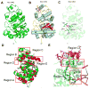
Structural diagrams were constructed using PyMOL. A. Overall structure of Nsa UNG shown in cartoon mode. B. Secondary structures of Nsa UNG. Helices are shown as blue column and beta strands are shown in red arrow. C. Active site of Nsa UNG. Nsa UNG structure is shown in cartoon mode and catalytic residues (Tyr 81, Asp 79, Gln 78, Asn168 and His230) are shown in licorice. D. Superimposition of Nsa UNG (in green) with human UNG (PDB: 1EMH, in red). The secondary structural differences are boxed in red, corresponding to structure-based sequence alignment shown in Fig. 2. E. Close-up view of interactions between Region B (in cyan) and Region C (in purple) in Nsa UNG. Residues involved in interactions are boxed in red.
Figure 4
Figure 4. Glycosylase activity of NsaUNG-deletion mutants
A. Purified Nsa UNG wt and mutant protein as shown in 12% SDS-PAGE gel. Lane1, protein standards; Lane 2, Nsa UNG wild type protein; Lane 3, Nsa UNG region B deletion mutant; Lane 4, Nsa UNG region C deletion mutant; B. The UDG assay profiles of of Nsa UNG-WT, Nsa UNG-deletion B, Nsa UNG-deletion C.
Figure 5
Figure 5. Representative Kinetic analysis of Nsa UNG-WT, Nsa UNG-D94A and hUNG
See Experimental Procedures under enzyme kinetic analysis for details. A. Nsa. UNG-WT. B. Nsa UNG-D94A. C. hUNG-WT. The assay for hUNG was performed similar to Nsa UNG except that the KCl concentration was 30 mM. Data are shown as average ± SD from three independent experiments.
Figure 6
Figure 6. Comparison of protein-DNA interactions between Nsa UNG and human UNG
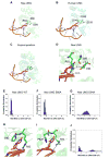
A. The interactions between Nsa UNG-R83 and modeled uracil-containing DNA (DNA model was taken from PDB 1EMH). B. The interactions between human UNG and Uracil-containing DNA (PDB 1EMH). C. Superimposition of the interactions between modeled Nsa UNG-DNA complex and human UNG-DNA complex. D. Close-up view of the networked salt-bridge in Nsa UNG that involves in DNA phosphate backbone interaction. The network of side chain interactions among R83, S86 and D94 are shown in licorice. Probability distribution of distance between R83 and DNA backbone in Nsa UNG-WT (E), Nsa UNG-S86A (F). Nsa UNG-D94A (G). R83-mediated interaction with DNA backbone in Nsa UNG-WT (H) and Nsa UNG-P82H (I). J. Probability distribution of distance between R83 and DNA backbone in Nsa UNG-P82H.
Figure 7
Figure 7. Structure of Nsa UNG-Y81G and its comparison with Nsa UNG-WT
A. The overall structure of Nsa UNG-Y81G. B. Superimposition of Nsa UNG-Y81G with Nsa UNG-WT. C. Close-up view of the local structural differences between Nsa UNG-Y81G and Nsa UNG-WT. The differences shown in red box are highlighted in licorice.
Figure 8
Figure 8. Interaction of Tyr residue with Phe residue in family 1 UNG enzymes
A. Human UNG (PDB 1UGH). B. E. coli UNG (PDB 1LQG). C. HSV UNG (PDB 1UDI). Protein structures are shown as cartoon, Tyr and Phe residues are shown as licorice.
Figure 9
Figure 9. Ugi inhibition and binding analysis
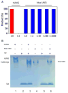
A. The reactions were performed as described under DNA golycosylase assay in Experimental Procedures with addition of indicated amount of Ugi protein. The concentrations of Nsa UNG versus Ugi are shown in molar ratio. B. Gel mobility shift analysis of binding between Ugi and hUNG/Nsa UNG [62]. After incubation of enzyme with/without Ugi, reaction products were separated on 15% native PAGE gel in Tris-glycing buffer (pH 8.5). Lane 1: human UNG (0.1 nmol); Lane 2: Ugi (1 nmol); lane 3: human UNG: Ugi = 1:10; Lane 4: Nsa UNG (0.1 nmol); Lane 5: Ugi (1 nmol); lane 6: Nsa UNG: Ugi = 1:10.
Figure 10
Figure 10. Potential interactions between Nsa UNG and Ugi and its comparison with human UNG
A. Superimposition between Nsa UNG and hUNG. B. Superimposition between Nsa UNG and human UNG in the presence of Ugi (Ugi was taken from PDB 1UGH). C. Horizontal 90 degree rotated of B to more clearly show the clash between Nsa UNG and Ugi. The electrostatic potential was calculated by Delphi 7.0 and visualized using UCSF Chimera. The positive potential region is colored in blue and the negative potential region is colored in red. The major differences of the Ugi interaction interface between Nsa UNG and hUNG are highlighted in yellow and red boxes. D. Modeled interactions between Nsa UNG and Ugi (PDB 1UGH). E. Interactions between human UNG and Ugi (PDB 1UGH). F. Close-up view of the superimposition between Nsa UNG and human UNG in the presence of Ugi.

References

    1. Lindahl T. Instability and decay of the primary structure of DNA. Nature. 1993;362:709–15. - PubMed
    1. Shapiro R. Damage to DNA caused by hydrolysis. In: Seeberg E, Kleppe K, editors. Chromosome Damage and Repair. Plenum Press; New York: 1981. pp. 3–18.
    1. Suzuki T, Yamaoka R, Nishi M, Ide H, Makino K. Isolation and characterization of a novel product, 2′-deoxyoxanosine, from 2′-deoxyguanosine, oligodeoxynucleotide and calf thymus DNA treated by nitrous-acid and nitric-oxide. J Am Chem Soc. 1996;118:2515–2516.
    1. Parikh SS, Putnam CD, Tainer JA. Lessons learned from structural results on uracil-DNA glycosylase. Mutat Res. 2000;460:183–99. - PubMed
    1. Krokan HE, Drablos F, Slupphaug G. Uracil in DNA--occurrence, consequences and repair. Oncogene. 2002;21:8935–48. - PubMed

Publication types

MeSH terms

LinkOut - more resources