Cytotoxic profiling of artesunic and betulinic acids and their synthetic hybrid compound on neurons and gliomas
- PMID: 28977877
- PMCID: PMC5617437
- DOI: 10.18632/oncotarget.18390
Cytotoxic profiling of artesunic and betulinic acids and their synthetic hybrid compound on neurons and gliomas
Abstract
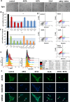
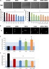
Gliomas are brain-born tumors with devastating impact on their brain microenvironment. Novel approaches employ multiple combinations of chemical compounds in synthetic hybrid molecules to target malignant tumors. Here, we report on the chemical hybridization approach exemplified by artesunic acid (ARTA) and naturally occurring triterpene betulinic acid (BETA). Artemisinin derived semisynthetic compound artesunic acid (ARTA) and naturally occurring triterpene BETA were used to synthetically couple to the hybrid compound termed 212A. We investigated the impact of 212A and its parent compounds on glioma cells, astrocytes and neurons. ARTA and BETA showed cytotoxic effects on glioma cells at micromolar concentrations. ARTA was more effective on rodent glioma cells compared to BETA, whereas BETA exhibited higher toxic effects on human glioma cells compared to ARTA. We investigated these compounds on non-transformed glial cells and neurons as well. Noteworthy, ARTA showed almost no toxic effects on astrocytes and neurons, whereas BETA as well as 212A displayed neurotoxicity at higher concentrations. Hence we compared the efficacy of the hybrid 212A with the combinational treatment of its parent compounds ARTA and BETA. The hybrid 212A was efficient in killing glioma cells compared to single compound treatment strategies. Moreover, ARTA and the hybrid 212A displayed a significant cytotoxic impact on glioma cell migration. Taken together, these results demonstrate that both plant derived compounds ARTA and BETA operate gliomatoxic with minor neurotoxic side effects. Altogether, our proof-of-principle study demonstrates that the chemical hybrid synthesis is a valid approach for generating efficacious anti-cancer drugs out of virtually any given structure. Thus, synthetic hybrid therapeutics emerge as an innovative field for new chemotherapeutic developments with low neurotoxic profile.
Keywords: artesunic acid; betulinic acid; cancer cytotoxicity; cell death; hybrid synthesis.
Conflict of interest statement
CONFLICTS OF INTERESTS The authors declare no competing financial conflicts of interest.
Figures


Similar articles
-
Chemical hybridization of sulfasalazine and dihydroartemisinin promotes brain tumor cell death.Sci Rep. 2021 Oct 21;11(1):20766. doi: 10.1038/s41598-021-99960-z. Sci Rep. 2021. PMID: 34675351 Free PMC article.
-
Sunitinib impedes brain tumor progression and reduces tumor-induced neurodegeneration in the microenvironment.Cancer Sci. 2015 Feb;106(2):160-70. doi: 10.1111/cas.12580. Epub 2015 Feb 15. Cancer Sci. 2015. PMID: 25458015 Free PMC article.
-
Synthesis and study of cytotoxic activity of 1,2,4-trioxane- and egonol-derived hybrid molecules against Plasmodium falciparum and multidrug-resistant human leukemia cells.Eur J Med Chem. 2014 Mar 21;75:403-12. doi: 10.1016/j.ejmech.2014.01.043. Epub 2014 Jan 31. Eur J Med Chem. 2014. PMID: 24561670
-
Novel anti-angiogenic therapies for malignant gliomas.Lancet Neurol. 2008 Dec;7(12):1152-60. doi: 10.1016/S1474-4422(08)70260-6. Lancet Neurol. 2008. PMID: 19007739 Review.
-
Current status of local therapy in malignant gliomas--a clinical review of three selected approaches.Pharmacol Ther. 2013 Sep;139(3):341-58. doi: 10.1016/j.pharmthera.2013.05.003. Epub 2013 May 18. Pharmacol Ther. 2013. PMID: 23694764 Review.
Cited by
-
The multifaceted NF-kB: are there still prospects of its inhibition for clinical intervention in pediatric central nervous system tumors?Cell Mol Life Sci. 2021 Sep;78(17-18):6161-6200. doi: 10.1007/s00018-021-03906-7. Epub 2021 Jul 31. Cell Mol Life Sci. 2021. PMID: 34333711 Free PMC article. Review.
-
Recent Developments in the Functionalization of Betulinic Acid and Its Natural Analogues: A Route to New Bioactive Compounds.Molecules. 2019 Jan 19;24(2):355. doi: 10.3390/molecules24020355. Molecules. 2019. PMID: 30669472 Free PMC article. Review.
-
Betulinic Acid-Brosimine B Hybrid Compound Has a Synergistic Effect with Imatinib in Chronic Myeloid Leukemia Cell Line, Modulating Apoptosis and Autophagy.Pharmaceuticals (Basel). 2023 Apr 13;16(4):586. doi: 10.3390/ph16040586. Pharmaceuticals (Basel). 2023. PMID: 37111343 Free PMC article.
-
TRPM8-regulated calcium mobilization plays a critical role in synergistic chemosensitization of Borneol on Doxorubicin.Theranostics. 2020 Aug 13;10(22):10154-10170. doi: 10.7150/thno.45861. eCollection 2020. Theranostics. 2020. PMID: 32929340 Free PMC article.
-
Oxidative Stress and Chronic Myeloid Leukemia: A Balance between ROS-Mediated Pro- and Anti-Apoptotic Effects of Tyrosine Kinase Inhibitors.Antioxidants (Basel). 2024 Apr 13;13(4):461. doi: 10.3390/antiox13040461. Antioxidants (Basel). 2024. PMID: 38671909 Free PMC article. Review.
References
-
- Eyupoglu IY, Buchfelder M, Savaskan NE. Surgical resection of malignant gliomas-role in optimizing patient outcome. Nat Rev Neurol. 2013;9:141–151. - PubMed
-
- Nishikawa R. Standard therapy for glioblastoma—a review of where we are. Neurol Med Chir (Tokyo) 2010;50:713–719. - PubMed
-
- Stupp R, Brada M, van den Bent MJ, Tonn JC, Pentheroudakis G. ESMO Guidelines Working Group. High-grade glioma: ESMO Clinical Practice Guidelines for diagnosis, treatment and follow-up. Ann Oncol / ESMO. 2014;25:iii93–101. - PubMed
-
- Wen PY, Kesari S. Malignant gliomas in adults. N Engl J Med. 2008;359:492–507. - PubMed
LinkOut - more resources
Full Text Sources
Other Literature Sources

