

Endoplasmic reticulum aminopeptidase 2 involvement in metastasis of oral cavity squamous cell carcinoma discovered by proteome profiling of primary cancer cells
- PMID: 28977897
- PMCID: PMC5617457
- DOI: 10.18632/oncotarget.18680
Endoplasmic reticulum aminopeptidase 2 involvement in metastasis of oral cavity squamous cell carcinoma discovered by proteome profiling of primary cancer cells
Abstract
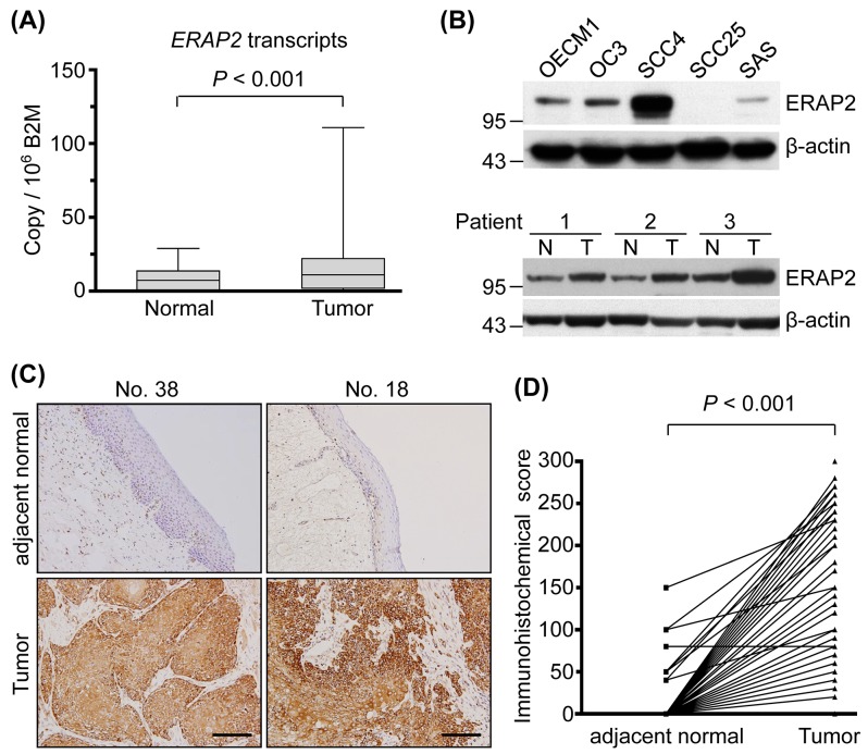
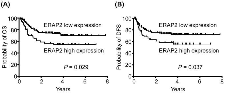
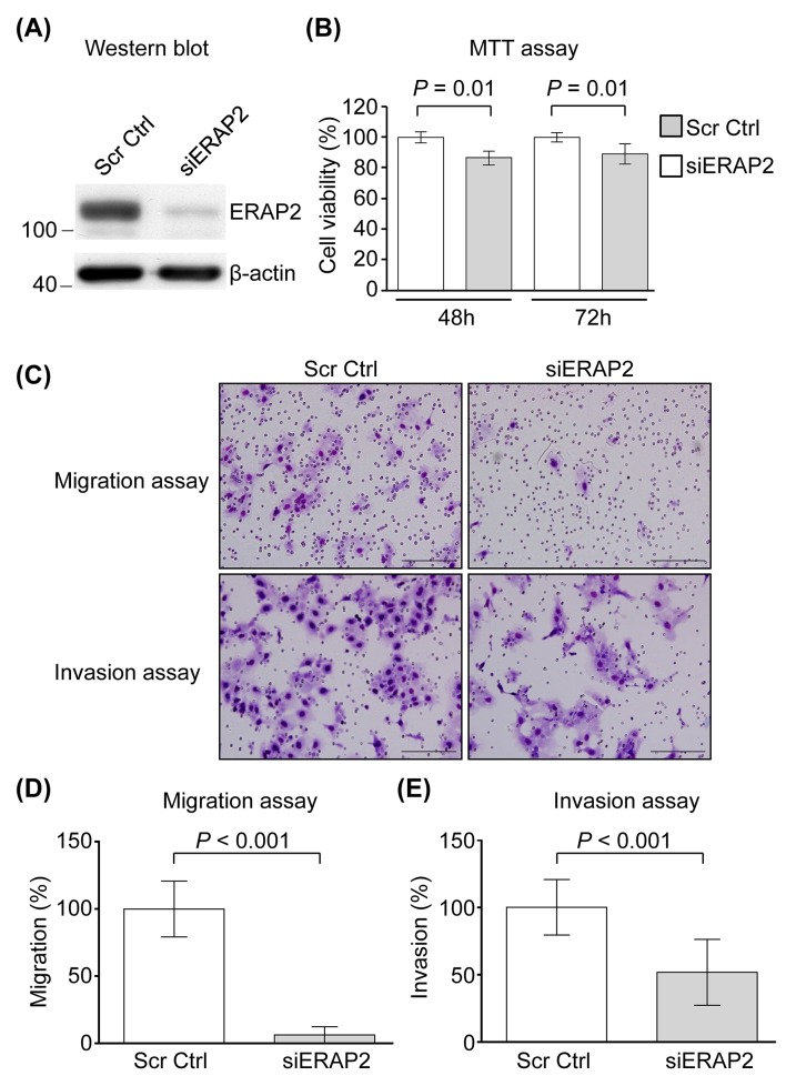
Oral cavity squamous cell carcinoma (OSCC) is a leading cause of cancer-related deaths worldwide and associated with poor prognosis and mortality. Discovery of proteins that can improve OSCC treatment is needed. Using comparative proteome profiling of primary cells derived from OSCC and adjacent noncancerous epithelium, endoplasmic reticulum aminopeptidases 2 (ERAP2) has been identified as an OSCC-associated protein. Compared with the adjacent normal tissues, ERAP2 levels were determined to be significantly elevated in OSCC tissues using quantitative real-time PCR and immunohistochemistry. Importantly, overexpression of ERAP2 was associated with positive N stage, advanced overall stage, positive perineural invasion, and tumor depth (P = 0.041, 0.015, 0.010, and 0.032, respectively). The overall survival rates of patients without and with the ERAP2 overexpression were 71.9% and 56.0%, respectively (P = 0.029). Furthermore, knockdown of ERAP2 inhibited the migration and invasion abilities of OSCC cells. Our results collectively show that ERAP2 overexpression is associated with the cervical metastasis and poorer prognosis of OSCC.
Keywords: ERAP2; OSCC; metastasis; prognosis; proteome.
Conflict of interest statement
CONFLICTS OF INTEREST None declared.
Figures
Similar articles
-
Secretome profiling of primary cells reveals that THBS2 is a salivary biomarker of oral cavity squamous cell carcinoma.J Proteome Res. 2014 Nov 7;13(11):4796-807. doi: 10.1021/pr500038k. Epub 2014 Apr 16. J Proteome Res. 2014. PMID: 24708169
-
Low-molecular-mass secretome profiling identifies HMGA2 and MIF as prognostic biomarkers for oral cavity squamous cell carcinoma.Sci Rep. 2015 Jul 3;5:11689. doi: 10.1038/srep11689. Sci Rep. 2015. PMID: 26138061 Free PMC article.
-
Overexpression of BST2 is associated with nodal metastasis and poorer prognosis in oral cavity cancer.Laryngoscope. 2014 Sep;124(9):E354-60. doi: 10.1002/lary.24700. Epub 2014 May 27. Laryngoscope. 2014. PMID: 24706327
-
Potential biomarkers for oral squamous cell carcinoma: proteomics discovery and clinical validation.Proteomics Clin Appl. 2015 Feb;9(1-2):86-97. doi: 10.1002/prca.201400091. Epub 2014 Dec 28. Proteomics Clin Appl. 2015. PMID: 25431113 Review.
-
Endoplasmic Reticulum Aminopeptidase 2, a common immunological link to adverse pregnancy outcomes and cancer clearance?Placenta. 2017 Aug;56:40-43. doi: 10.1016/j.placenta.2017.03.012. Epub 2017 Mar 18. Placenta. 2017. PMID: 28343731 Free PMC article. Review.
Cited by
-
The association of XRCC1 polymorphism with osteosarcoma risk, clinicopathologic features, and prognosis in a Chinese Han population.Cancer Manag Res. 2018 Oct 25;10:4959-4967. doi: 10.2147/CMAR.S177452. eCollection 2018. Cancer Manag Res. 2018. PMID: 30464596 Free PMC article.
-
Unveiling the impact of ERAP1 and ERAP2 on migration, angiogenesis and ER stress response.Front Cell Dev Biol. 2025 Mar 28;13:1564649. doi: 10.3389/fcell.2025.1564649. eCollection 2025. Front Cell Dev Biol. 2025. PMID: 40226591 Free PMC article. Review.
-
The Role of Antigen Processing and Presentation in Cancer and the Efficacy of Immune Checkpoint Inhibitor Immunotherapy.Cancers (Basel). 2021 Jan 4;13(1):134. doi: 10.3390/cancers13010134. Cancers (Basel). 2021. PMID: 33406696 Free PMC article. Review.
-
ERAP2 as a potential biomarker for predicting gemcitabine response in patients with pancreatic cancer.Aging (Albany NY). 2022 Oct 8;14(19):7941-7958. doi: 10.18632/aging.204324. Epub 2022 Oct 8. Aging (Albany NY). 2022. PMID: 36214762 Free PMC article.
-
Endoplasmic Reticulum Associated Aminopeptidase 2 (ERAP2) Is Released in the Secretome of Activated MDMs and Reduces in vitro HIV-1 Infection.Front Immunol. 2019 Jul 16;10:1648. doi: 10.3389/fimmu.2019.01648. eCollection 2019. Front Immunol. 2019. PMID: 31379846 Free PMC article.
References
-
- Jemal A, Bray F, Center MM, Ferlay J, Ward E, Forman D. Global cancer statistics. CA Cancer J Clin. 2011;61:69–90. - PubMed
-
- Cooper JS, Pajak TF, Forastiere AA, Jacobs J, Campbell BH, Saxman SB, Kish JA, Kim HE, Cmelak AJ, Rotman M, Machtay M, Ensley JF, Chao KS, et al. Postoperative concurrent radiotherapy and chemotherapy for high-risk squamous-cell carcinoma of the head and neck. N Engl J Med. 2004;350:1937–1944. - PubMed
-
- Hsu CW, Yu JS, Peng PH, Liu SC, Chang YS, Chang KP, Wu CC. Secretome profiling of primary cells reveals that THBS2 is a salivary biomarker of oral cavity squamous cell carcinoma. J Proteome Res. 2014;13:4796–4807. - PubMed
-
- Wu CC, Chu HW, Hsu CW, Chang KP, Liu HP. Saliva proteome profiling reveals potential salivary biomarkers for detection of oral cavity squamous cell carcinoma. Proteomics. 2015;15:3394–3404. - PubMed
LinkOut - more resources
Full Text Sources
Other Literature Sources
