. 2017 Jun 27;8(37):61742-61760.
doi: 10.18632/oncotarget.18687. eCollection 2017 Sep 22.

Overexpression of stathmin plays a pivotal role in the metastasis of esophageal squamous cell carcinoma

Affiliations

Overexpression of stathmin plays a pivotal role in the metastasis of esophageal squamous cell carcinoma

Gaijing Han et al. Oncotarget. .

Abstract

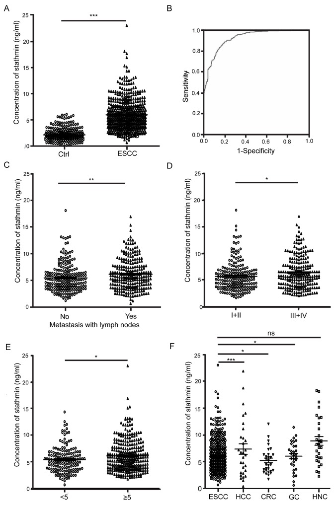
Purpose: Esophageal squamous cell carcinoma (ESCC) is a serious malignant tumor that affects human health. We analyzed the correlation between serum stathmin level and ESCC and elucidated the molecular mechanisms of stathmin's promotion of ESCC cell invasion and metastasis.

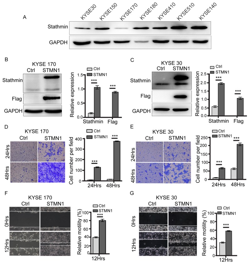
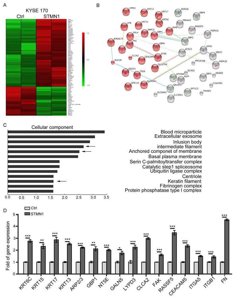
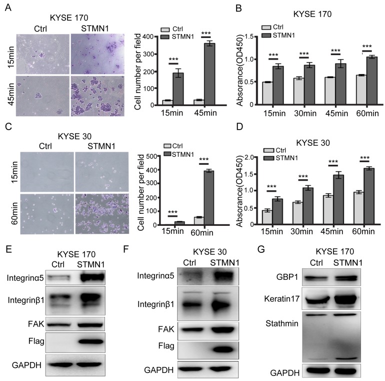
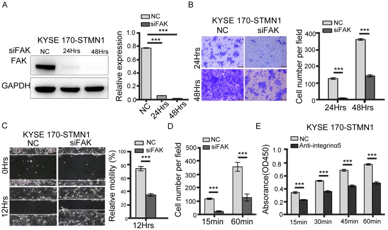
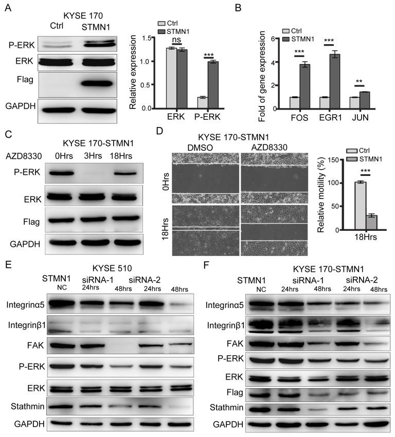
Methods: Stathmin level in ESCC and healthy control serum were detected by enzyme-linked immunosorbent assay (ELISA), and the clinical parameters were analyzed. We established ESCC cells with stathmin overexpression or knockdown and then evaluated the effects of stathmin on invasion and metastasis in ESCC. Differentially expressed genes were analyzed by Human Transcriptome Array and confirmed by RT-PCR. The expression levels of the integrin family, focal adhesion kinase (FAK) and extracellular signal-regulated kinase (ERK) were detected by immunoblotting.

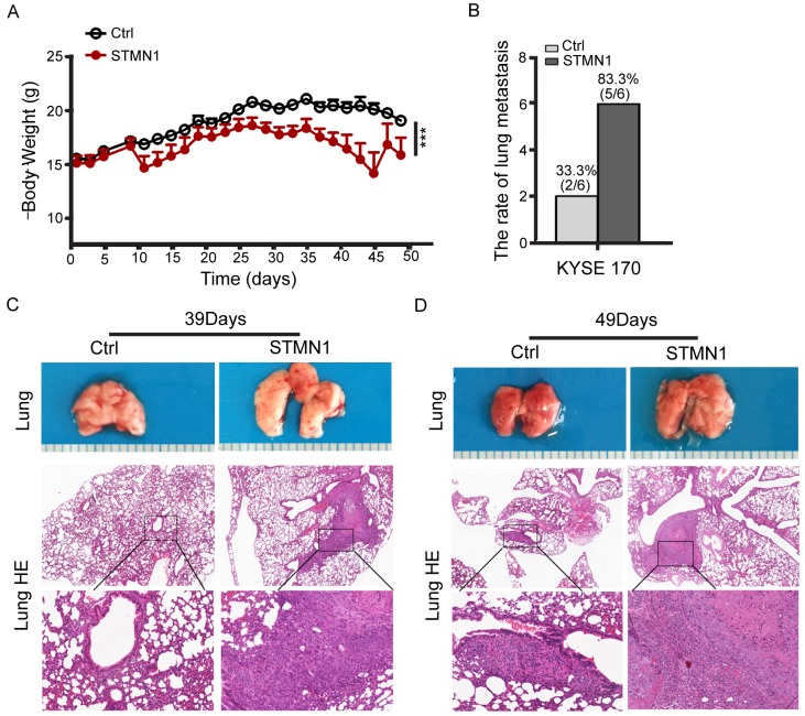
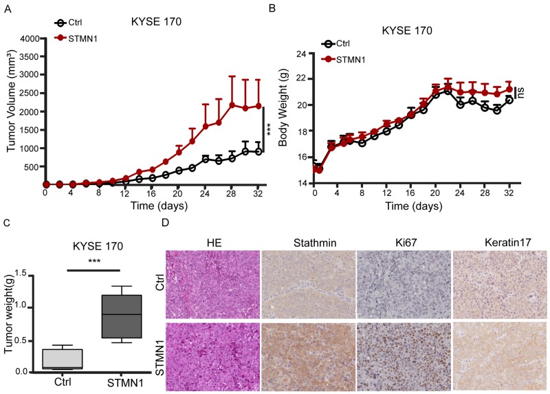
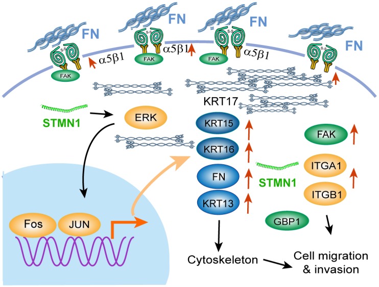
Results: Serum levels of stathmin were significantly higher in ESCC than in control serum and associated with lymph node metastasis, tumor stage and size. Furthermore, we found that stathmin promoted migration and invasion of ESCC cells in vitro and in vivo. In addition, we confirmed that the activation of the integrinα5β1/FAK/ERK pathway is increased in stathmin-overexpression cells and accelerates cell motility by enhancing cell adhesion ability.

Conclusion: Stathmin may predict a potential metastasis biomarker for ESCC.

Keywords: ERK; ESCC; FAK; integrinα5β1; stathmin.


Conflict of interest statement

CONFLICTS OF INTEREST The authors have declared that no competing interests exist.

Figures

Figure 1
Figure 1. Clinical significance of serum stathmin in ESCC
(A) The concentration of stathmin in ESCC patients was significantly higher than that in healthy controls (P<0.0001). (B) ROC curve analysis of the sensitivity and specificity of serum stathmin in ESCC and healthy controls (P<0.005). (C) Stathmin levels in ESCC lymph node metastasis were significantly higher than those in non-metastatic (P<0.05). (D) Stathmin levels in ESCC stage III+IV were higher than those in stage I+II (P<0.005). (E) Comparison of stathmin levels among different cancers, include ESCC, hepatocellular carcinoma (HCC), colorectal cancer (CRC), gastric cancer (GC) and head and neck cancer (HNC).
Figure 2
Figure 2. Stathmin promoted ESCC cell invasion and migration
(A) Stathmin expression in seven ESCC cell lines was examined by western blotting. GAPDH was used as a loading control. (B, C) Immunoblotting was used to analyze the stathmin protein level in KYSE 170 and KYSE 30 cells. Control (Ctrl) represents KYSE 170 or KYSE 30 cells transfected with the control plasmid; STMN1 represents KYSE 170 or KYSE 30 cells transfected with STMN1-plasmid. (D, E) The transwell invasion system demonstrated an enhanced invasion capacity of the KYSE 170 and KYSE 30 stathmin-overexpression groups compared with controls. Images of invading cells were captured by phase contrast microscopy at 200× magnification. The y-axis represents the number of invading cells. (F, G) A wound-healing assay was performed to investigate the migratory potential of KYSE 170 and KSYE 30 cells after stathmin levels changed. In the quantitative migration assay results, the y-axis represents the migration rate relative to that of control cells. Stathmin overexpression significantly promoted the migration ability of both cell lines. All assays were replicated, and results are presented as the mean±SD (*, P<0.05).
Figure 3
Figure 3. Gene expression changes in stathmin-overexpressing ESCC cells
(A) Heatmap representation of 90 genes showing significant differential expression between the STMN1 group and the control group. A color scale for the normalized expression data is shown on the right side of the microarray heatmap (green represents downregulated genes, whereas red represents upregulated genes). (B) The gene changes were functionally analyzed using the online tool STRING. (C) The upregulated genes were functionally classified based on their cellular components using the DAVID functional annotation clustering tool. (D) The mRNA levels of candidate genes were determined using real-time PCR. The data represent the mean±SD of relative mRNA levels versus control cells. (*, P<0.05; **, P<0.01; ***, P<0.001).
Figure 4
Figure 4. Stathmin increased ESCC cell adhesion to FN by promoting integrinα5β1/FAK expression
(A, C) Adhesion experiments showed that the level of cell adhesion to the FN-coated slides was significantly increased in the STMN1 group compared with that in the control group. (B, D) Adhesion experiments using FN-coated 96-well plates showed that the absorbance value of the STMN1 group was significantly higher than that of the control group. (E, F) The protein levels of integrinα5β1 and FAK were much higher in the STMN1 group than in the control group. (G) Immunoblotting identified differential keratin17 and GBP1 protein expression between theSTMN1 group and Ctrl group (***, P<0.001).
Figure 5
Figure 5. FAK knockdown inhibited metastasis of stathmin-overexpressing cells
(A) Western blotting showed that FAK-specific siRNA decreased FAK expression. (B) The transwell assay revealed that the invasion number of the FAK-knockdown group was significantly decreased compared with that of the NC group. (C) The wound-healing assay was performed to detect the migratory potential of FAK-knockdown KYSE 170-STMN1 cells and showed that the motility of the FAK-knockdown group was decreased compared with that of the NC group. (D) The cell adhesion assay to investigate cell adhesion ability showed that the level of cell adhesion to FN was lower in the FAK-knockdown group than in the NC group. (E) Cells were incubated with 10 ug/ml antibody against integrinα5 45min before plating on FN. Histograms represent the absorbance (OD450) (***, P<0.001).
Figure 6
Figure 6. Stathmin overexpression increased ESCC cell lung metastasis in vivo
(A) ESCC cells were implanted into mouse tail veins, and body weight was measured every two days. Body weight was significantly lower in mice injected with STMN1 cells than in the control group (P<0.001). (B) Statistical results of lung metastasis. (C) After 39 days, the STMN1 cell-injected mice had developed metastatic nodules, whereas the control group had not. (D) After 49 days, both groups of ESCC cell-injected mice developed metastatic nodules, although the lung metastasis area was larger in the STMN1-injected mice.
Figure 7
Figure 7. Stathmin overexpression increased xenografted tumor growth
(A, C) Control cells and STMN1 cells were orthotopically inoculated into nude mice. Tumor volume and tumor weight significantly increased in mice injected with STMN1 group compared with the control group. (B) Body weight was measured every two days after injection, and no significant difference was observed between the two groups. (D) IHC staining showed strong staining in the tissue sections for stathmin, Ki67 and keratin 17 in the STMN1 group.
Figure 8
Figure 8. Stathmin regulated ERK activation
(A) Western blot analysis showed that the protein level of P-ERK in the STMN1 group was significantly higher than that of the control group. (B) RT-PCR analysis of the mRNA levels of ERK downstream transcription factors such as FOS, EGR1, and JUN revealed significantly higher levels in the STMN1group than in the control group. (C) KYSE 170-STMN1 cells were treated with ERK inhibitors AZD8330; western blot analysis showed that ERK phosphorylation was inhibited. (D) The effects of ERK phosphorylation on the migration ability of KYSE 170-STMN1 cells were analyzed by wound-healing assay. The results showed that inhibition of ERK phosphorylation in KYSE 170-STMN1 cells markedly reduced cell motility. (E) Stathmin expression in KYSE 510 and (F) KYSE 170-STMN1 cells was knocked down by two different STMN1-specific siRNAs (siRNA-1 and siRNA-2), and the activation of the integrinα5β1/FAK/ERK pathway was measured by immunoblotting (ns, P>0.05; *, P<0.05; ***, P<0.001).
Figure 9
Figure 9. Illustration of the signaling pathway for ESCC cell migration induced by stathmin overexpression
Overexpression of stathmin increased the number of cellular adhesion molecules and the level cytokeratin 17 of intermediate filaments, promoting cell invasion and migration via the FN/integrinα5β1/FAK signaling pathway. The red arrows indicate the upregulated genes.

Similar articles

Cited by

References

    1. Torre LA, Bray F, Siegel RL, Ferlay J, Lortet-Tieulent J, Jemal A. Global cancer statistics, 2012. CA Cancer J Clin. 2015;65:87–108. - PubMed
    1. Zhu ML, Shi TY, Hu HC, He J, Wang M, Jin L, Yang YJ, Wang JC, Sun MH, Chen H, Zhao KL, Zhang Z, Chen HQ, et al. Polymorphisms in the ERCC5 gene and risk of esophageal squamous cell carcinoma (ESCC) in eastern Chinese populations. PLoS One. 2012;7:e41500. - PMC - PubMed
    1. Qiu H, Mao Y, Gu Y, Zhu J, Wang Y, Zeng J, Huang N, Liu Q, Yang Y. The potential of photodynamic therapy to treat esophageal candidiasis coexisting with esophageal cancer. J Photochem Photobiol B. 2014;130:305–309. - PubMed
    1. Gupta GP, Massagué J. Cancer metastasis: building a framework. Cell. 2006;127:679–695. - PubMed
    1. Worni M, Martin J, Gloor B, Pietrobon R, D'Amico TA, Akushevich I, Berry MF. Does surgery improve outcomes for esophageal squamous cell carcinoma? An analysis using the surveillance epidemiology and end results registry from 1998 to 2008. J Am Coll Surg. 2012;215:643–651. - PMC - PubMed

LinkOut - more resources