. 2017 Jun 27;8(37):61846-61860.
doi: 10.18632/oncotarget.18705. eCollection 2017 Sep 22.

2-Acetylamino-3-[4-(2-acetylamino-2-carboxyethylsulfanylcarbonylamino) phenyl carbamoylsulfanyl] propionic acid, a glutathione reductase inhibitor, induces G2/M cell cycle arrest through generation of thiol oxidative stress in human esophageal cancer cells

Affiliations

2-Acetylamino-3-[4-(2-acetylamino-2-carboxyethylsulfanylcarbonylamino) phenyl carbamoylsulfanyl] propionic acid, a glutathione reductase inhibitor, induces G2/M cell cycle arrest through generation of thiol oxidative stress in human esophageal cancer cells

Xia Li et al. Oncotarget. .

Abstract

Esophageal squamous cell carcinoma (ESCC) is a highly malignant cancer with poor response to both of chemotherapy and radiotherapy. 2-Acetylamino-3-[4-(2-acetylamino-2-carboxyethylsulfanylcarbonylamino) phenyl carbamoylsulfanyl] propionic acid (2-AAPA), an irreversible inhibitor of glutathione reductase (GR), is able to induce intracellular oxidative stress, and has shown anticancer activity in many cancer cell lines. In this study, we investigated the effects of 2-AAPA on the cell proliferation, cell cycle and apoptosis and aimed to explore its mechanism of action in human esophageal cancer TE-13 cells. It was found that 2-AAPA inhibited growth of ESCC cells in a dose-dependent manner and it did not deplete reduced glutathione (GSH), but significantly increased the oxidized form glutathione (GSSG), resulting in decreased GSH/GSSG ratio. In consequence, significant reactive oxygen species (ROS) production was observed. The flow cytometric analysis revealed that 2-AAPA inhibited growth of esophageal cancer cells through arresting cell cycle in G2/M phase, but apoptosis-independent mechanism. The G2/M arrest was partially contributed by down-regulation of protein expression of Cdc-25c and up-regulation of phosphorylated Cdc-2 (Tyr15), Cyclin B1 (Ser147) and p53. Meanwhile, 2-AAPA-induced thiol oxidative stress led to increased protein S-glutathionylation, which resulted in α-tubulin S-glutathionylation-dependent depolymerization of microtubule in the TE-13 cells. In conclusion, we identified that 2-AAPA as an effective thiol oxidative stress inducer and proliferation of TE-13 cells were suppressed by G2/M phase cell cycle arrest, mainly, through α-tubulin S-glutathionylation-mediated microtubule depolymerization. Our results may introduce new target and approach for esophageal cancer therapy through generation of GR-mediated thiol oxidative stress.

Keywords: S-glutathionylation; cell cycle arrest; glutathione reductase; microtubule depolymerization; oxidative stress.


Conflict of interest statement

CONFLICTS OF INTEREST No potential conflicts of interest were disclosed.

Figures

Figure 1
Figure 1. 2-AAPA induced cytotoxicity in ESCC cells
The ESCC cells ((A), TE-13; (B), KYSE-450; (C), KYSE-510) were treated with various concentrations of 2-AAPA for 2 or 3 days. Cell survival rates were determined by the MTT assay. The data are presented as the mean ± SD of three independent experiments.
Figure 2
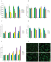
Figure 2. Intracellular GR activity reduction, thiol oxidative stress generation and protein S-glutathionylation induced by 2-AAPA
(A) GR activity in the control and 2-AAPA-treated TE-13 cells. The data are presented as unit/mg protein. (B, C & D) Evaluation of intracellular GSH, GSSG, and GSH/GSSG ratio in the control and 2-AAPA-treated TE-13 cells. (E) ROS production in the control and 2-AAPA-treated TE-13 cells. The data are presented as fluorescence intensity. (F) 2-AAPA increased protein S-glutathionylation in TE-13 cells. The cells were treated with the indicated concentrations of 2-AAPA for 1 h, followed by fixation, and fluorescent staining as described in Materials and Methods. The data are derived from one of the three independent experiments. Cells were viewed under a fluorescent microscope. *, P < 0.05 compared with control group. All the data are expressed as the mean ± SD of three independent experiments.
Figure 3
Figure 3. Effect of 2-AAPA on cell cycle distribution in TE-13 cells
(A) The histograms of the TE-13 cells treated with various concentrations of 2-AAPA for 24 h and 48 h are presented. (B & C) The bar presentations reflects the quantification of cell cycle distribution at 24 h and 48 h, respectively. Results are presented as the mean ± SD of three independent experiments. *, P < 0.05 indicates statistical significance in 2-AAPA treated groups as compared to the control.
Figure 4
Figure 4. Effect of 2-AAPA on the induction of apoptosis in TE-13 cells
TE-13 cells were treated with different concentrations of 2-AAPA for 24 h, 48 h and 72 h and then stained with Annexin V and PI. (A) Apoptotic assays were performed by flow cytometry. (B) The percentages of apoptotic cells are presented for 24 h, 48 h and 72 h treatments. Results are presented as the mean ± SD of three independent experiments.
Figure 5
Figure 5. Identification of proteins potentially regulated by 2-AAPA in TE-13 cells
(A) Assessment of the possible signaling pathways involved in G2/M phase cell cycle arrest and apoptosis. TE-13 cells were treated with 2-AAPA at the indicated concentrations for 48 h and 72 h, followed by the Western blot analysis for detection of Cdc-2, Cdc-25c, p-Cdc-2, Cyclin B1, p-Cyclin B1, p21, ATM, p-ATM, p53, p-p53, Bcl-2 and Bax expressions. β-actin was used as a loading control. The data are derived from one of the three independent experiments. (B and C) TE-13 cells were treated the same as in (A). Protein S-glutathionyaltion on α/β-tubulin was examined by reciprocal immunmoprecipitation and western blotting analysis as indicated.
Figure 6
Figure 6. 2-AAPA induces microtubule depolymerization in TE-13 cells
TE-13 cells were treated with the indicated concentrations of 2-AAPA for 5 h, followed by fixation, permeabilization and indirect immunofluorescent analysis with an anti-α-tubulin-FITC. Nuclei were stained with DAPI. Taxol and vincristine sulfate were employed as positive controls for microtubule stabilization and microtubule depolymerization, respectively. Fluorescent images were captured by an Olympus Fluoview FV1200 microscope. The data are derived from one of the three independent experiments.
Figure 7
Figure 7. Effect of 2-AAPA on cell morphology in TE-13 cells
TE-13 cells were treated with 40 and 60 μM of 2-AAPA. Some of the cells undergoing morphological changes are marked by red circles.

Similar articles

Cited by

References

    1. Peng X, Xue H, Lu L, Shi P, Wang J, Wang J. Accumulated promoter methylation as a potential biomarker for esophageal cancer. Oncotarget. 2017;8:679–691. doi: 10.18632/oncotarget.13510. - DOI - PMC - PubMed
    1. Torre LA, Bray F, Siegel RL, Ferlay J, Lortet-Tieulent J, Jemal A. Global cancer statistics, 2012. CA Cancer J Clin. 2015;65:87–108. - PubMed
    1. Ren QG, Yang SL, Hu JL, Li PD, Chen YS, Wang QS. Evaluation of HO-1 expression, cellular ROS production, cellular proliferation and cellular apoptosis in human esophageal squamous cell carcinoma tumors and cell lines. Oncol Rep. 2016;35:2270–2276. - PubMed
    1. Hsieh MS, Yang PW, Wong LF, Lee JM. The AXL receptor tyrosine kinase is associated with adverse prognosis and distant metastasis in esophageal squamous cell carcinoma. Oncotarget. 2016;7:36956–36970. doi: 10.18632/oncotarget.9231. - DOI - PMC - PubMed
    1. Lin Y, Totsuka Y, He Y, Kikuchi S, Qiao Y, Ueda J, Wei W, Inoue M, Tanaka H. Epidemiology of esophageal cancer in Japan and China. J Epidemiol. 2013;23:233–242. - PMC - PubMed