. 2017 Jun 12;8(40):67227-67240.
doi: 10.18632/oncotarget.18438. eCollection 2017 Sep 15.

Intrinsic remote conditioning of the myocardium as a comprehensive cardiac response to ischemia and reperfusion

Affiliations

Intrinsic remote conditioning of the myocardium as a comprehensive cardiac response to ischemia and reperfusion

Noemi Pavo et al. Oncotarget. .

Abstract

We have previously shown that distal anterior wall ischemia/reperfusion induces gene expression changes in the proximal anterior myocardial area, involving genes responsible for cardiac remodeling. Here we investigated the molecular signals of the ischemia non-affected remote lateral and posterior regions and present gene expression profiles of the entire left ventricle by using our novel and straightforward method of 2D and 3D image reconstruction. Five or 24h after repetitive 10min ischemia/reperfusion without subsequent infarction, pig hearts were explanted and myocardial samples from 52 equally distributed locations of the left ventricle were collected. Expressional changes of seven genes of interest (HIF-1α; caspase-3, transcription factor GATA4; myocyte enhancer factor 2C /MEF2c/; hexokinase 2 /HK2/; clusterin /CLU/ and excision repair cross-complementation group 4 /ERCC4/) were measured by qPCR. 2D and 3D gene expression maps were constructed by projecting the fold changes on the NOGA anatomical mapping coordinates. Caspase-3, GATA4, HK2, CLU, and ERCC4 were up-regulated region-specifically in the ischemic zone at 5 h post ischemia/reperfusion injury. Overexpression of GATA4, clusterin and ERCC4 persisted after 24 h. HK2 showed strong up-regulation in the ischemic zone and down-regulation in remote areas at 5 h, and was severely reduced in all heart regions at 24 h. These results indicate a quick onset of regulation of apoptosis-related genes, which is partially reversed in the late phase of ischemia/reperfusion cardioprotection, and highlight variations between ischemic and unaffected myocardium over time. The NOGA 2D and 3D construction system is an attractive method to visualize expressional variations in the myocardium.

Keywords: LV remodelling; NOGA mapping; cardioprotection; gene expression; ischemia/reperfusion.


Conflict of interest statement

CONFLICTS OF INTEREST None.

Figures
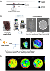
Figure 1
Figure 1. Study design and principle of NOGA-guided image-omics
(A) Timeline of the protocol and the three groups. Gene expression profiles of the whole LV was determined either without intervention (Group Control), or 5h (Group I/R-5h) or 24h (Group I/R-24h) after 3×10 min I/R by repetitive inflation/deflation of an intracoronary balloon placed in the mid part of the porcine left anterior descending coronary artery. (B) Method of the myocardial sampling: the explanted heart (without the right ventricular part) was placed on a 50 mL Falcon tube, and 52 myocardial biopsies were collected with a skin biopsy needle with an equal distribution of the sampling locations throughout the entire left ventricle. The biopsy samples were labelled in accordance to the biopsy location, and transferred immediately into RNALater. (C) Schematic illustration of the image-omics of the gene expression maps. Using mapping principles of the myocardial viability map, sampling locations were detected in in vitro epicardial (surface) NOGA-mapping, using the location point record principles. The voltage values recorded by NOGA in the distinct locations were replaced by the respective values of fold changes in gene expressions gathered from the excised tissue samples. (D) Delineation of the ischemic and remote areas on the 5h polar map images with transposition of the same area onto the control and 24h polar maps. Gene expression values of the corresponding ischemic and remote areas of the animals in control, 5h and 24h groups were collectively compared for statistical evaluations.
Figure 2
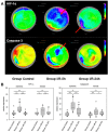
Figure 2. Spatiotemporal 2D bulls-eye display of HIF-1α, and caspase-3 gene expression of the entire left ventricle after repetitive ischemia/reperfusion (r-I/R)
(A) Time-dependent presentation of the different gene expression patterns of HIF-1α, and caspase-3, of the LV of animals in groups control, I/R-5h and I/R-24h after repetitive (3 times) 10 min I/R without consecutive myocardial infarction. Temporary overexpression of HIF-1α and caspase-3 at 5h (red arrow) was detected. Expression levels of HIF-1α and caspase-3 were reduced to baseline levels after 24h, except for part of the remote area with HIF-1α upregulation. Pink and blue colors represent up-regulation of genes; green represents baseline values, while yellow and red areas show down-regulation of the respective genes. (B) Boxplots of values of the ischemic and remote zone. *: p<0.05, **: p>0.01, ***: p<0.001. n.s.: not significant.
Figure 3
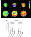
Figure 3. Spatiotemporal 2D bulls-eye display of GATA4 and myocyte enhancer factor 2C (MEF2c) gene expression of the entire left ventricle after repetitive ischemia/reperfusion (r-I/R)
(A) Time-dependent presentation of the different gene expression patterns of GATA4 and MEF2c of the LV of animals in groups control, I/R-5h and I/R-24h after repetitive (3 times) 10 min I/R without consecutive myocardial infarction. Mildly increasing upregulation of GATA-4 in the ischemic area at 5 and 24h (red arrows) was detected. Only mild upregulation of MEFC2 in the ischemic area was encountered. Pink and blue colors represent up-regulation of genes; green represents baseline values, while yellow and red areas show down-regulation of the respective genes. (B) Boxplots of values of the ischemic and remote zone. *: p<0.05, **: p>0.01, ***: p<0.001. n.s.: not significant.
Figure 4
Figure 4. Image-omics (2D and 3D modeling) of the repetitive ischemia/reperfusion (I/R) induced gene expression pattern of hexokinase 2 (HK2)
(A) Representative 3D (top row) and 2D bulls-eye maps (bottom row) of the left ventricle (LV) showing the expression patterns of HK-2. Marked upregulation of HK2 was found in the ischemia-affected apical myocardial region and the border zone with concomitant downregulation in the remote myocardial area (yellow arrow) at 5h. At 24h, HK2 expression was severely downregulated in all myocardial regions. (B) Boxplots of values of the ischemic and remote zone. *: p<0.05, **: p>0.01, ***: p<0.001.
Figure 5
Figure 5. Image-omics (2D and 3D modeling) of the repetitive ischemia/reperfusion (I/R) induced gene expression pattern of clusterin (CLU)
(A) Representative 3D (top row) and 2D bulls-eye maps (bottom row) of the left ventricle (LV) showing the expression pattern of CLU. Moderate up-regulation in part of the ischemic zone was detected at 5h (red arrow), with a high degree of variation within the remote myocardium. (B) Boxplots of values of the ischemic and remote zone. *: p<0.05, **: p>0.01, ***: p<0.001. n.s.: not significant.
Figure 6
Figure 6. Image-omics (2D and 3D modeling) of the repetitive ischemia/reperfusion (I/R) induced gene expression pattern of excision repair cross-complementation group 4 (ERCC4)
(A) Representative 3D (top row) and 2D bulls-eye maps (bottom row) of the left ventricle (LV) showing the expression pattern of ERCC4. Moderate up-regulation is shown in the ischemic zone at both 5h and 24h (red arrows), with little changes between the two time points. (B) Boxplots of values of the ischemic and remote zone. *: p<0.05, **: p>0.01, ***: p<0.001. n.s.: not significant.

References

    1. Pavo N, Zimmermann M, Pils D, Mildner M, Petrasi Z, Petnehazy O, Fuzik J, Jakab A, Gabriel C, Sipos W, Maurer G, Gyongyosi M, Ankersmit HJ. Long-acting beneficial effect of percutaneously intramyocardially delivered secretome of apoptotic peripheral blood cells on porcine chronic ischemic left ventricular dysfunction. Biomaterials. 2014;35:3541–3550. - PubMed
    1. Pavo N, Lukovic D, Zlabinger K, Zimba A, Lorant D, Goliasch G, Winkler J, Pils D, Auer K, Jan Ankersmit H, Giricz Z, Baranyai T, Sárközy M, et al. Sequential activation of different pathway networks in ischemia-affected and non-affected myocardium, inducing intrinsic remote conditioning to prevent left ventricular remodeling. Sci Rep. 2017;7:43958. - PMC - PubMed
    1. Murry CE, Jennings RB, Reimer KA. Preconditioning with ischemia: a delay of lethal cell injury in ischemic myocardium. Circulation. 1986;74:1124–1136. - PubMed
    1. Bolli R. Preconditioning: a paradigm shift in the biology of myocardial ischemia. Am J Physiol Heart Circ Physiol. 2007;292:H19–H27. - PMC - PubMed
    1. Ferdinandy P, Schulz R, Baxter GF. Interaction of cardiovascular risk factors with myocardial ischemia/reperfusion injury, preconditioning, and postconditioning. Pharmacol Rev. 2007;59:418–458. - PubMed