. 2017 Jun 29;8(40):67790-67804.
doi: 10.18632/oncotarget.18850. eCollection 2017 Sep 15.

Immunotherapy of WAP-TNP mice with early stage mammary gland tumors

Affiliations

Immunotherapy of WAP-TNP mice with early stage mammary gland tumors

Michael Bruns et al. Oncotarget. .

Abstract

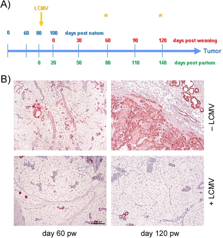
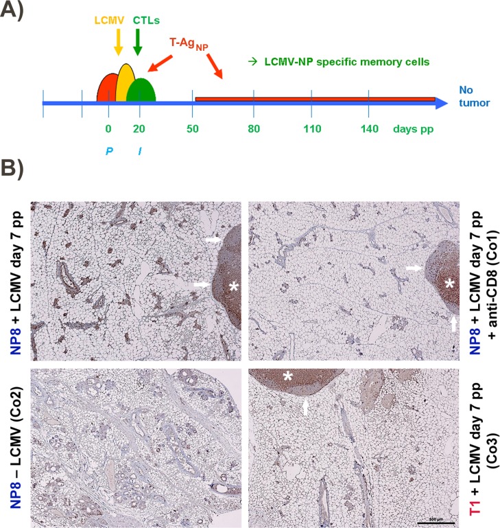
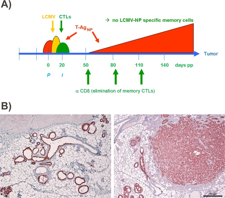
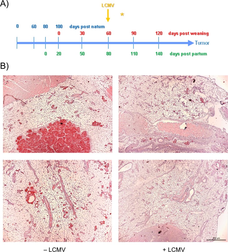
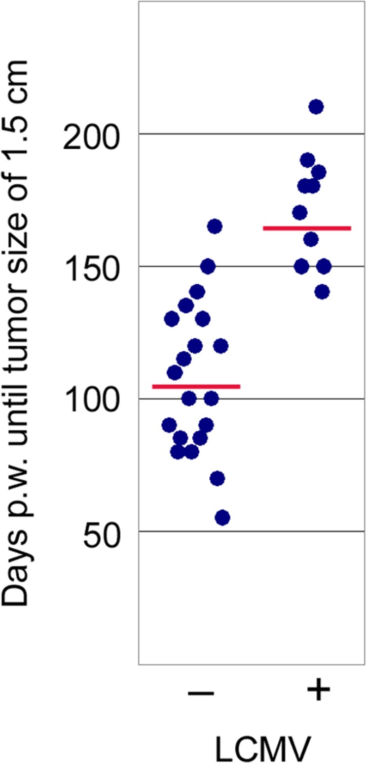
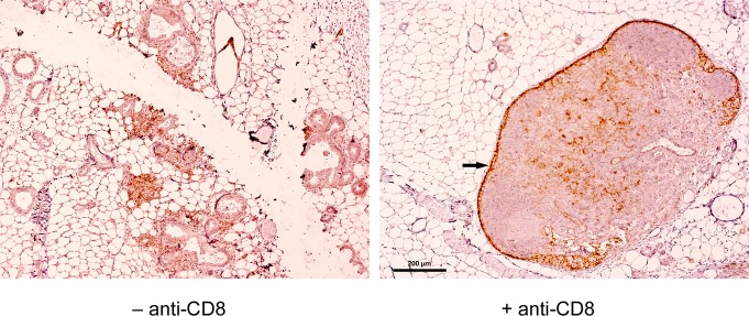
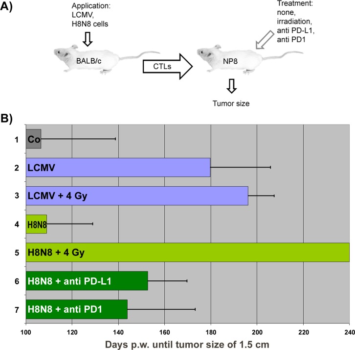
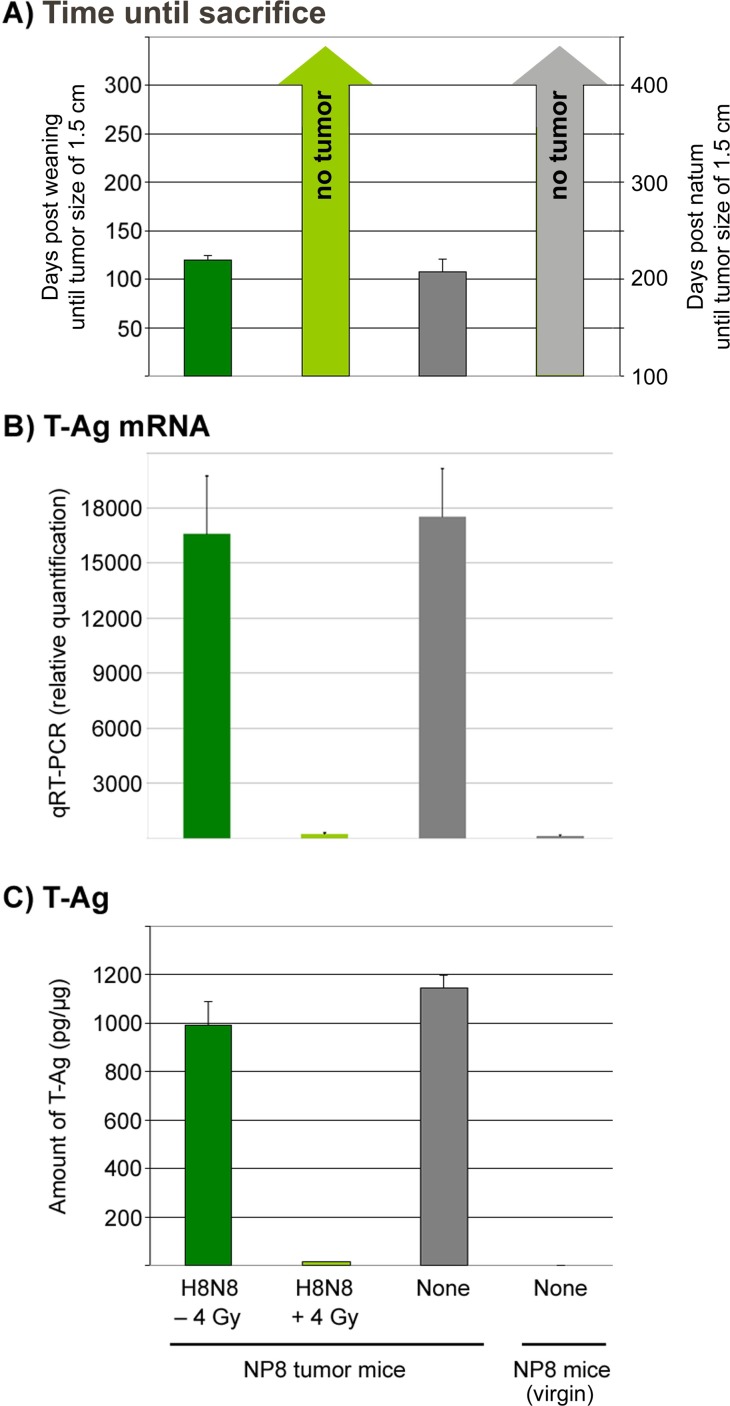
The SV40 transgenic BALB/c mouse based WAP-T/WAP-TNP model for triple-negative breast cancer allows the analysis of parameters influencing immunotherapeutic approaches. Except for WAP-TNP tumors expressing the immune-dominant LCMV NP-epitope within SV40 T-antigen (T-AgNP) which is not expressed by T-Ag of WAP-T tumors, the tumors are extremely similar. Comparative anti-PD1/PD-L1 immunotherapy of WAP-T and WAP-TNP mice supported the hypothesis that the immunogenicity of tumor antigen T-cell epitopes strongly influences the success of immune checkpoint blockade therapy, with highly immunogenic T-cell epitopes favoring rapid CTL exhaustion. Here we analyzed the immune response in NP8 mice during early times of tumor development. LCMV infection of lactating NP8 mice induced lifelong tumor protection by memory CTLs. Immunization with LCMV after involution and appearance of T-AgNP expressing parity-induced tumor progenitor cells could not cure the mice, as memory CTLs became exhausted. However, immunization significantly prolonged the time of tumor outgrowth. Elimination of exhausted CTLs and of immunosuppressive cells by sub-lethal γ-irradiation, followed by adoptive transfer of NP-epitope specific CTLs into NP8 tumor mice with early lesions, completely prevented tumor outgrowth, when lymphocytes obtained after injection of weakly immunogenic NP8 tumor-derived cells into BALB/c mice were transferred. Transfer of lymphocytes obtained after infection of BALB/c mice with highly immunogenic LCMV into such mice delayed tumor outgrowth for a significant period, but could not prevent it. We conclude that eliminating exhausted CTLs and immune-suppressive cells followed by transfer or generation of low-avidity tumor antigen-specific CTLs might be a promising approach for curative tumor immunotherapy.

Keywords: CTL exhaustion; LCMV NP T-cell epitope; SV40 T-antigen; adoptive transfer of low/high avidity CTLs; transgenic breast cancer mouse model.


Conflict of interest statement

CONFLICTS OF INTEREST The authors declare no potential conflicts of interest.

Figures

Figure 1
Figure 1. Analyses of mammary glands at days 60 and 120 pw
(A) Time-line for acute LCMV infection of NP8 mice at day 7 pp. (B) Observation at different time points after infections; the upper panels represent the tissues of control mice, the lower panels those of infected mice. Bar, 200 μm.
Figure 2
Figure 2
(A) Proposed mode of protection of NP8 mice against tumor development by infection with LCMV at day 7 pp. The semi-ovals in red, yellow, and green, represent the consecutively slightly retarded increases and decreases of SV40 T-AgNP production, LCMV infection, and LCMV NP-epitope specific CTL activation; P, partum, I, begin of involution. (B) Enrichment of memory CTLs within mammary glands of NP8 mice 120 days pw, after infection with LCMV at day 7 pp. The lymph nodes seen here partially served as internal controls, where CD3+ cells could also be localized in two of the three control experiments (e.g. Co1 and Co3) within the paracortex (*), whereas (as expected) no T cells could be visualized in the B cell areas of the secondary follicles (arrows). Bar, 500 μm.
Figure 3
Figure 3. Scheme for treatment of LCMV infected NP8 tumor mice with anti-CD8 antibodies
(A) To prove that memory CTLs are responsible for the healthy state of NP8 mice infected with LCMV at day 7 pp, the mice received anti-CD8 antibodies at days 50, 80, and 110, respectively, for the deletion of LCMV-NP specific CTLs. (B) T-Ag positive hyperplastic lesions in a mammary gland not carrying a tumor (left panel) and mammary carcinomas in various stages (right panel) from treated animals harvested and prepared 150 days pw. Bar, 200 μm.
Figure 4
Figure 4. Therapeutic effect of LCMV immunization starting early in tumor growth
(A) scheme; (B) immune-histochemistry of T-Ag. NP8 mice were infected at day 60 pw and analyzed 14 days later (right panel); uninfected animals served as negative controls (left panel). Bar, 500 μm.
Figure 5
Figure 5. Comparison of tumor development in NP8 mice infected at day 60 pw [right dots, n = 10; means = 172 days, with a 95% confidence that the population mean (μ) falls between 156 and 187 days] with that of uninfected NP8 mice [left dots, n = 20; means = 107 days, with a 95% confidence that μ falls between 94 and 120 days]; y-axis shows time until tumors had grown to a size of 1.5 cm in diameter; p-valueinfection vs control < 0.00001 significance at p < 0.01
Figure 6
Figure 6. Presence of PD-L1+ lymphocytes in the mammary glands of NP8 mice when infected at day 60 pw, which were eliminated in mice treated before with anti-CD8 antibodies; the dark staining of cells within the capsules of lymph nodes (arrow on the right picture) was caused by an abundant presence of macrophages (Wanger et al. unpublished), which are not removable by anti-CD8 antibodies and therefore served as an internal control
In addition, other PD-L1+ cells, which could represent further macrophages, T helper cells, NK cells, or regulatory T cells, are visible inside of the lymph nodes. Bar, 200 μm.
Figure 7
Figure 7. Therapeutic efficiencies after different immunologic treatments of NP8 tumor mice
(A) Experimental outline of experiments for curative immunotherapy of NP8 mice with early stage tumors. (B) Adoptive transfer of differently generated NP-epitope specific CTLs to differently pretreated NP8 mice during an early time of breast cancer development and their influence on tumor outgrowth until tumors reached a size of about 1.5 cm; in brackets p-valuestreatment vs control for significance: 1) Untreated NP8 mice, 2) Transfer of LCMV activated CTLs to NP8 mice [p = 0.000036 at p < 0.01], 3) Transfer of LCMV activated CTLs to irradiated NP8 mice [p < 0.00001 at p < 0.01], 4) Transfer of H8N8 activated CTLs to NP8 mice [p = 0.913833; not significant], 5) Transfer of H8N8 activated CTLs to irradiated NP8 mice [p < 0.00001 at p < 0.01], 6) Transfer of H8N8 activated CTLs to anti-PD-L1 treated NP8 mice [p = 0.002805 at p < 0.01], 7) Transfer of H8N8 activated CTLs to anti-PD1 treated NP8 mice [p = 0.09422 at p < 0.05].
Figure 8
Figure 8. Quantification of mRNAs and proteins for T-Ag either in non-irradiated tumor mice treated with H8N8 cells (dark green columns) or in irradiated tumor mice treated with H8N8 cells (light green columns) in comparison to untreated tumor mice (dark grey columns) or untreated non-induced (virgin) NP8 mice (light grey columns)
(A) Days post weaning (for tumor mice) or alternatively post natum (for virgin) mice until tumor sizes reached 1.5 cm or until day 300/400 of sacrifice. (B) Relative quantities of mRNA for T-Ag calculated by qRT-PCR. (C) Amount of pg T-Ag per μg of mammary gland tissues measured by ELISA.

Similar articles

References

    1. Farkona S, Diamandis EP, Blasutig IM. Cancer immunotherapy: the beginning of the end of cancer? BMC Med. 2016;14:73–91. - PMC - PubMed
    1. Bruns M, Wanger J, Utermohlen O, Deppert W. An inducible transgenic mouse breast cancer model for the analysis of tumor antigen specific CD8+ T-cell responses. Oncotarget. 2015;6:38487–38503. doi: 10.18632/oncotarget.5750. - DOI - PMC - PubMed
    1. Bruns M, Wanger J, Schumacher U, Deppert W. T-cell epitope strength in WAP-T mouse mammary carcinomas is an important determinant in PD1/PD-L1 immune checkpoint blockade therapy. Oncotarget. 2016;7:64543–64559. doi: 10.18632/oncotarget.11620. - DOI - PMC - PubMed
    1. Jannasch K, Dullin C, Heinlein C, Krepulat F, Wegwitz F, Deppert W, Alves F. Detection of different tumor growth kinetics in single transgenic mice with oncogene-induced mammary carcinomas by flat-panel volume computed tomography. Int J Cancer. 2009;125:62–70. - PubMed
    1. Deppert W, Bruns M. Cancer immunotherapy: weak beats strong. Aging (Albany NY) 2016;8:2607–2608. doi: 10.18632/aging.101134. - DOI - PMC - PubMed