. 2017 Jul 10;8(40):68012-68025.
doi: 10.18632/oncotarget.19137. eCollection 2017 Sep 15.

Native glycan fragments detected by MALDI-FT-ICR mass spectrometry imaging impact gastric cancer biology and patient outcome

Affiliations

Native glycan fragments detected by MALDI-FT-ICR mass spectrometry imaging impact gastric cancer biology and patient outcome

Thomas Kunzke et al. Oncotarget. .

Abstract

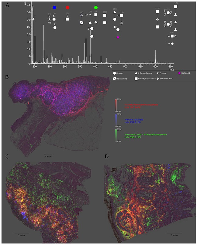
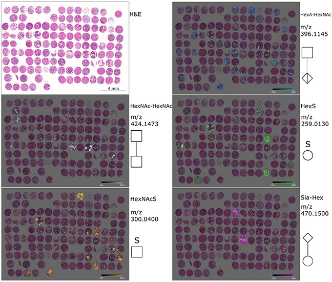
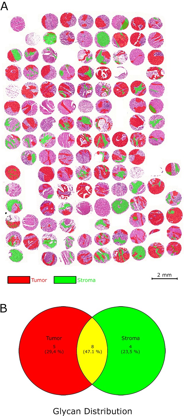
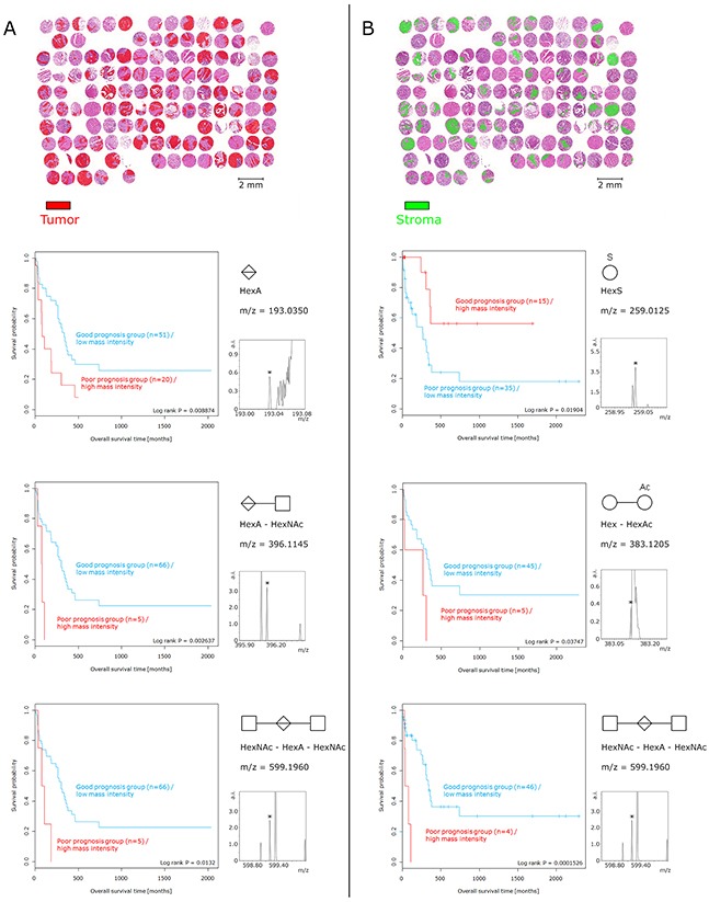
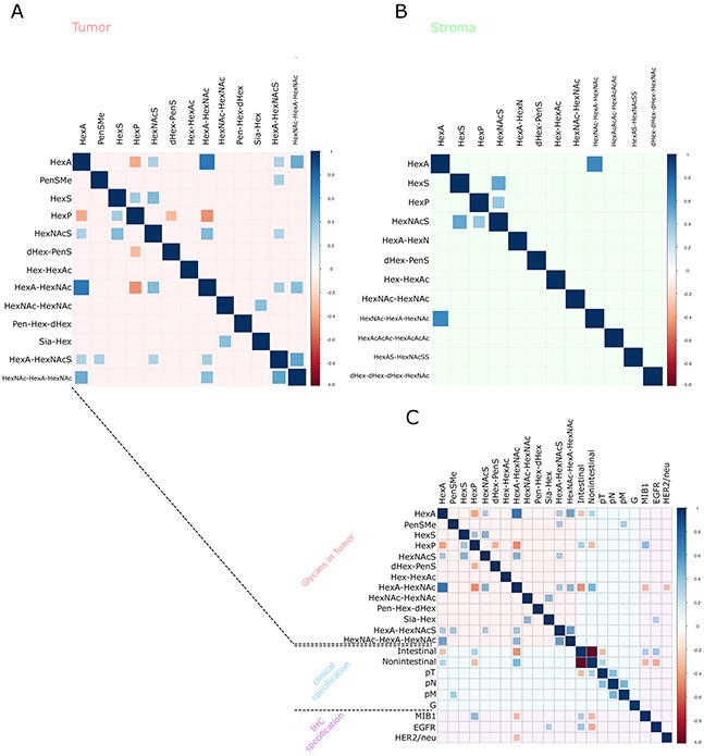
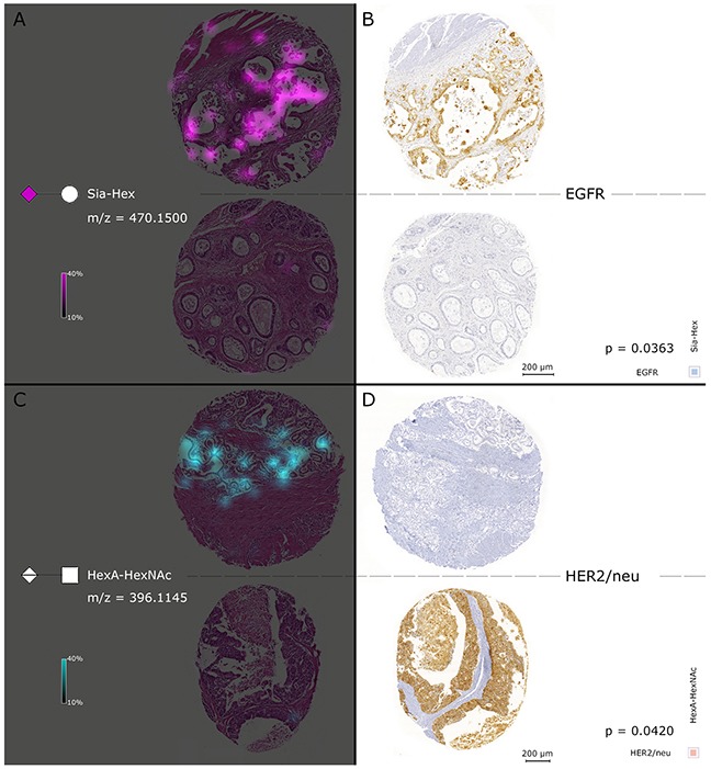
Glycosylation in cancer is a highly dynamic process that has a significant impact on tumor biology. Further, the attachment of aberrant glycan forms is already considered a hallmark of the disease state. Mass spectrometry has become a prominent approach to analyzing glycoconjugates. Specifically, matrix-assisted laser desorption/ionisation -mass spectrometric imaging (MALDI-MSI) is a powerful technique that combines mass spectrometry with histology and enables the spatially resolved and label-free detection of glycans. The most common approach to the analysis of glycans is the use of mass spectrometry adjunct to PNGase F digestion and other chemical reactions. In the current study, we perform the analysis of formalin-fixed, paraffin-embedded (FFPE) tissues for natively occurring bioactive glycan fragments without prior digestion or chemical reactions using MALDI-FT-ICR-MSI. We examined 106 primary resected gastric cancer patient tissues in a tissue microarray and correlated native-occurring fragments with clinical endpoints, therapeutic targets such as epidermal growth factor receptor (EGFR) and HER2/neu expressions and the proliferation marker MIB1. The detection of a glycosaminoglycan fragment in tumor stroma regions was determined to be an independent prognostic factor for gastric cancer patients. Native glycan fragments were significantly linked to the expression of EGFR, HER2/neu and MIB1. In conclusion, we are the first to report the in situ detection of native-occurring bioactive glycan fragments in FFPE tissues that influence patient outcomes. These findings highlight the significance of glycan fragments in gastric cancer tumor biology and patient outcome.

Keywords: MALDI; formalin-fixed paraffin-embedded tissue; gastric cancer; glycans; mass spectrometry imaging.


Conflict of interest statement

CONFLICTS OF INTEREST The authors declare no conflicts of interest.

Figures

Figure 1
Figure 1. Detectable native glycan fragments and whole gastric cancer tissue section ion map
(A) Detectable glycan fragments were in the mass range of 190–660 m/z and are shown as symbols with numbers. The explanation of the symbols used (according to GlycoWorkbench) can be found in the caption in the right bottom corner. The numbers refer to Table 1. (B-D) Ion map of N-acetylhexosamine sulphate, hexose sulphate, and hexuronic acid N-acetylhexosamine in whole tissue sections from a gastric cancer patient. Every tissue section corresponds to an individual patient and highlights altered specific distribution of each glycan fragment.
Figure 2
Figure 2. Ion maps of different glycan fragments analyzed in the gastric cancer tissue microarray
The H&E figure serves a histological overview. Ion map distribution patterns of the native glycan fragments HexNAc-HexNAc, HexNAcS, HexA-HexAc, HexS and Sia-Hex are specific for patients and tissue compartments. The measured m/z values as well as the symbols (according to GlycoWorkbench) are displayed on the right side in each case.
Figure 3
Figure 3. Regions of interests (virtual microdissection) separating tumor cells and tumor stroma
(A) The gastric cancer tissue microarray was divided into specific cell compartments by defining the regions of interests in the tumor and tumor stroma areas. (B) Venn-diagram illustrating the distribution of glycan fragments in the regions of interests.
Figure 4
Figure 4. Survival analysis of glycan fragments
Tumor cell regions and tumor stroma regions were considered individually. (A) Presence and abundance of glycan fragments detected only in tumor cell regions. The high abundance of HexA, HexA–HexNAc, and HexNAc–HexA–HexNAc in tumor cells resulted in poor prognosis. (B) With respect to the tumor stroma regions, increased intensities of Hex–HexAc and HexNAc–HexA–HexNAc corresponded to an unfavorable patient prognosis. In contrast, a high abundance of HexS in tumor stroma regions was associated with a positive patient prognosis. The relative intensity of small peaks (pictured) served as basis for the prognostic calculation.
Figure 5
Figure 5. Correlation-Plots
(A, B) The glycan fragment intensities measured and the internal mass correlations. Plot (A) details the tumor cell regions and Plot (B) shows the tumor stroma regions. Plot (C) contains plot (A) in addition to the clinical and immunohistochemical staining results. The sizes of the squares are dependent on the Spearman's rank correlation coefficient. Blue squares correspond to positive correlations and red squares correspond to negative correlations. Insignificant correlation results (p > 0.05) are indicated by an empty square.
Figure 6
Figure 6. Simultaneous consideration of MALDI FT-ICR-MSI and results of the EGFR and HER2/neu immunohistochemical staining
The left cores in (A) show the Sia-Hex ion map of two individual patients. Sia-Hex was positively correlated with EGFR. High abundances of Sia-Hex were observed in accord with high EGFR expression, as indicated in the immunohistochemical staining with EGFR in the two right cores in (B). In contrast, (C) contains the HexA–HexNAc ion map, which was negatively correlated with HER2/neu expression. High HexA–HexNAc mass intensities were accompanied by low expression of HER2/neu, which can be observed in (D).

Similar articles

Cited by

References

    1. Dube DH, Bertozzi CR. Glycans in cancer and inflammation--potential for therapeutics and diagnostics. Nat Rev Drug Discov. 2005;4:477–88. doi: 10.1038/nrd1751. - DOI - PubMed
    1. Fuster MM, Esko JD. The sweet and sour of cancer: glycans as novel therapeutic targets. Nat Rev Cancer. 2005;5:526–42. doi: 10.1038/nrc1649. - DOI - PubMed
    1. Pinho SS, Reis CA. Glycosylation in cancer: mechanisms and clinical implications. Nat Rev Cancer. 2015;15:540–55. doi: 10.1038/nrc3982. - DOI - PubMed
    1. Kearse KP, Hart GW. Lymphocyte activation induces rapid changes in nuclear and cytoplasmic glycoproteins. Proc Natl Acad Sci U S A. 1991;88:1701–5. - PMC - PubMed
    1. Hakomori S, Kannagi R. Glycosphingolipids as tumor-associated and differentiation markers. J Natl Cancer Inst. 1983;71:231–51. - PubMed