Cross-talk between p21-activated kinase 4 and ERα signaling triggers endometrial cancer cell proliferation
- PMID: 28978098
- PMCID: PMC5620238
- DOI: 10.18632/oncotarget.19188
Cross-talk between p21-activated kinase 4 and ERα signaling triggers endometrial cancer cell proliferation
Abstract
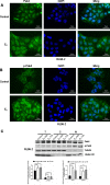
Cross-talk between estrogen receptor alpha (ERα) and signal transduction pathways plays an important role in the progression of endometrial cancer (EC). Here, we show that 17β-estradiol (E2) stimulation increases p21-activated kinase 4 (Pak4) expression and activation in ER-positive EC cells. The estrogen-induced Pak4 activation is mediated via the PI3K/AKT pathway. Estrogen increases Pak4 and phosphorylated-Pak4 (p-Pak4) nuclear accumulation, and Pak4 in turn enhances ERα trans-activation. Depletion or functional inhibition of Pak4 abrogates EC cell proliferation induced by E2, whereas overexpression of Pak4 rescues cell proliferation decreased by inhibiting the estrogen pathway. Pak4 knockdown decreases cyclin D1 expression and induces G1-S arrest. Importantly, Pak4 suppression inhibits E2 induced EC tumor growth in vivo, in a mouse xenograft model. These data demonstrate that estrogen stimulation increases Pak4 expression and activation, which in turn enhances ERα transcriptional activity and ERα-dependent gene expression, resulting in increased proliferation of EC cells. Thus inhibition of Pak4-ERα signaling may represent a novel therapeutic strategy against endometrial carcinoma.
Keywords: cross-talk; endometrial carcinoma; estrogen receptor alpha (ERα); p21-activated kinase 4 (Pak4); proliferation.
Conflict of interest statement
CONFLICTS OF INTEREST No potential conflicts of interest were disclosed.
Figures


Similar articles
-
Roles of estrogen receptor α in endometrial carcinoma (Review).Oncol Lett. 2023 Oct 25;26(6):530. doi: 10.3892/ol.2023.14117. eCollection 2023 Dec. Oncol Lett. 2023. PMID: 38020303 Free PMC article. Review.
-
p21-Activated Kinases 1, 2 and 4 in Endometrial Cancers: Effects on Clinical Outcomes and Cell Proliferation.PLoS One. 2015 Jul 28;10(7):e0133467. doi: 10.1371/journal.pone.0133467. eCollection 2015. PLoS One. 2015. PMID: 26218748 Free PMC article.
-
Benzopyran derivative CDRI-85/287 induces G2-M arrest in estrogen receptor-positive breast cancer cells via modulation of estrogen receptors α- and β-mediated signaling, in parallel to EGFR signaling and suppresses the growth of tumor xenograft.Steroids. 2013 Nov;78(11):1071-86. doi: 10.1016/j.steroids.2013.07.004. Epub 2013 Jul 26. Steroids. 2013. PMID: 23891847
-
Targeting estrogen receptor-α reduces adrenocortical cancer (ACC) cell growth in vitro and in vivo: potential therapeutic role of selective estrogen receptor modulators (SERMs) for ACC treatment.J Clin Endocrinol Metab. 2012 Dec;97(12):E2238-50. doi: 10.1210/jc.2012-2374. Epub 2012 Oct 16. J Clin Endocrinol Metab. 2012. PMID: 23074235
-
Targeting fatty acid synthase in breast and endometrial cancer: An alternative to selective estrogen receptor modulators?Endocrinology. 2006 Sep;147(9):4056-66. doi: 10.1210/en.2006-0486. Epub 2006 Jun 29. Endocrinology. 2006. PMID: 16809439 Review.
Cited by
-
Targeting PAK4 Inhibits Ras-Mediated Signaling and Multiple Oncogenic Pathways in High-Risk Rhabdomyosarcoma.Cancer Res. 2021 Jan 1;81(1):199-212. doi: 10.1158/0008-5472.CAN-20-0854. Epub 2020 Nov 9. Cancer Res. 2021. PMID: 33168646 Free PMC article.
-
Estrogen induces cell proliferation by promoting ABCG2-mediated efflux in endometrial cancer cells.Biochem Biophys Rep. 2018 Oct 23;16:74-78. doi: 10.1016/j.bbrep.2018.10.005. eCollection 2018 Dec. Biochem Biophys Rep. 2018. PMID: 30377671 Free PMC article.
-
The Role of the p21-Activated Kinase Family in Tumor Immunity.Int J Mol Sci. 2025 Apr 20;26(8):3885. doi: 10.3390/ijms26083885. Int J Mol Sci. 2025. PMID: 40332759 Free PMC article. Review.
-
The significance of PAK4 in signaling and clinicopathology: A review.Open Life Sci. 2022 Jun 20;17(1):586-598. doi: 10.1515/biol-2022-0064. eCollection 2022. Open Life Sci. 2022. PMID: 35800076 Free PMC article. Review.
-
Roles of estrogen receptor α in endometrial carcinoma (Review).Oncol Lett. 2023 Oct 25;26(6):530. doi: 10.3892/ol.2023.14117. eCollection 2023 Dec. Oncol Lett. 2023. PMID: 38020303 Free PMC article. Review.
References
-
- Siegel RL, Miller KD, Jemal A. Cancer statistics, 2017. CA Cancer J Clin. 2017;67:7–30. - PubMed
-
- Torre LA, Bray F, Siegel RL, Ferlay J, Lortet-Tieulent J, Jemal A. Global cancer statistics, 2012. CA Cancer J Clin. 2015;65:87–108. - PubMed
-
- Chen W, Zheng R, Baade PD, Zhang S, Zeng H, Bray F, Jemal A, Yu XQ, He J. Cancer statistics in China, 2015. CA Cancer J Clin. 2016;66:115–32. - PubMed
-
- Wright JD, Barrena Medel NI, Sehouli J, Fujiwara K, Herzog TJ. Contemporary management of endometrial cancer. Lancet. 2012;379:1352–60. - PubMed
-
- Morice P, Leary A, Creutzberg C, Abu-Rustum N, Darai E. Endometrial cancer. Lancet. 2016;387:1094–108. - PubMed
LinkOut - more resources
Full Text Sources
Other Literature Sources
Research Materials

