. 2017 Jun 1;8(40):68769-68779.
doi: 10.18632/oncotarget.18332. eCollection 2017 Sep 15.

A cerebrospinal fluid microRNA signature as biomarker for glioblastoma

Affiliations

A cerebrospinal fluid microRNA signature as biomarker for glioblastoma

Johnny C Akers et al. Oncotarget. .

Abstract

Purpose: To develop a cerebrospinal fluid (CSF) miRNA diagnostic biomarker for glioblastoma.

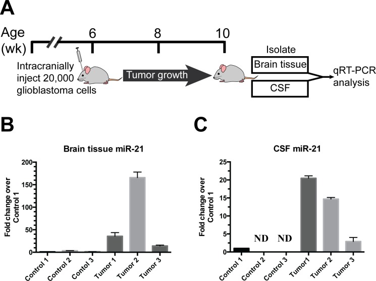
Experimental design: Glioblastoma tissue and matched CSF from the same patient (obtained prior to tumor manipulation) were profiled by TaqMan OpenArray® Human MicroRNA Panel. CSF miRNA profiles from glioblastoma patients and controls were created from three discovery cohorts and confirmed in two validation cohorts.

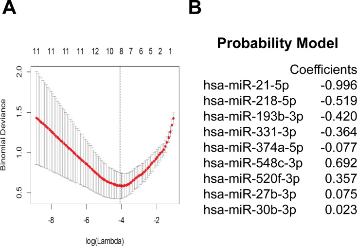
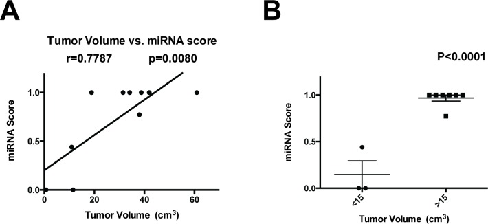
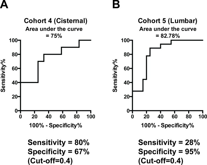
Results: miRNA profiles from clinical CSF correlated with those found in glioblastoma tissues. Comparison of CSF miRNA profiles between glioblastoma patients and non-brain tumor patients yielded a tumor "signature" consisting of nine miRNAs. The "signature" correlated with glioblastoma tumor volume (p=0.008). When prospectively applied to cisternal CSF, the sensitivity and specificity of the 'signature' for glioblastoma detection were 67% and 80%, respectively. For lumbar CSF, the sensitivity and specificity of the signature were 28% and 95%, respectively. Comparable results were obtained from analyses of CSF extracellular vesicles (EVs) and crude CSF.

Conclusion: We report a CSF miRNA signature as a "liquid biopsy" diagnostic platform for glioblastoma.

Keywords: CSF; extracellular vesicle; liquid biopsy.


Conflict of interest statement

CONFLICTS OF INTEREST The authors declare that there is no conflict of interest.

Figures

Figure 1
Figure 1. miRNA analysis of matched glioblastoma tumor and CSF samples
miRNA profile of matched glioblastoma tumor and CSF EV samples were analyzed using the TaqMan OpenArray platform. A. Venn diagrams indicating the unique and shared detectable miRNAs between tumor tissue and CSF EVs. B. Correlation between miRNA profiles of matched glioblastoma specimens and CSF. For each patient, CT values of shared miRNAs in tumor specimen were plotted against CT values from CSF EVs. Pearson correlation coefficient was then calculated for each patient. The correlations were highly significant for all matched pairs of tumor and CSF specimens. C. Venn diagrams comparing the miRNA profile of crude CSF versus CSF EV. > 95% of miRNA found in CSF EVs were also represented in the crude CSF.
Figure 2
Figure 2. Identification of miRNA signature
Differentially expressed miRNAs between glioblastoma and non-oncologic CSF samples were selected from miRNA qPCR array based on FDR < 2 and log(fold-change) > 2 and cross-validated using multiple cohorts. A. 28 candidate miRNAs was used to train a classifier with LASSO using a using cross-validated minimum deviance as the model selection criterion, B. yielding a 9 miRNA signature.
Figure 3
Figure 3. Correlation of miRNA score with tumor volume
A. The tumor volume of 11 patients in Cohort 3 was plotted against the CSF miRNA signature score, and the Pearson correlation coefficient was calculated. B. Glioblastoma < 15 cc's in volume showed a lowered miRNA signature score relative to those with > 15cc’s.
Figure 4
Figure 4. Validation of miRNA signature
A. Performance of the 9-miRNA signature using crude cisternal CSF from an independent collection of prospectively collected samples. B. Performance of the 9-miRNA signature using crude lumbar CSF from an independent collection of prospectively collected samples.
Figure 5
Figure 5. Direct release of miR-21 from glioblastoma xenograft in vivo
A. 20,000 human glioblastoma stem cells were intracranially injected into nude mice. 4 weeks later, brain tissues and murine CSFs were collected from tumor bearing mice and age-matched nude mice without the xenograft injection. B. Human miR-21 levels were elevated in the brain tissue of patient derived glioblastoma xenograft bearing mice and undetectable in mice without xenograft implant. C. Human miR-21 levels were elevated in the CSF of patient derived glioblastoma xenograft bearing mice and undetectable in mice without xenograft implant.

References

    1. Bartek J, Jr, Ng K, Bartek J, Fischer W, Carter B, Chen CC. Key concepts in glioblastoma therapy. J Neurol Neurosurg Psychiatry. 2012;83:753–60. https://doi.org/10.1136/jnnp-2011-300709 - DOI - PubMed
    1. Ng K, Kim R, Kesari S, Carter B, Chen CC. Genomic profiling of glioblastoma: convergence of fundamental biologic tenets and novel insights. J Neurooncol. 2012;107:1–12. - PubMed
    1. Vuorinen V, Hinkka S, Färkkilä M, Jääskeläinen J. Debulking or biopsy of malignant glioma in elderly people - a randomised study. Acta Neurochir (Wien) 2003;145:5–10. - PubMed
    1. Gonda DD, Warnke P, Sanai N, Taich Z, Kasper EM, Chen CC. The value of extended glioblastoma resection: insights from randomized controlled trials. Surg Neurol Int. 2013;4:110. - PMC - PubMed
    1. Air EL, Leach JL, Warnick RE, McPherson CM. Comparing the risks of frameless stereotactic biopsy in eloquent and noneloquent regions of the brain: a retrospective review of 284 cases. J Neurosurg. 2009;111:820–24. - PubMed