. 2017 Oct 3;21(1):110-125.
doi: 10.1016/j.celrep.2017.09.028.

Post-transcriptional Inhibition of Hsc70-4/HSPA8 Expression Leads to Synaptic Vesicle Cycling Defects in Multiple Models of ALS

Affiliations

Post-transcriptional Inhibition of Hsc70-4/HSPA8 Expression Leads to Synaptic Vesicle Cycling Defects in Multiple Models of ALS

Alyssa N Coyne et al. Cell Rep. .

Abstract

Amyotrophic lateral sclerosis (ALS) is a synaptopathy accompanied by the presence of cytoplasmic aggregates containing TDP-43, an RNA-binding protein linked to ∼97% of ALS cases. Using a Drosophila model of ALS, we show that TDP-43 overexpression (OE) in motor neurons results in decreased expression of the Hsc70-4 chaperone at the neuromuscular junction (NMJ). Mechanistically, mutant TDP-43 sequesters hsc70-4 mRNA and impairs its translation. Expression of the Hsc70-4 ortholog, HSPA8, is also reduced in primary motor neurons and NMJs of mice expressing mutant TDP-43. Electrophysiology, imaging, and genetic interaction experiments reveal TDP-43-dependent defects in synaptic vesicle endocytosis. These deficits can be partially restored by OE of Hsc70-4, cysteine-string protein (Csp), or dynamin. This suggests that TDP-43 toxicity results in part from impaired activity of the synaptic CSP/Hsc70 chaperone complex impacting dynamin function. Finally, Hsc70-4/HSPA8 expression is also post-transcriptionally reduced in fly and human induced pluripotent stem cell (iPSC) C9orf72 models, suggesting a common disease pathomechanism.

Keywords: C9orf72; Drosophila; RNA processing; TDP-43; amyotrophic lateral sclerosis; endocytosis; iPSC; neuromuscular junction; synaptic vesicle cycle; translation.


Figures

Figure 1
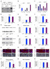
Figure 1. hsc70-4 mRNA is a translation target of TDP-43G298S and Hsc70-4 protein expression is reduced at synaptic terminals of TDP-43 expressing animals
(A) Immunoprecipitation (IP) of TDP-43 expressed in motor neurons. Genotypes and antibodies used for IP indicated on top. Antibodies used for western blot (WB) indicated on right. (B) qPCR for hsc70-4 mRNA in TDP-43 complexes. (C) qPCR for hsc70-4 mRNA in soluble and insoluble TDP-43 complexes. (D–F) qPCR for hsc70-4 mRNA in input (D), RNP (E), and polysomes (F) from flies expressing TDP-43WT and TDP-43G298S in motor neurons compared to controls. (G) WB for Hsc70-4 levels in whole larvae expressing TDP-43 in motor neurons. Genotypes indicated on bottom. Actin was used as loading control. (H) Quantification of Hsc70-4 protein levels from WBs represented as a ratio to w1118 controls. (I) qPCR for hsc70-4 mRNA in whole larvae of animals expressing TDP-43WT or TDP-43G298S in motor neurons versus controls. (J) WB for Hsc70-4 levels in VNCs of TDP-43 expressing larvae. Genotypes indicated on bottom. Actin was used as loading control. (K) Quantification of Hsc70-4 protein levels from WBs represented as a ratio to w1118 controls. (L) qPCR for hsc70-4 mRNA in VNCs of animals expressing TDP-43WT or TDP-43G298S in motor neurons versus controls. (M–O) Single confocal sections of synaptic boutons in NMJ preparations immunostained for Hsc70-4 and the neuronal membrane marker Hrp from larvae expressing TDP-43WT (N), and TDP-43G298S (O) compared to w1118 controls (M). Antibodies as indicated on left. (P) Quantification of Hsc70-4 intensity in synaptic boutons normalized to bouton area. Note p<0.001 for TDP-43WT vs TDP-43G298S. (Q) qPCR for hsc70-4 mRNA in NMJ preparations of animals expressing TDP-43WT or TDP-43G298S in motor neurons versus controls. (R) Quantification of Hsc70-4 intensity in muscle normalized to muscle area. Scale bars (M–O) 5 μm, 1 μm.
Figure 2
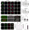
Figure 2. Hsc70/HSPA8 expression is reduced in mutant TDP-43 expressing mouse primary motor neurons, at synaptic terminals of mouse NMJs, and in TDP-43G298S human iPSC neurons
(A–D) Representative fluorescence images of cell bodies from primary motor neurons transfected with expression constructs for GFP (A–A‴) or GFP-tagged TDP-43WT (B–B‴), TDP-43Q331K (C–C‴), or TDP-43M337V (D–D‴) indicated on left. Antibodies and stains indicated on top. (E–H) Representative fluorescence images of growth cones from primary motor neurons expressing GFP (E–E′) or GFP-tagged TDP-43WT (F–F′), TDP-43Q331K (G–G′), or TDP-43M337V (H–H′) as indicated on left. Antibodies indicated on top. (I–J) Quantification of fluorescent intensity (a.u.) of Hsc70 in the cell body (I) and growth cones (J). (K–L) Epifluorescent images of mouse NMJs immunostained for Hsc70/HSPA8 and AChR. Genotypes indicated on left and antibodies indicated on top. (M) Quantification of Hsc70/HSPA8 intensity from NMJs. (N) Confocal images of control and TDP-43G298S human iPSC motor neurons labeled with DAPI, the dendritic marker Map2, and Hsc70/HSPA8. Genotypes indicated on left and antibodies indicated on top. (O) Quantification of Hsc70/HSPA8 intensity in the soma and dendrites of control and TDP-43G298S human iPSC motor neurons normalized to control. Symbols represent mean of 25–30 neurons from each line (controls) or differentiation (TDP-43) and indicate differentiation pairs. (P) qPCR for Hsc70/HSPA8 mRNA in control and TDP-43G298S human iPSC motor neurons. Scale bars (A, N) 10 μm, (K) 20 μm.
Figure 3
Figure 3. TDP-43 expression results in defects in SV endocytosis that are suppressed by Hsc70-4 in a variant dependent manner
(A) Motor neuron expression of TDP-43WT or TDP-43G298S results in increased larval turning time, which is mitigated by OE of Hsc70-4. (B–C) Motor neuron expression of TDP-43 variants WT (B) or G298S (C) leads to reduced lifespan. OE of Hsc70-4 increases lifespan for both TDP-43WT (B) and TDP-43G298S (C). (D) Representative electrophysiology traces of EJPs. Genotypes indicated on top. (E–G) EJP amplitude (E), mEJP amplitude (F), and quantal content (G) measurements from electrophysiology recordings. Genotypes indicated on bottom. (H–P) Confocal images of FM1-43 dye uptake in synaptic boutons of Drosophila larvae after 5 min of stimulation in HL-3 saline containing 90 mM KCl and 2 mM Ca2+. Genotypes indicated on top and left. (Q) Quantification of FM1-43 dye uptake normalized to total FM1-43 uptake area. Note an 18 ± 1.8% reduction in FM1-43 dye uptake for TDP-43G298S, Hsc70-4 OE compared to w1118 controls (p<0.001). Scale bar (D) 10 μm.
Figure 4
Figure 4. Factors involved in Hsc70-4 independent and dependent steps of SVC modulate TDP-43 toxicity
(A) Diagram of the SV cycle. (B–F) Motor neuron expression of TDP-43 WT or G298S results in increased larval turning time, which is enhanced upon OE of the Csp J domain mutant CspH45Q (E), and suppressed upon genomic expression of Csp (D) or OE of dynamin (shibire, F). OE (B) or reduction in auxilin (Auxilin RNAi, C) does not alter TDP-43 mediated locomotor defects. (G–O) Confocal images of FM1-43 dye uptake after 5 min of stimulation in HL-3 saline containing 90 mM KCl and 2 mM Ca2+. Genotypes indicated on top and left. (P) Quantification of FM1-43 dye uptake normalized to total FM1-43 uptake area. Scale bar (G) 10 μm.
Figure 5
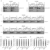
Figure 5. TDP-43WT insolubility is reduced by OE of Hsc70-4 and Hsc70-4 insolubility is increased in an age dependent manner
(A–B) Solubility studies of third instar larvae show the distribution of TDP-43WT (A) and TDP-43G298S (B) in low salt (LS), sarkosyl (Sark), and Urea containing fractions alone and in the context of Hsc70-4 OE. (C) Solubility studies of third instar larvae show Hsc70-4 distribution in LS, Sark, and Urea Fractions. (D) Solubility studies of 7 day old adults show Hsc70-4 distribution in LS, Sark, and Urea Fractions. (E) Quantification of TDP-43WT and TDP-43G298S levels in LS, Sark, and Urea fractions normalized to input. (F) Quantification of Hsc70-4 levels in LS, Sark, and Urea fractions from larval fractionations normalized to input. (G) Quantification of Hsc70-4 levels in LS, Sark, and Urea fractions from adult fractionations normalized to input.
Figure 6
Figure 6. C9orf72 repeat expansions cause reduced Hsc70-4/HSPA8 expression and defects in SVC
(A) WB for Hsc70-4 levels in VNCs of G4C2 expressing larvae. Genotypes indicated on bottom. Actin was used as loading control. (B) Quantification of Hsc70-4 protein levels from WBs represented as a ratio to G4C2 3X controls. (C) qPCR for hsc70-4 mRNA in VNCs from animals expressing G4C2 36X in motor neurons versus G4C2 3X controls. (D) qPCR for hsc70-4 mRNA in NMJ preparations from animals expressing G4C2 36X in motor neurons versus G4C2 3X controls. (E–F) Single confocal sections of synaptic boutons in NMJ preparations immunostained for Hsc70-4 and the neuronal membrane marker Hrp from larvae expressing G4C2 36X (F) compared to G4C2 3X controls (E). Antibodies indicated on left. (G) Quantification of Hsc70-4 intensity in synaptic boutons normalized to bouton area. (H) Quantification of Hsc70-4 intensity in muscle normalized to muscle area. (I) Confocal images of control and C9 ALS human iPSC motor neurons immunostained for DAPI, the dendritic marker Map2, and Hsc70/HSPA8. Genotypes indicated on left and antibodies indicated on top. (J) Quantification of Hsc70/HSPA8 intensity in the soma and dendrites of control and C9 ALS human iPS motor neurons. (K) qPCR for Hsc70/HSPA8 mRNA in control and C9 ALS human iPS motor neurons. (L) Confocal images of FM1-43 dye uptake at Drosophila NMJs after 5 min of stimulation in HL-3 saline containing 90 mM KCl and 2 mM Ca2+. Genotypes indicated on top and left. (M) Quantification of FM1-43 dye uptake normalized to total FM1-43 uptake area. Scale bars (E–F) 5 μm, 1 μm, (I, L) 10 μm.
Figure 7
Figure 7. A model for TDP-43 and Hsc70-4 interactions
(A) In controls, TDP-43 does not sequester mRNA targets or protein partners leading to normal levels of mRNA translation and synaptic proteins at the NMJ. As a result, SVC occurs as normal. (B) Motor neuron expression of TDP-43 results in decreased synaptic expression of Hsc70-4 and defects in SV endocytosis. Mutant TDP-43 sequesters hsc70-4 mRNA in insoluble complexes and inhibits its translation. Note: post-transcriptional reduction in Hsc70 expression and SVC deficits are also present in C9orf72 models of ALS.

Similar articles

Cited by

References

    1. Alami NH, Smith RB, Carrasco MA, Williams LA, Winborn CS, Han SS, Kiskinis E, Winborn B, Freibaum BD, Kanagaraj A, et al. Axonal transport of TDP-43 mRNA granules is impaired by ALS-causing mutations. Neuron. 2014;81:536–543. - PMC - PubMed
    1. Braun JE, Wilbanks SM, Scheller RH. The cysteine string secretory vesicle protein activates Hsc70 ATPase. J Biol Chem. 1996;271:25989–25993. - PubMed
    1. Bronk P, Nie Z, Klose MK, Dawson-Scully K, Zhang J, Robertson RM, Atwood HL, Zinsmaier KE. The multiple functions of cysteine-string protein analyzed at Drosophila nerve terminals. J Neurosci. 2005;25:2204–2214. - PMC - PubMed
    1. Bronk P, Wenniger JJ, Dawson-Scully K, Guo X, Hong S, Atwood HL, Zinsmaier KE. Drosophila Hsc70-4 is critical for neurotransmitter exocytosis in vivo. Neuron. 2001;30:475–488. - PubMed
    1. Burgoyne RD, Morgan A. Cysteine string protein (CSP) and its role in preventing neurodegeneration. Semin Cell Dev Biol. 2015;40:153–159. - PMC - PubMed

MeSH terms