. 2017 Oct 3;21(1):246-258.
doi: 10.1016/j.celrep.2017.09.029.

Omega-3 Fatty Acids Modulate TRPV4 Function through Plasma Membrane Remodeling

Affiliations

Omega-3 Fatty Acids Modulate TRPV4 Function through Plasma Membrane Remodeling

Rebeca Caires et al. Cell Rep. .

Abstract

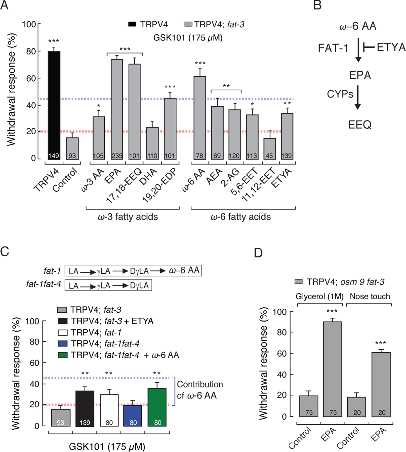
Dietary consumption of ω-3 polyunsaturated fatty acids (PUFAs), present in fish oils, is known to improve the vascular response, but their molecular targets remain largely unknown. Activation of the TRPV4 channel has been implicated in endothelium-dependent vasorelaxation. Here, we studied the contribution of ω-3 PUFAs to TRPV4 function by precisely manipulating the fatty acid content in Caenorhabditis elegans. By genetically depriving the worms of PUFAs, we determined that the metabolism of ω-3 fatty acids is required for TRPV4 activity. Functional, lipid metabolome, and biophysical analyses demonstrated that ω-3 PUFAs enhance TRPV4 function in human endothelial cells and support the hypothesis that lipid metabolism and membrane remodeling regulate cell reactivity. We propose a model whereby the eicosanoid's epoxide group location increases membrane fluidity and influences the endothelial cell response by increasing TRPV4 channel activity. ω-3 PUFA-like molecules might be viable antihypertensive agents for targeting TRPV4 to reduce systemic blood pressure.

Keywords: 17,18-epoxyeicosatetraenoic acid; Caenorhabditis elegans; TRP channels; TRPV4; atomic force microscopy; eicosapentaenoic acid; endothelial cells; fatty acids; neurons; polyunsaturated fatty acids.


Conflict of interest statement

Conflict of Interest

The authors declare no competing financial interests.

Figures

Figure 1.
Figure 1.
GSK101 elicits withdrawal responses in rat TRPV4-expressing worms. (A) Schematic representation of the withdrawal responses after addition of GSK101 drop in front of freely moving worms. (B) GSK101 dose-response profile for wild-type (WT [N2]) and TRPV4-expressing worms. (C) Inhibition of GSK101-mediated withdrawal responses in TRPV4 worms by HC067047 (2 µM). (D) Withdrawal responses elicited by 4α-Phorbol in WT and TRPV4 worms. (E) Withdrawal responses elicited by 1 M glycerol and nose touch in WT, osm9, and TRPV4; osm9 strains. Bars are mean ± SEM, the number of worms tested during 3 assays sessions is indicated inside the bars. The asterisks indicate values significantly different from control. ***p < 0.001, **p < 0.01, and *p < 0.05. See also Figure S1.
Figure 2.
Figure 2.
PUFAs are required for TRPV4 function in C. elegans. (A) Fatty acid desaturase (FAT) and elongase enzymes (ELO) synthesize long PUFAs, and cytochrome P450 (CYPs) generates eicosanoid derivatives, adapted from (Watts, 2009). LA, linolenic acid; γLA, γ-linolenic acid; DγLA, dihomo-linolenic acid; ω-6 AA, arachidonic acid; EET, epoxy-eicosatrienoic acid; ALA, α-linolenic acid; STA, stearidonic acid; ω-3 AA; EPA, eicosapentaenoic acid; EEQ, 17’18’-epoxy eicosatetraenoic acid. (B) Withdrawal responses elicited by GSK101 in WT, TRPV4, TRPV4; fat-3, and TRPV4; fat-3 worms supplemented with PUFAs. (C) Withdrawal responses elicited by 4α-Phorbol in WT, TRPV4, and TRPV4; fat-3 worms. (D) Withdrawal responses elicited by GSK101 in TRPV4 and TRPV4; fat-4 worms. (E) Withdrawal responses elicited by 1 M glycerol and nose touch in TRPV4; osm9 and TRPV4; osm9fat-3 strains. (F) Representative micrographs of TRPV4::GFP and TRPV4::GFP; fat-3 ASH neurons. (G) Box plots show the mean, median, and the 75th to 25th percentiles of the fluorescence intensity analysis from images in (F). The number of neurons imaged during 2 sessions is indicated below the boxes. (H) Schematic representation of the phospholipid synthesis. (I) GSK101 withdrawal responses after knocking down the expression of mboa-6 in TRPV4 worms. Bars are mean ± SEM, the number of worms tested during 3 assays sessions is indicated inside the bars. The asterisks indicate values significantly different from control. ***p < 0.001 and ns: no significant. See also Figure S2.
Figure 3.
Figure 3.
EPA and 17,18-EEQ fully restore TRPV4 function in C. elegans. (A) GSK101-mediated withdrawal responses of TRPV4 and TRPV4; fat 3 mutants after worms were fed with specified PUFAs and eicosanoid derivatives (200 µM). Dotted red and blue lines represent the 20% and 45% thresholds for positive and intermediate responses, respectively. (B) Schematic representation of the effect of ETYA (non-metabolizable analogue of ω-6 AA) in worms. (C) Top inset, ω-6 PUFAs present in fat-1 and fat-1fat-4. Bottom, withdrawal responses elicited by GSK101 in TRPV4; fat-3, TRPV4; fat-3 supplemented with ETYA, TRPV4; fat-1, TRPV4; fat-1fat-4, and TRPV4; fat-1fat-4 supplemented with ω-6 AA. (D) Withdrawal responses elicited by 1 M glycerol and nose touch in TRPV4; osm9fat-3 mutants after being fed with EPA (200 µM). Bars are mean ± SEM, the number of worms tested during 3 assays sessions is indicated inside the bars. The asterisks indicate values significantly different from control. ***p < 0.001, **p < 0.01, and *p < 0.05. See also Figure S3.
Figure 4.
Figure 4.
EPA supplementation enhances TRPV4 activity in HMVEC. (A) Representative whole-cell patch-clamp recordings (+80 mV) of control and EPA (100 µM)-treated HMVEC challenged with GSK101 (100 nM) and HC067047 (10 µM). (B) Box plots show the mean, median, standard deviation, and standard error of the mean from TRPV4 currents (IGSK101- IHC / pF) obtained by whole-cell patch-clamp recordings (+80 mV) of control, EPA-, and ω-6 AA-treated HMVEC. (C) Left, representative current-voltage relationships determined by whole-cell patch-clamp recording of control and EPA (100 µM)-treated HMVEC challenged with GSK101 (100 nM) in the presence of 5 mM Ca2+. Right, bar graph of peak currents (at +80 mV) relative to the currents after 5 min of exposure to GSK101 (I max/I 5 min). Bars are mean ± SEM. (D) HMVEC were challenged with isosmotic (IB, 320 mOsm), hyposmotic (HB, 240 mOsm), and GSK101 (100 nM) solutions and analyzed for their responses using Ca2+ imaging (Fluo-4 AM); color bar indicates relative change in fluorescence intensity. Control and EPA (100–300 µM)-treated HMVEC were analyzed from 5 independent preparations. (E) Representative traces corresponding to normalized (ΔF/F) intensity changes of individual cells shown in (D). (F) Area under the curve of control and EPA-treated HMVEC challenged with hyposmotic buffer. Bars are mean ± SEM. The number of endothelial cells measured is indicated below the boxes and inside the bars. The asterisks indicate values significantly different from control. ***p < 0.001 and **p < 0.01. See also Figure S4.
Figure 5.
Figure 5.
EPA supplementation increases ω-3 fatty acid eicosanoid derivatives in HMVEC and does not affect TRPV4 expression and trafficking. (A) EPA and ω-6 AA content in control and EPA (100 µM)-treated HMVEC, as determined by LC-MS. (B) ω-6 AA, EPA, and DHA eicosanoid derivatives content in control and EPA-treated HMVEC, as determined by LC-MS. (C) TRPV4 expression levels detected in control and EPA-treated HMVEC by immunostaining. (D-E) Western blots with anti-TRPV4 antibody in control and EPA-treated HMVEC from total protein extracts (D) and membrane protein fractions (E). Normalized relative intensities (RI) against total protein present in the PVDF membranes (Figure S5) are denoted. Similar results were observed in at least five independent Western blots. See also Figure S5.
Figure 6.
Figure 6.
ω-3 PUFAs contribute to membrane fluidity and bending stiffness. (A) Thermotropic characterization of the DPPC/PUFA systems using differential scanning calorimetry; control (Tm = 41.68 ºC), ω-6 AA (41.04 ºC), EPA (40.85 ºC), 5,6-EET (39.36 ºC), and 17,18-EEQ (37.85 ºC). (B) Effect of DPPC/PUFAs on melting temperatures (∆Tm) with respect to DPPC membranes. We plotted ΔTm absolute magnitude to better illustrate the effect. Experiments were performed from two independent preparations. Bars are mean obtained during two liposome assay sessions. (C) Top, schematic representation of the atomic force microscopy setup on HMVEC. Bottom, representative force distance traces acquired at 10 µm/s and magnification of the force step for control and EPA (300 µM)-treated cells. (D) Box plots show the mean, median, and the 75th to 25th percentiles analysis from tether forces of control and EPA-treated cells. The number of endothelial cells measured during two sessions is indicated below the boxes. The asterisks indicate values significantly different from control. ***p < 0.001. See also Figure S6.
Figure 7.
Figure 7.
Proposed model by which ω-3 fatty acids enhance human endothelial cells response. Plasma membranes with reduced levels of ω-3 fatty acid displays low number of channels available for activation (A) as well as a reduced number of active channels after stimulation (B). ω-3 fatty acids enriched plasma membranes increase the number of channels available for activation (C) as well as the number of active channels after stimulation (D).

References

    1. Barden AE, Mas E, and Mori TA (2016). n-3 Fatty acid supplementation and proresolving mediators of inflammation. Curr Opin Lipidol 27, 26–32. - PubMed
    1. Bargmann CI (2006). Chemosensation in C. elegans. In WormBook, Jorgensen E, ed. (The C. elegans Research Community, WormBook, , http://www.wormbook.org/.), pp. 1–29. - PMC - PubMed
    1. Basak S, Schmandt N, Gicheru Y, and Chakrapani S (2017). Crystal structure and dynamics of a lipid-induced potential desensitized-state of a pentameric ligand-gated channel. Elife 6. - PMC - PubMed
    1. Bavi N, Cortes DM, Cox CD, Rohde PR, Liu W, Deitmer JW, Bavi O, Strop P, Hill AP, Rees D, et al. (2016). The role of MscL amphipathic N terminus indicates a blueprint for bilayer-mediated gating of mechanosensitive channels. Nat Commun 7, 11984. - PMC - PubMed
    1. Brenner S (1974). The genetics of Caenorhabditis elegans. Genetics 77, 71–94. - PMC - PubMed

MeSH terms