Case Reports
. 2017 Oct 4:2017:bcr2017221024.
doi: 10.1136/bcr-2017-221024.

Hydrocele of the canal of Nuck presenting as a sausage-shaped mass

Affiliations
Case Reports

Hydrocele of the canal of Nuck presenting as a sausage-shaped mass

Ana Filipa Ferreira et al. BMJ Case Rep. .

Abstract

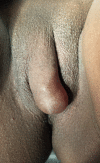
A 23-year-old woman presented with a painless vulval swelling. On physical examination, a soft fluctuant sausage-shaped mass was found, measuring approximately 4 cm, extending from the right inguinal region to the labia majora. Ultrasound revealed a well-defined hypoechoic elongated mass, septated, extending from the superficial inguinal canal to labia majora. Sonographic findings were consistent with the diagnosis of a hydrocele of the canal of Nuck. Surgical exploration revealed an elongated cystic lesion with a total length of 13 cm, mucous component and internal septations. Histopathological examination of the surgical specimen confirmed the suspected diagnosis.

Keywords: General Surgery; Obstetrics And Gynaecology; Pathology; Radiology.


Conflict of interest statement

Competing interests: None declared.

Figures

Figure 1
Figure 1
Illustration of the canal of Nuck.
Figure 2
Figure 2
Swelling of the right labia majora.
Figure 3
Figure 3
Longitudinal ultrasonographic image using high frequency transvaginal transducer (5–12 MHz), showing a well-defined hypoechoic elongated cyst with 5.5 cm of long axis with septations.
Figure 4
Figure 4
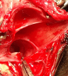
Intraoperative photograph. Surgery revealed a cystic lesion with mucous component and a smooth internal surface with septations.
Figure 5
Figure 5
Microscopic aspect of the lesion. (A) The cyst was lined with a single layer of flat cells, without atypia. Underlying this layer was a dense collagenous fibrous tissue and a thin band of inflammatory cells, mostly histiocytic cells (H&E 400×). (B) Mesothelium origin of the cells covering the cyst was confirmed by negative immunostaining with BerEp4 (BerEp4 400×).

References

    1. Wei BP, Castles L, Stewart KA. Hydrocele of the canal of Nuck. ANZ J Surg 2002;72:603–5. 10.1046/j.1445-2197.2002.02466.x - DOI - PubMed
    1. Heer J, McPheeters R, Atwell AE, et al. . Hydrocele of the canal of Nuck. West J Emerg Med 2015;16:786–7. 10.5811/westjem.2015.6.27582 - DOI - PMC - PubMed
    1. Kono R, Terasaki H, Murakami N, et al. . Hydrocele of the canal of Nuck: a case report with magnetic resonance hydrography findings. Surg Case Rep 2015;1:86 10.1186/s40792-015-0086-5 - DOI - PMC - PubMed
    1. Matsumoto T, Hara T, Hirashita T, et al. . Laparoscopic diagnosis and treatment of a hydrocele of the canal of Nuck extending in the retroperitoneal space: a case report. Int J Surg Case Rep 2014;5:861–4. 10.1016/j.ijscr.2014.08.016 - DOI - PMC - PubMed
    1. Pandey A, Jain S, Verma A, et al. . Hydrocele of the canal of Nuck – rare differential for vulval swelling. Indian J Radiol Imaging 2014;24:175–7. 10.4103/0971-3026.134408 - DOI - PMC - PubMed

Publication types