. 2017 Dec;16(12):2138-2152.
doi: 10.1074/mcp.RA117.000065. Epub 2017 Oct 4.

Calcineurin-mediated Dephosphorylation of Acetyl-coA Carboxylase is Required for Pheromone Biosynthesis Activating Neuropeptide (PBAN)-induced Sex Pheromone Biosynthesis in Helicoverpa armigera

Affiliations

Calcineurin-mediated Dephosphorylation of Acetyl-coA Carboxylase is Required for Pheromone Biosynthesis Activating Neuropeptide (PBAN)-induced Sex Pheromone Biosynthesis in Helicoverpa armigera

Mengfang Du et al. Mol Cell Proteomics. 2017 Dec.

Abstract

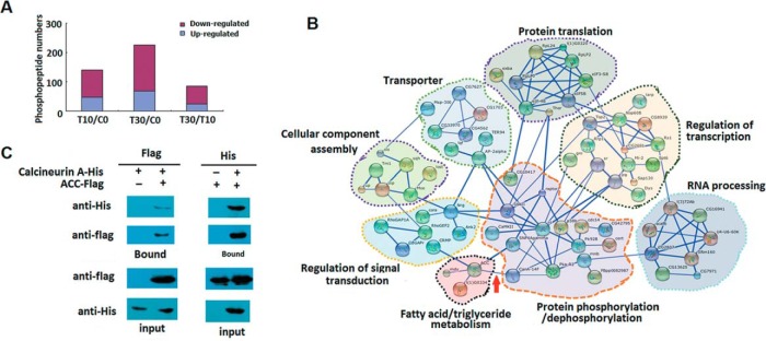
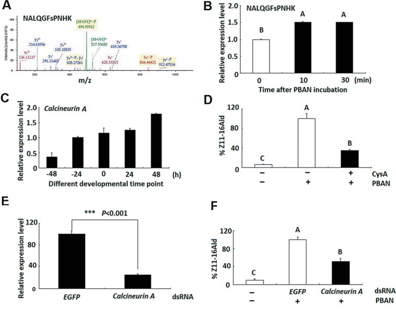
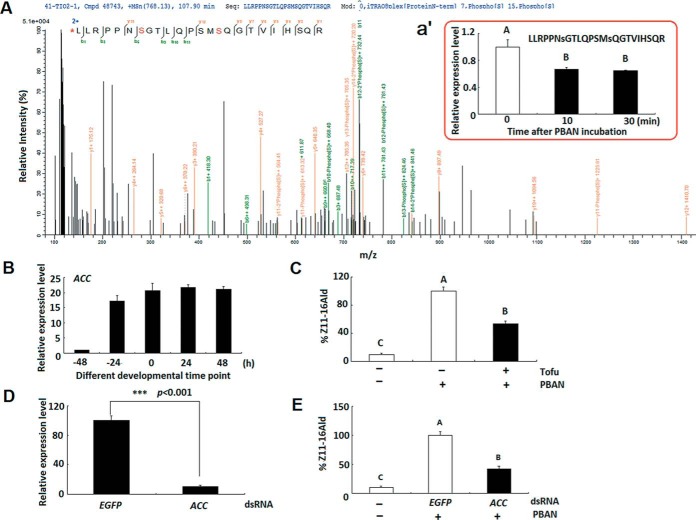
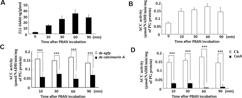
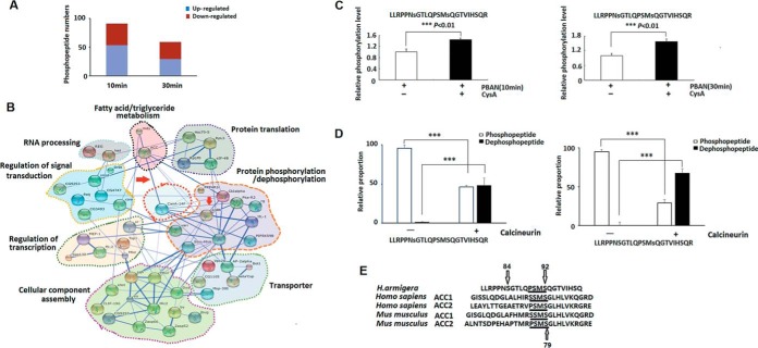
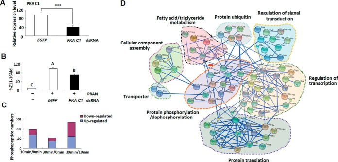
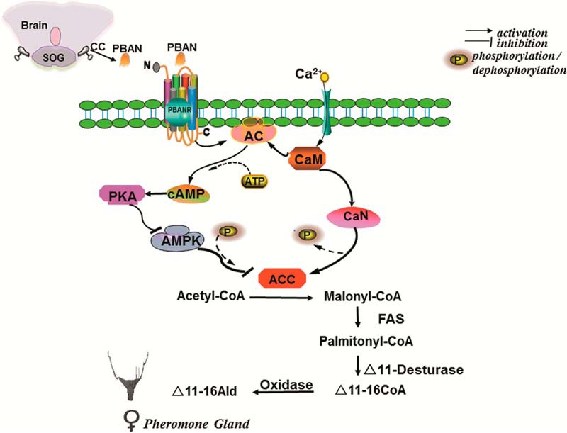
Chemical signaling plays a critical role in the behavior and physiology of many animals. Female insects, as many other animals, release sex pheromones to attract males for mating. The evolutionary and ecological success of insects therefore hinges on their ability to precisely mediate (including initiation and termination) pheromone biosynthesis. Pheromone biosynthesis activating neuropeptide (PBAN) acts directly on pheromone glands to regulate sex pheromone production using Ca2+ and cyclic-AMP as secondary messengers in the majority of species. However, the molecular mechanism downstream of the secondary messengers has not yet been elucidated in heliothine species. The present study shows that calcineurin, protein kinase A (PKA) and acetyl-coA carboxylase (ACC) are key components involved in PBAN-induced sex pheromone biosynthesis in Helicoverpa armigera using PBAN-dependent phosphoproteomics in combination with transcriptomics. RNAi-mediated knockdown and inhibitor assay demonstrated that calcineurin A is required for PBAN-induced ACC activation and sex pheromone production. Calcineurin-dependent phosphoproteomics and in vitro calcineurin phosphorylation assay further revealed that calcineurin regulated ACC activity by dephosphorylating ser84 and ser92. In addition, PKA-dependent phosphoproteomics and activity analysis revealed that PKA reduces the activity of AMP-activated protein kinase (AMPK), a negative regulator of ACC by phosphorylating the conserved ser92. Taken together, our findings indicate that calcineurin acts as the downstream signal of PBAN/G-protein receptor/Ca2+ to activate ACC through dephosphorylation while inactivating AMPK via PKA to reduce ACC phosphorylation, thus facilitating calcineurin activation of ACC.


Conflict of interest statement

We declare no conflicts of interest

Figures

Fig. 1.
Fig. 1.
Characterization of the PBAN-dependent phosphoproteome. A, Differentially expressed phosphopeptides after PBAN treatment. B, PBAN-mediated signal network. Protein interaction networks of the differentially expressed proteins induced by PBAN were constructed by using string website (https://string-db.org/) with high confidence (>0.7). Arrow represents interaction between calcineurin A and ACC. C, Interaction of ACC with calcineurin.
Fig. 2.
Fig. 2.
Calcineurin positively regulates sex pheromone biosynthesis in H. armigera PGs. A, Calcineurin MS spectrum identified in the PBAN-regulated phosphoproteome. B, Differentially expressed phosphopeptides identified in the PBAN-regulated phosphoproteome. C, Developmental expression profile of calcineurin. D, Effect of calcineurin inhibitor on sex pheromone biosynthesis. Bars indicate the mean ± S.D. of three biological replicates for independent experimental animals (n ≥ 12). Statistically significant differences are denoted by different capital letters (ANOVA and Tukey's test, p < 0.01). E, Effects of CN dsRNA on calcineurin transcript level. Significant comparisons are marked with *** (p < 0.001) as determined by Student's t test. F, Effect of calcineurin knockdown on sex pheromone biosynthesis. Bars indicate the mean ± S.D. of three biological replicates for independent experimental animals (n ≥12). Statistically significant differences are denoted by different capital letters (ANOVA and Tukey's test, p < 0.01).
Fig. 3.
Fig. 3.
ACC positively regulates sex pheromone biosynthesis in H. armigera PGs. A, ACC MS spectrum identified in the PBAN-regulated phosphoproteome. (a') Differentially expressed phosphopeptides identified in the PBAN-regulated phosphoproteome. B, Developmental expression profile of ACC. C, Effect of ACC inhibitor on sex pheromone biosynthesis. Bars indicate the mean ± S.D. of three biological replicates for independent experimental animals (n ≥ 12). Statistically significant differences are denoted by different capital letters (ANOVA and Tukey's test, p < 0.01). D, Effects of ACC dsRNA on ACC transcript level. Significance of comparisons are marked with *** (p < 0.001) as determined by Student's t test. E, Effect of ACC knockdown on sex pheromone biosynthesis. Bars indicate the mean ± S.D. of three biological replicates for independent experimental animals (n ≥12). Statistically significant differences are denoted by different capital letters (ANOVA and Tukey's test, p < 0.001).
Fig. 4.
Fig. 4.
Calcineurin positively regulates ACC activity in H. armigera PGs. A, Effects of PBAN treatment on sex pheromone production. Newly emerged females were decapitated, and maintained for 24 h to allow depletion of endogenous PBAN. PGs were then dissected and incubated in Grace's Insect Medium. After 1 h of incubation, the medium was replaced by the fresh Grace's Insect Medium containing 10 pmol PBAN. PG samples were collected at different time points of PBAN and subjected to GC/MS for Z11–16 Ald measurement. B, Effects of PBAN treatment on ACC activity. ACC activity was shown as μmol NADH/min/mg of PG protein. C, Effects of calcineurin A knockdown on ACC activity. ACC activity was shown as μmol NADH/min/mg of PG protein. Bars indicate the mean ± S.D. of three biological replicates for independent experimental animals (n ≥30). Statistically significant differences are denoted with *** (p < 0.01) or ** (p < 0.05) as determined by Student's t test. D, Effects of calcineurin inhibitor CysA on ACC activity. Ck represents control females that were incubated with PBAN only. ACC activity was shown as μmol NADH/min/mg of PG protein. Bars indicate the mean ± S.D. of three biological replicates for independent experimental animals (n ≥ 30). Statistically significant differences are denoted with *** (p < 0.01) as determined by Student's t test.
Fig. 5.
Fig. 5.
Calcineurin positively regulates ACC activity by dephosphorylating ser84 and ser92. A, Differentially expressed phosphopeptides identified in the calcineurin-regulated phosphoproteome. B, Calcineurin-regulated signaling network. Protein interaction networks of the differentially expressed proteins regulated by calcineurin were constructed by using string website (https://string-db.org/) with high confidence (>0.7). Arrow represents interaction between calcineurin A and marker proteins in two pathways (fatty acid synthesis pathway (ACC) and cAMP/PKA pathway (PKA regulatory subunit II)). C, Effect of calcineurin inhibitor treatment on ACC dephosphorylation in vivo. Small s represents phosphorylation serine site. Bars indicate the mean ± S.D. of three biological replicates for independent experimental animals (n ≥ 300). Statistically significant differences are denoted with ** (p < 0.01) as determined by Student's t test. D, Effects of calcineurin treatment on ACC dephosphorylation. Small s represents phosphorylation serine site. Bars indicate the mean ± S.D. of three biological replicates. Statistically significant differences are denoted with *** (p < 0.001) as determined by Student's t test. E, Sequence alignment of ACC consensus activity residues.
Fig. 6.
Fig. 6.
Identification of PKA-regulated phosphopeptides in H. armigera PGs. A, Effect of PKA C 1 dsRNA on PKA C1 transcript levels. Bars indicate the mean ± S.D. of three biological replicates. Statistically significant differences are denoted with *** (p < 0.001) as determined by Student's t test. B, Effect of PKA C 1 dsRNA on sex pheromone biosynthesis. Bars indicate the mean ± S.D. of three biological replicates for independent experimental animals (n ≥ 12). Statistically significant differences are denoted by different capital letter (ANOVA and Tukey's test, p < 0.001). C, Differentially expressed phosphopeptides identified in the PKA-regulated phosphoproteome. D, PKA-mediated signal network. Protein interaction networks of the differentially expressed proteins induced by PKA were constructed by using string website (https://string-db.org/) with high confidence (>0.7). Arrow represents interaction between AMPK and ACC.
Fig. 7.
Fig. 7.
PKA regulates sex pheromone biosynthesis by inhibiting AMPK activity in H. armigera PGs. A, Effect of PBAN stimulation on AMPK activity. Bars indicate the mean ± S.D. of three biological replicates for independent experimental animals (n ≥ 30). Statistically significant differences are denoted by different capital letters (ANOVA and Tukey's test, p < 0.01). B, Effect of PKA activator on AMPK activity. Bars indicate the mean ± S.D. of three biological replicates for independent experimental animals (n ≥30). Statistically significant differences are denoted with *** (p < 0.001) as determined by Student's t test. C, RNAi-mediated knockdown of PKAc1 on AMPK activity. Bars indicate the mean ± S.D. of three biological replicates for independent experimental animals (n ≥ 20). Statistically significant differences are denoted by different capital letter (ANOVA and Tukey's test, p < 0.01). D, Effect of PKA activator on ACC activity. Bars indicate the mean ± S.D. of three biological replicates for independent experimental animals (n ≥30). Statistically significant differences are denoted by different capital letters (ANOVA and Tukey's test, p < 0.01). E, Effect of AMPK activator on ACC activity. Bars indicate the mean ± S.D. of three biological replicates for independent experimental animals (n ≥ 30). Statistically significant differences are denoted by different capital letters (ANOVA and Tukey's test, p < 0.01). F, Effect of AMPK activator on sex pheromone production. Bars indicate the mean ± S.D. of three biological replicates for independent experimental animals (n ≥ 20). Statistically significant differences are denoted by different capital letters (ANOVA and Tukey's test, p < 0.01).
Fig. 8.
Fig. 8.
Schematic model of the PBAN signal transduction pathway in H. armigera PGs (adapted from Jurenka & Rafaeli, 2011) (14). PBAN is produced in the suboesophageal ganglion (SOG) and released into the hemolymph through the corpora cardiaca (CC) to reach its target organ the PG and bind to its G-Protein coupled receptor PBAN-GPCR. Binding initiates the activation of adenylate cyclase (AC) through the G subunit of the G-Protein. In turn, an influx of extracellular calcium and an increase in intracellular cAMP levels lead to the activation of calcineurin and a concomitant inhibition of AMPK. Both these signals lead to a facilitated dephosphorylation of acetyl-CoA carboxylase with a concomitant increase in sex pheromone biosynthesis.

Similar articles

Cited by

References

    1. Rafaeli A. (2002) Neuroendocrine control of pheromone biosynthesis in moths. Int. Rev. Cytol. 213, 49–91 - PubMed
    1. Engelmann F. (1960) Mechanisms controlling reproduction in two viviparous cockroaches (Blattaria). Ann. N.Y. Acad. Sci. 89, 516–536
    1. Riddiford L. M., and Williams C. M. (1971) Role of corpora cardiaca in the behavior of saturniid moths I. Release of sex pheromone. Biol. Bull. 140, 1–7 - PubMed
    1. Raina A. K., and Klun J. A. (1984) Brain factor control of sex pheromone production in the female corn earworm moth. Science 225, 531–533 - PubMed
    1. Raina A. K., Jaffe H., Kempe T. G., Keim P., Blacher R. W., Fales H. M. C., Riley T., Klun J. A., Ridgway R. L., and Hayes D. K. (1989) Identification of an neuropeptide hormone that regulates sex pheromone production in female moths. Science 244, 796–798 - PubMed