Review
. 2017 Oct;69(4):497-564.
doi: 10.1124/pr.117.014050.

International Union of Basic and Clinical Pharmacology. CII: Pharmacological Modulation of H2S Levels: H2S Donors and H2S Biosynthesis Inhibitors

Affiliations
Review

International Union of Basic and Clinical Pharmacology. CII: Pharmacological Modulation of H2S Levels: H2S Donors and H2S Biosynthesis Inhibitors

Csaba Szabo et al. Pharmacol Rev. 2017 Oct.

Abstract

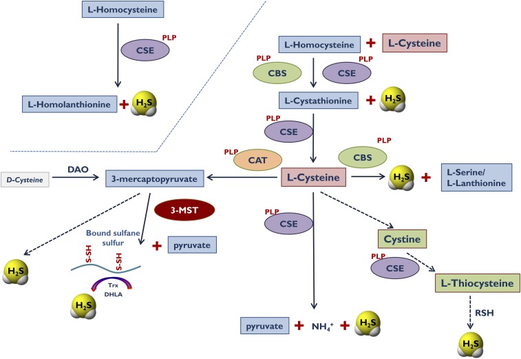
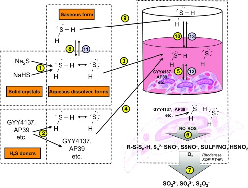
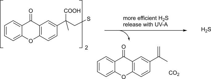
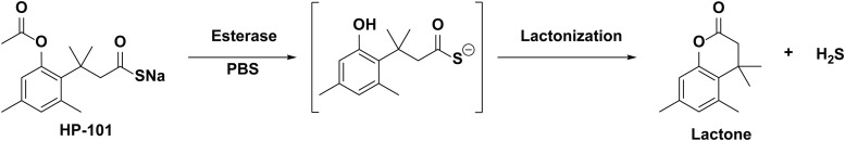
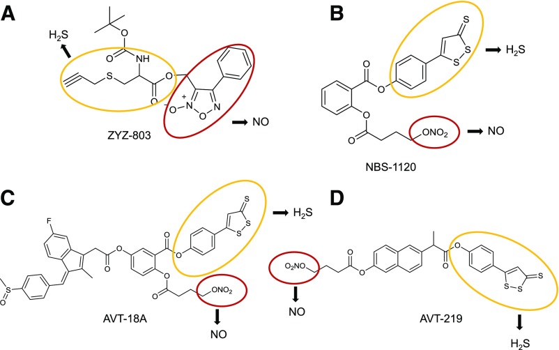
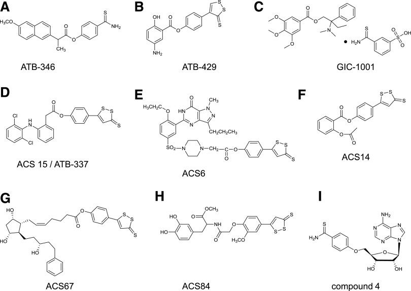
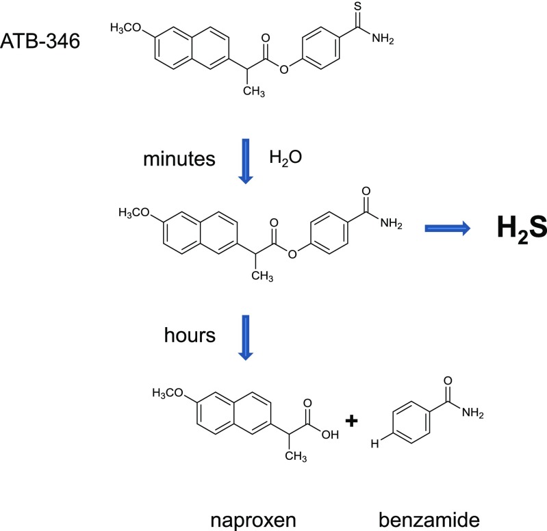
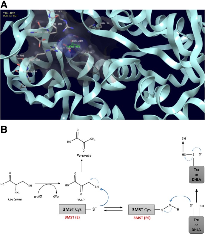
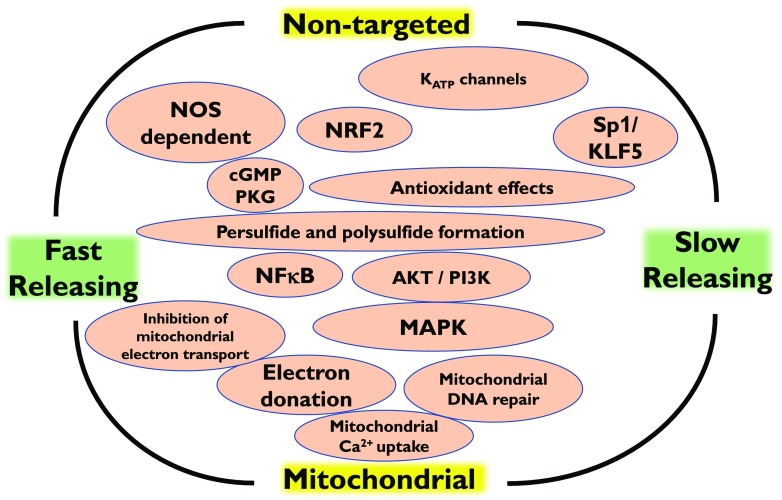
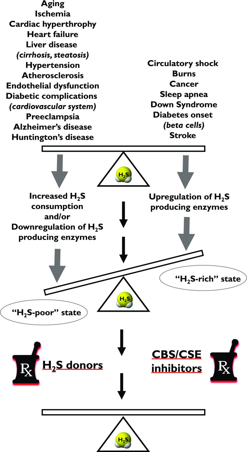
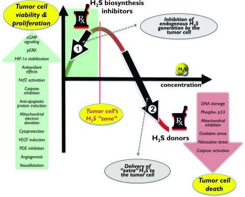
Over the last decade, hydrogen sulfide (H2S) has emerged as an important endogenous gasotransmitter in mammalian cells and tissues. Similar to the previously characterized gasotransmitters nitric oxide and carbon monoxide, H2S is produced by various enzymatic reactions and regulates a host of physiologic and pathophysiological processes in various cells and tissues. H2S levels are decreased in a number of conditions (e.g., diabetes mellitus, ischemia, and aging) and are increased in other states (e.g., inflammation, critical illness, and cancer). Over the last decades, multiple approaches have been identified for the therapeutic exploitation of H2S, either based on H2S donation or inhibition of H2S biosynthesis. H2S donation can be achieved through the inhalation of H2S gas and/or the parenteral or enteral administration of so-called fast-releasing H2S donors (salts of H2S such as NaHS and Na2S) or slow-releasing H2S donors (GYY4137 being the prototypical compound used in hundreds of studies in vitro and in vivo). Recent work also identifies various donors with regulated H2S release profiles, including oxidant-triggered donors, pH-dependent donors, esterase-activated donors, and organelle-targeted (e.g., mitochondrial) compounds. There are also approaches where existing, clinically approved drugs of various classes (e.g., nonsteroidal anti-inflammatories) are coupled with H2S-donating groups (the most advanced compound in clinical trials is ATB-346, an H2S-donating derivative of the non-steroidal anti-inflammatory compound naproxen). For pharmacological inhibition of H2S synthesis, there are now several small molecule compounds targeting each of the three H2S-producing enzymes cystathionine-β-synthase (CBS), cystathionine-γ-lyase, and 3-mercaptopyruvate sulfurtransferase. Although many of these compounds have their limitations (potency, selectivity), these molecules, especially in combination with genetic approaches, can be instrumental for the delineation of the biologic processes involving endogenous H2S production. Moreover, some of these compounds (e.g., cell-permeable prodrugs of the CBS inhibitor aminooxyacetate, or benserazide, a potentially repurposable CBS inhibitor) may serve as starting points for future clinical translation. The present article overviews the currently known H2S donors and H2S biosynthesis inhibitors, delineates their mode of action, and offers examples for their biologic effects and potential therapeutic utility.


Figures

Fig. 1.
Fig. 1.
Pathways of H2S generation in mammalian cells. Cystathionine-β-synthase (CBS; EC 4.2.1.22), cystathionine-γ-lyase (CSE; 4.4.1.1), and 3-mercaptopyruvate sulfurtransferase (3-MST; EC.2.8.1.2) are the three principal enzymes that contribute to the endogenous production of H2S. CBS and CSE are components of the reverse transsulfuration pathway, a biochemical pathway responsible for the conversion of methionine to cysteine, and catalyze a multitude of reactions that yield H2S, including the conversion of l-homocysteine to l-homolanthionine (by CSE), the conversion of l-homocysteine and l-cysteine to l-cystathionine (by CBS and CSE), the conversion of l-cystathionine to l-cysteine (by CSE), the conversion of l-cysteine to pyruvate and ammonia (by CSE), and the conversion of l-cysteine to l-serine and l-lanthinonine (by CBS). An additional pathway involves the CSE-dependent conversion of cystine to l-thiocystenine, which, in turn, produces H2S via thiol-dependent reactions. The third H2S-producing enzyme, 3-MST, is part of the cysteine catabolism pathway and uses 3-mercaptopyruvate (3-MP) as a substrate. 3-MST works in tandem with aspartate aminotransferase that also possesses cysteine aminotransferase activity (CAT) activity, generating 3-MP from cysteine via a series of reductions that first involve the generation of bound sulfane sulfur. 3-MP, in addition to acting as a substrate of 3-MST, can also produce H2S spontaneously. In some cells and tissues, d-cysteine can also be a significant substrate for H2S production; it is converted to 3-MP by d-amino acid oxidase (DAO). Pyridoxal 5′-phosphate (PLP) is a cofactor for CSE, CBS, and CAT.
Fig. 2.
Fig. 2.
H2S delivery to cell in culture. H2S and HS are immediately generated when rapid-release H2S donors (i.e., sulfide salts) are dissolved in aqueous stock solutions (1). Likewise, when H2S donors (e.g., GYY4137, AP39 etc.) are dissolved in solution, some H2S and HS can already begin to form (the extent of which depends on the chemical properties of the donor) (2). When stock solutions are added to the cell culture medium, these species (H2S-donor molecules, H2S and HS) are delivered, first into the medium (3,4) and from there into the cultured cells (5). Some donors themselves are hydrophilic and may not have high cell permeability; these donors are likely to remain extracellular, and the H2S produced from them will enter the cells. Other H2S donors may enter the cells more readily (some of them may be cell-compartment-specific, e.g., AP39 sequesters into the mitochondria and delivers H2S preferentially to the mitochondrial component). Intracellularly, production is via glutathione-dependent conversion mechanisms. Intracellularly, H2S will react with various molecules (proteins, thiols, nitric oxide, reactive oxygen species) to create a mixture of biologically active species (polysulfides, persulfides, hybrid S/N compounds). Some of these reactions, e.g., with proteins and thiols, will already occur extracellularly in the cell culture medium (not shown) (6). Thus the cellular effects of H2S donors are produced by a complex array of interactions and biological actions induced by multiple species. H2S decomposition products (sulfite, sulfate, thiosulfate) are also produced via enzymatic and nonenzymatic processes (7). Another way to deliver H2S is by bubbling H2S into aqueous solutions (for instance, the method was used to produce IK-1001) (8). This solution, then, can be added to cells the same way as the other H2S delivery approaches (3). One can also supply H2S gas into the cell culture headspace, which, in turn, dissolves in the culture medium (9, 10) and delivers H2S and HS to the cells. As soon as the H2S donors are dissolved in the stock solution, H2S starts to escape through diffusion into the air (11). Loss of H2S will also occur through diffusion of H2S from the cells into the culture medium (12) and then into the headspace (13).
Fig. 3.
Fig. 3.
Chemical composition of SG-1002.
Fig. 4.
Fig. 4.
Structures of naturally occurring H2S donors and derivatives of naturally occurring compounds modified to release H2S. Diallyl sulfide (DAS; A), diallyl disulfide (DADS; B), diallyl trisulfide (DATS; C), S-allylcysteine (SAC; D), S-propargyl-l-cysteine (SPRC, also known as ZYZ802;E) thioglycine (TG; F), l-thiovaline (TV; G), thiocarbamate-functionalized carbonyl sulfide/H2S donor (TCO-1; G).
Fig. 5.
Fig. 5.
Pathways of H2S generation and mechanisms of action of polysulfide diallyl trisulfate (DATS) in mammalian cells. (1) H2S production via glutathione-dependent conversion mechanisms. This group of processes can involve several different mechanisms, including a carbon nucleophilic attack as well as various thiol-disulfide exchange reactions (not shown); (2) H2S production via reactions with protein-SH groups; (3) H2S production via upregulation of CSE and/or via stimulation of CSE activity. In these processes, H2S is produced from the endogenous substrates of CSE, l-cysteine/l-homocysteine, and DATS stimulates this reaction; (4) H2S production via the oxidoreductase function of catalase. An additional, indirect mechanism (5) involves redox mechanisms. DATS elevates the cell’s antioxidant pools, and this attenuates the oxidative degradation of H2S, in effect elevating the biologically available pools of H2S.
Fig. 6.
Fig. 6.
Thioacetimide releases H2S by hydrolysis.
Fig. 7.
Fig. 7.
Structure of Lawesson’s reagent and structurally related compounds, including the slow-release H2S donor GYY4137. Lawesson’s reagent (A; 2,4-bis(4-methoxyphenyl)-2,4-dithioxo-1,3,2,4-dithiadiphosphetane), GYY4137 (B; P-(4-methoxyphenyl)-P-4-morpholinyl-phosphinodithioic acid), FW1256 (C; 3-dihydro-2-phenyl-2 sulfanylenebenzo[d] [1,3,2]oxazaphosphole).
Fig. 8.
Fig. 8.
Commonly used compounds to deliver H2S. Structures shown are used either as stand-alone donors or are attached to known pharmacophores to give H2S-releasing properties in these structures, thus creating “combination donors.” DTT (A; 1,2,dithiole-3-thione), ADT-OH (B; 5-(4-hydroxyphenyl)-3H-1,2-dithiole-3-thione), or ADT (C; 5-(4-methoxyphenyl) -3H-1,2-dithiole-3-thione).
Fig. 9.
Fig. 9.
Spontaneous H2S release from ADT-OH (5-(4-hydroxyphenyl)-3H-1,2-dithiole-3-thione). It has been suggested that the rates of H2S release from ADT-OH are increased when the compound is incubated with biologic material; however, the mechanisms responsible for the enhanced release remain uncharacterized.
Fig. 10.
Fig. 10.
GYY4137 releases H2S upon hydrolysis. Two molecules of H2S are released per molecule of GYY4137.
Fig. 11.
Fig. 11.
Structures of thiol-activated H2S donors. N-(Benzoylthio)benzamide derivates (A), NSHD-1 (D), and NHSD-2 (E) are N-mercapto-based donors, Compound 8 is a perthiol-based donor (B), 4-carboxy-phenyl-isothiocyanate (4CPI; C), TAGDD-1 is a geminaldithiol-dithiol based compound (F). Cysteine-triggered H2S release from S-aroylthiooximes (G) have half-lives between 8 and 82 minutes depending on the substitution of the S-aroylthiohydroxylamine ring.
Fig. 12.
Fig. 12.
H2S release from FeIISH complexes.
Fig. 13.
Fig. 13.
Mechanism of H2S release from TAGDD-1. The reaction is initiated by a reversible thiol exchange between TAGDD-1 and cysteine to generate S-acetyl cysteine and gem-dithiol. S-acetyl cysteine undergoes a fast S- to-N-acyl transfer to form N-acetylcysteine and drive the equilibrium. Meanwhile, the gem-dithiol releases H2S spontaneously in aqueous solution to yield benzaldehyde.
Fig. 14.
Fig. 14.
Mechanisms of H2S release from S-aroylthiooximes (SATOs). Two pathways could lead to H2S generation from SATOs. The first involving addition of the cysteine thiol to the SATO acyl group followed by rapid SN-acyl transfer (Pathway A). The arylidenethiooxime would form, which could decompose to generate a ketone or aldehyde along with H2S and NH3. The second pathway involves a hydrolysis step to generate the S-aroylthiohydroxylamine and the ketone or aldehyde used to make the SATO (Pathway B). The fast reaction between SATOs and cysteine to yield H2S (t1/2 approximately 8−82 minutes) compared with the hydrolysis rate (t1/2 ranging from 45–250 hours) rules out pathway B as the mechanism of H2S release mechanism for most SATOs under the conditions tested in Foster et al., 2014.
Fig. 15.
Fig. 15.
Mechanisms of H2S release from pH-controlled donors. Protonation of phosphonamidothioates at neutral or slightly acidic pH yields the corresponding phosphorothiols. This process facilitates the release of H2S if a nucleophilic carboxylate is presented at a suitable position. The formation of the five-membered ring could be the driving force for H2S release.
Fig. 16.
Fig. 16.
Controlled-release and targeted H2S donors. JK-2 (A) and JK-1 (C) are based on a phosphonamidothioate template and generate H2S in a pH-controlled manner, ΒW-HP-101 is an esterase cleavable H2S donor (B), SPD-1 (D) and SPD-2 (E) are photoactivatable H2S donors, Donors depicted in (F) are thiocarbamate-based donors that generate H2S after exposure to ROS (mainly H2O2). AP39 (G) is a mitochondria-targeted donor consisting of a mitochondria-targeting motif (triphenylphosphonium) coupled to an H2S-donating moiety (dithiolethione; DTT) by an aliphatic linker.
Fig. 17.
Fig. 17.
Photoactivated H2S release from SPD-2.
Fig. 18.
Fig. 18.
Mechanism of H2S release from esterase-cleaved prodrugs. Prodrugs of this category release H2S upon cleavage of an ester group, followed by lactonization. H2S release rates can be changed by 1) modifying the ester group (acyl moiety) and thus altering susceptibility to esterase and 2) altering structural features that are crucial for the lactonization rate. BW-HP-101 is shown as an example.
Fig. 19.
Fig. 19.
(A) ZYZ-803 is a derivative of S-propyl-l-cysteine and is thiol activated. (B) NBS-1120 is NOSH-aspirin (also designated as NOSH-1). (C) AVT-18A is a sulindac derivative. (D) AVT-219 is a naproxen derivative.
Fig. 20.
Fig. 20.
Combined (hybrid) H2S-donating derivatives of clinically used drugs. ATB-346 naproxen-benzamide conjugate (Α), ATB-429 is a mesalamine-ADT conjugate (Β), GIC-1001 is trimebutine 3-thiocarbamoylbenzene-sulfonate (C), ATB-337 (also known as ACS15) is a diclophenac-benzamide conjugate (D), ACS-6 is a sildenafil-ADT conjugate (E), ACS14 is an aspirin-ADT conjugate (F), ACS67 is a latanoprost-ADT conjugate (G) and ACS84 is an l-DOPA-ADT conjugate (H), compound 4 is an analog of adenosine (I).
Fig. 21.
Fig. 21.
Proposed mechanism of H2S release and breakdown of ATB-346. The release of H2S from ATB-346 occurs at a very low pace when the drug is dissolved in an aqueous solution. The rate of H2S release is enhanced in the presence of tissue or in the presence of reducing agents (dithiothreitol, l-cysteine, or glutathione).
Fig. 22.
Fig. 22.
Stereoview of PAG-hCSE active site (A) and superimposed PAG complexes (B). PAG, PLP, and nitrate ion are shown in a thick line. hCSE⋅PAG, methionine γ‐lyase⋅PAG, and CsdB⋅PAG are colored green, gray, and pink, respectively. Residues interacting with PAGs and nitrate ion are shown. (C): Stereoview of the 2Fo ‐ Fc simulated annealing omit map of PAG, Tyr114 from hCSE⋅PLP⋅PAG. All atoms within 3.5 Å of PAG and Tyr114 were omitted prior to refinement. The map was contoured at a level of 1.0σ. Reproduced with permission from Sun et al., 2009.
Fig. 23.
Fig. 23.
Multiple modes of AOAA’s action in cancer cells. By directly inhibiting CBS and CSE activity, by suppressing H2S formation through the 3-MST pathway via inhibition of CAT, and by inhibiting a variety of transaminases (including GOT1, a key enzyme of the malate/aspartate shuttle), AOAA acts as an inducer of “synthetic lethality” in cancer cells. CBS-derived and 3-MST-derived H2S supports mitochondrial electron transport and cancer cell bioenergetics by donating electrons at complex II, by stimulating ATP synthase, and by inhibiting intramitochondrial adenyl cyclase (this latter effect is not shown on this scheme). By inhibiting CBS and CAT, AOAA suppresses this bioenergetic pathway. The malate-aspartate shuttle translocates electrons that are produced in glycolysis across the semipermeable inner membrane of the mitochondrion to support oxidative phosphorylation. These electrons enter the electron transport chain at complex I. The shuttle system is required because the mitochondrial inner membrane is impermeable to NADH (a primary reducing equivalent of the electron transport chain). In humans, the cytoplasmic enzyme (GOT1) is one of the key enzymes in the malate shuttle: it functions to catalyze the interconversion of aspartate and α-ketoglutarate to oxaloacetate and glutamate using pyridoxal phosphate as a cofactor. By inhibiting GOT, AOAA reduces the transfer of electron donors to the mitochondria, thereby suppressing cancer cell bioenergetics. By the simultaneous inhibition of H2S production and various transaminases, AOAA interferes with key pathways of cancer cell mitochondrial function.
Fig. 24.
Fig. 24.
Inhibition of CBS by AOAA. Docking simulation of PLP in the active center of CBS free (blue) or bound to AOAA (red) (left). Catalytic residues Tyr223 and Gly307 are also shown. Part of heme is visible in the lower right-hand side in dark blue. Residues in proximity to the AOAA-PLP complex are shown (right).
Fig. 25.
Fig. 25.
(A) 3-MST is a 32.8-kDa protein comprising an N-terminal catalytically inactive domain and a C-terminal catalytically active domain. The catalytic site, Cys247, is redox-active and is oxidized to form sulfenyl cysteine. The sulfenyl cysteine is then reduced to the active form by thioredoxin (Trx). The catalytic site, Cys247, serves as an intrasubunit redox-sensing switch. (B) The production of H2S by 3-MST in the presence of Trx or DHLA. 3-MST reacts with 3MP to produce H2S via a persulfide intermediate. Trx or DHLA accepts a sulfur atom from a persulfide intermediate that is attacked by another thiol and releases H2S.
Fig. 26.
Fig. 26.
An overview of the cellular signaling processes elicited by H2S donors. The cellular effects of H2S donors can be different, depending on the rate of the H2S release and the targeted versus nontargeted nature of the donor. For example, mitochondrially targeted H2S donors preferentially activate mitochondrial processes (e.g., protection against mitochondrial oxidative stress, or facilitation of mitochondrial DNA repair processes, or electron donation to the mitochondrial electron transport chain) and have lesser effect on cytoplasmatic signaling pathways. High concentrations of mitochondrial H2S donors may also suppress mitochondrial electron transport by inhibiting mitochondrial Complex IV. When fast-acting H2S donors are applied to cells or animals, the initial high H2S concentration may be sufficient to inhibit mitochondrial Complex IV to induce a short-lasting chemical hypoxia, which, in turn, may stimulate compensatory (preconditioning type) processes. Fast-acting H2S donors tend to be more potent activators of cGMP-dependent processes than slow-release H2S donors. Fast-acting H2S donors also tend to exert their action in cooperation with NO synthase-dependent signaling processes. Please note that the downstream pathways activated by the various H2S donors have not yet been characterized in a systematic manner.
Fig. 27.
Fig. 27.
Therapeutic effects of H2S donors and H2S biosynthesis inhibitors: a simplified overview. Some pathophysiological states are associated with H2S deficiency; this can be corrected by H2S donors (a form of replacement therapy) (left side). Other pathophysiological states are associated with H2S overproduction; this can be corrected by H2S biosynthesis inhibitors (right side). The scheme represents an oversimplification for a number of reasons. For example, the same pathophysiological condition can manifest itself with both H2S overproduction and H2S deficiency. In diabetes, the pancreatic beta cell destruction is linked to H2S overproduction; diabetes can also elevate H2S levels in the liver, with pathophysiological consequences. At the same time, the cardiovascular consequences of diabetes include vascular H2S deficiency, which contributes to vascular complications. In addition, in some diseases (e.g., cancer or burn injury), both systemic H2S biosynthesis inhibition and H2S donation can exert beneficial effects through different sets of biologic actions.
Fig. 28.
Fig. 28.
Mechanisms underlying the therapeutic effects of H2S biosynthesis inhibitor (left side) and H2S donors (right side) in cancer. Because of the bell-shaped pharmacological profile of H2S, both H2S biosynthesis inhibition and H2S donation can exert therapeutic effects. Low concentrations of H2S that are produced endogenously by CBS, CSE, and/or 3-MST can support tumor growth and tumor angiogenesis through a variety of pathways shown in the green arrow. Pharmacological inhibition of these responses (depicted by arrow #1) can be of therapeutic benefit, either on its own, or to sensitize the tumor cell to standard anticancer therapies. On the other hand, high concentrations of H2S can be cytostatic or cytotoxic through a variety of pathways shown in the red arrow. Thus therapeutic administration of H2S (depicted by arrow #2), which induces high concentrations of H2S in the tumor cell can be used to induce anticancer effects and/or to potentiate anticancer chemo-or radiotherapy.

Similar articles

Cited by

References

    1. Abe K, Kimura H. (1996) The possible role of hydrogen sulfide as an endogenous neuromodulator. J Neurosci 16:1066–1071. - PMC - PubMed
    1. Abdel-Salam E, Abdel-Meguid I, Korraa S. (2013) Assessment of immune function in Down syndrome patients. The Egyptian Journal of Medical Human Genetics 14:307–310.
    1. Abeles RH, Walsh CT. (1973) Acetylenic enzyme inactivators. Inactivation of gamma-cystathionase, in vitro and in vivo, by propargylglycine. J Am Chem Soc 95:6124–6125. - PubMed
    1. Abou-Hamdan A, Guedouari-Bounihi H, Lenoir V, Andriamihaja M, Blachier F, Bouillaud F. (2015) Oxidation of H2S in mammalian cells and mitochondria. Methods Enzymol 554:201–228. - PubMed
    1. Agrawal N, Banerjee R. (2008) Human polycomb 2 protein is a SUMO E3 ligase and alleviates substrate-induced inhibition of cystathionine β-synthase sumoylation. PLoS One 3:e4032. - PMC - PubMed

Publication types