

Review
doi: 10.1523/JNEUROSCI.3218-16.2017.
Are the Neural Correlates of Consciousness in the Front or in the Back of the Cerebral Cortex? Clinical and Neuroimaging Evidence
Affiliations
- PMID: 28978697
- PMCID: PMC5628406
- DOI: 10.1523/JNEUROSCI.3218-16.2017
Item in Clipboard
Review
Are the Neural Correlates of Consciousness in the Front or in the Back of the Cerebral Cortex? Clinical and Neuroimaging Evidence
J Neurosci.
.
Abstract
The role of the frontal cortex in consciousness remains a matter of debate. In this Perspective, we will critically review the clinical and neuroimaging evidence for the involvement of the front versus the back of the cortex in specifying conscious contents and discuss promising research avenues.Dual Perspectives Companion Paper: Should a Few Null Findings Falsify Prefrontal Theories of Conscious Perception?, by Brian Odegaard, Robert T. Knight, and Hakwan Lau.
Keywords: consciousness; frontal cortex; lesion studies; neuroimaging; stimulation studies.
Copyright © 2017 the authors 0270-6474/17/379603-11$15.00/0.
Figures
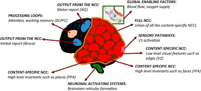
Definition of the NCC. Content-specific NCC (red) directly contribute to phenomenal distinctions (e.g., low-level visual features, faces, or places) within consciousness. The full NCC (orange) is constituted by the union of all the content-specific NCC. Background conditions to the NCC encompass neural processes that enable or modulate the activity of the full NCC and thus influence the level of consciousness (green), including global enabling factors, such as blood flow or oxygen supply, and neuronal activating systems, such as brainstem reticular formation; neural processes that modulate the activity of only some content-specific NCC, including processing loops involving attention or working memory (beige), sensory pathways activating primary sensory cortices (pink), and outputs from the NCC (blue) involved in task-related verbal or motor reports. V1, Primary visual cortex; V2, secondary visual cortex; PPA, parahippocampal place area; M1, primary motor cortex.
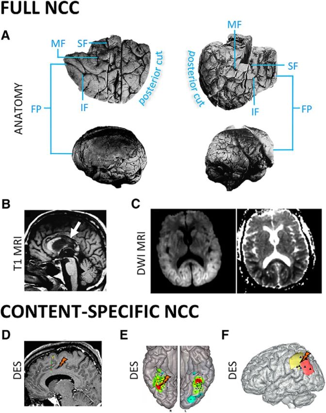
Clinical evidence. Full NCC. A, Complete bilateral prefrontal lobectomy does not noticeably change the level of consciousness. Top row, Bilateral views of the resected left and right frontal lobes (Brickner, 1936). Bottom row, Postmortem lateral views of both hemispheres (Brickner, 1952). B, Anoxic lesions of posterior corpus callosum predict permanent VS after head trauma (Kampfl et al., 1998). C, Lesions of posterior corpus callosum, with restricted diffusion extending to parieto-temporo-occipital regions, predict permanent coma after anoxic brain damage (Bianchi and Sims, 2008). Content-specific NCC. D, A recent study suggests that intrusive thoughts can be elicited by electrical stimulation of anterior cingulate cortex (Popa et al., 2016). Eliciting any experience is, however, far more common when stimulating posterior than anterior cortical structures (Selimbeyoglu and Parvizi, 2010). E, F, Direct electrical brain stimulation (DES) supports a causal role for different parts of the posterior cortex in specifying conscious content, for example, the right FFA in contributing to face percepts (Rangarajan et al., 2014) (E) and the parietal cortex contributing the feeling of intention to move (F) (Desmurget et al., 2013). SF, Superior frontal sulcus; MF, middle frontal sulcus; IF, inferior frontal sulcus; DWI, diffusion weighted imaging.
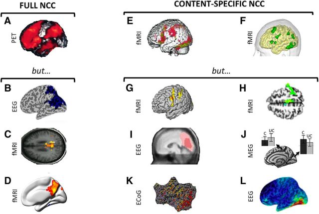
Neuroimaging, Forward inference. Full NCC. A, Between-state paradigm contrasting brain activity during NREM sleep and wakefulness (Kajimura et al., 1999) shows a relative deactivation of frontoparietal cortices. B, When subjects are awoken from NREM sleep and asked if they experienced anything before being awakened, EEG data during dream experiences show reduced low-frequency activity (1–10 Hz) compared with dreamless sleep in a posterior parieto-occiptal “hot zone” (Siclari et al., 2014). C, D, Directly comparing patients in an MCS with patients in a VS reveals differences in connectivity restricted to posterior cortex. C, Vanhaudenhuyse et al. (2010). D, Wu et al. (2015). Content-specific NCC. E, F, Tasks involving reporting seen versus unseen stimuli highlight differences in frontoparietal cortices: E, Binocular rivalry (Lumer et al., 1998); F, Visual word masking tasks (Dehaene et al., 2001). G, When conscious visual perception is dissociated from behavior (i.e., button press), only differences in activity in occipital and parietal regions remain (Frässle et al., 2014). H, Conscious perception of weak somatosensory stimuli correlates with cortical changes in BOLD signal restricted to contralateral rolandic and parietal areas (Meador et al., 2017). I, J, An early “visual awareness negativity” ∼200 ms in posterior temporal and occipital areas is found in two masking paradigms: I, Koivisto and Revonsuo (2010); J, Andersen et al. (2016). C, Conscious stimulus; UC, unconscious stimulus. K, Visual one-back paradigm in patients implanted with subdural electrode arrays. The visual cortex (right of the dashed white line) responds rapidly to the seen stimulus (red), whereas frontal regions are modulated by the task (yellow) (Noy et al., 2015). L, A within-state no-task experiment (Fig. 1D), contrasting EEG activity during REM sleep dreams with and without faces, highlighted the fusiform gyrus as content-specific NCC (Siclari et al., 2014).
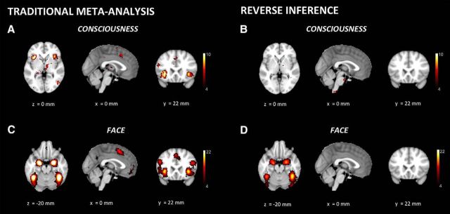
Neuroimaging, Reverse inference. A, When using Neurosynth for a traditional meta-analysis, computing the probability that different brain regions are active when the topics of a study include consciousness, parts of frontal cortex show up. B, When using Neurosynth in reverse inference mode, computing the probability that consciousness is included within the topics of a study, given the activation of different parts of the brain, frontal cortex disappears. The key term “conscious” was used on the Neurosynth website to extract both “forward” meta-analysis and reverse inference analysis steps in A, B. C, The same frontal areas that identified in a traditional meta-analysis for consciousness also appear activated in a traditional meta-analysis for faces. D, In contrast, reverse inference for faces no longer identifies frontal cortex activity but rather locates the activation predicting highest probability for face percepts in the right FFA. The key term “faces” was used on the Neurosynth website to extract both “forward” meta-analysis and reverse inference analysis steps in C, D. x, y, z values represent MNI coordinates, and a color scale is used for Z values.
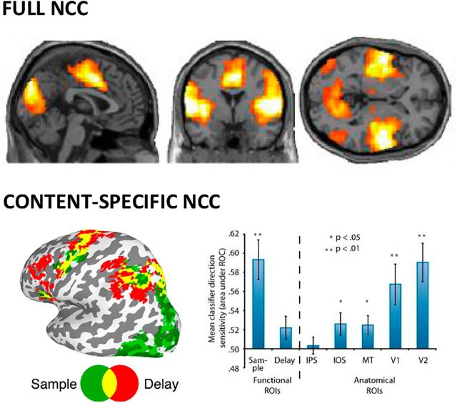
Neuroimaging, Predictive approaches. Full NCC, Machine learning approaches applied to fMRI resting state show that temporo-parieto-occipital connectivity best differentiates patients in MCS versus VS (Demertzi et al., 2015). Content-specific NCC, The contents of a working memory task can best be decoded from the back of the brain (posterior green ROI, activated during sample period), but not from the front of the brain (red fronto-parietal ROI, activated during delay period) (Emrich et al., 2013). Right side panel, Classification accuracy to identify conscious contents is much higher for posterior Sample ROI than for fronto-parietal Delay ROI (left part of panel, before dashed line). Classification accuracy is also higher in occipital areas compared with parietal cortex (right part of panel, after dashed line). IPS, Intraparietal sulcus; IOS, intraoccipital sulcus; MT, area MT; V1, primary visual cortex; V2, secondary visual cortex; ROI, region of interest.
References
-
- Avidan MS, Jacobsohn E, Glick D, Burnside BA, Zhang L, Villafranca A, Karl L, Kamal S, Torres B, O'Connor M, Evers AS, Gradwohl S, Lin N, Palanca BJ, Mashour GA (2011) Prevention of intraoperative awareness in a high-risk surgical population. N Engl J Med 365:591–600. 10.1056/NEJMoa1100403 - DOI - PubMed
Publication types
MeSH terms
Grants and funding
LinkOut - more resources
Full Text Sources
Other Literature Sources