. 2017 Nov 30;91(24):e01078-17.
doi: 10.1128/JVI.01078-17. Print 2017 Dec 15.

Processing of Potato Spindle Tuber Viroid RNAs in Yeast, a Nonconventional Host

Affiliations

Processing of Potato Spindle Tuber Viroid RNAs in Yeast, a Nonconventional Host

Dillon Friday et al. J Virol. .

Abstract

Potato spindle tuber viroid (PSTVd) is a circular, single-stranded, noncoding RNA plant pathogen that is a useful model for studying the processing of noncoding RNA in eukaryotes. Infective PSTVd circles are replicated via an asymmetric rolling circle mechanism to form linear multimeric RNAs. An endonuclease cleaves these into monomers, and a ligase seals these into mature circles. All eukaryotes may have such enzymes for processing noncoding RNA. As a test, we investigated the processing of three PSTVd RNA constructs in the yeast Saccharomyces cerevisiae Of these, only one form, a construct that adopts a previously described tetraloop-containing conformation (TL), produces circles. TL has 16 nucleotides of the 3' end duplicated at the 5' end and a 3' end produced by self-cleavage of a delta ribozyme. The other two constructs, an exact monomer flanked by ribozymes and a trihelix-forming RNA with requisite 5' and 3' duplications, do not produce circles. The TL circles contain nonnative nucleotides resulting from the 3' end created by the ribozyme and the 5' end created from an endolytic cleavage by yeast at a site distinct from where potato enzymes cut these RNAs. RNAs from all three transcripts are cleaved in places not on path for circle formation, likely representing RNA decay. We propose that these constructs fold into distinct RNA structures that interact differently with host cell RNA metabolism enzymes, resulting in various susceptibilities to degradation versus processing. We conclude that PSTVd RNA is opportunistic and may use different processing pathways in different hosts.IMPORTANCE In higher eukaryotes, the majority of transcribed RNAs do not encode proteins. These noncoding RNAs are responsible for mRNA regulation, control of the expression of regulatory microRNAs, sensing of changes in the environment by use of riboswitches (RNAs that change shape in response to environmental signals), catalysis, and more roles that are still being uncovered. Some of these functions may be remnants from the RNA world and, as such, would be part of the evolutionary past of all forms of modern life. Viroids are noncoding RNAs that can cause disease in plants. Since they encode no proteins, they depend on their own RNA and on host proteins for replication and pathogenicity. It is likely that viroids hijack critical host RNA pathways for processing the host's own noncoding RNA. These pathways are still unknown. Elucidating these pathways should reveal new biological functions of noncoding RNA.

Keywords: RNA conformation; RNA processing; host functions; mRNA degradation; noncoding RNA; viroids.


Figures

FIG 1
FIG 1
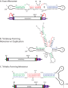
In vivo constructs and corresponding structural motifs used to express PSTVd in yeast. The left and right arms of PSTVd are indicated schematically, while the nucleotides of the CCR upper and lower strands are given explicitly. Nucleotides shown in green (nt 80 to 87), red (nt 90 to 99), and blue (nt 102 to 110) are part of the upper CCR (UCCR), which is duplicated to a different extent in each construct. (A) Monomer construct. The construct is flanked by 5′ hammerhead (HH) and 3′ hairpin (HP) ribozymes, which cleave at the curved arrows. Upon ligation, the construct should fold into the wild-type extended rod containing a loop E motif. (B) GNRA tetraloop (TL) construct. 3′ duplication of UCCR nucleotides 80 to 96 (green) allows formation of a metastable structure in which nucleotides 93 to 106 form a stem capped by the tetraloop; this stem contains the position 95/96 processing cleavage site (arrowhead). This construct contains a 3′ delta ribozyme (δ). (C) Trihelix-forming monomeric (triH) construct. The triH RNA has the same 5′ end as the TL RNA; its 3′ end duplicates nucleotides 80 to 110 (green, red, and blue). The two complete copies of the UCCR hybridize to form the trihelix (top), which has two potential position 95/96 cleavage sites (arrowheads).
FIG 2
FIG 2
Transcription of constructs in yeast and corresponding ligation-monitoring RT-PCR scheme. Once the PSTVd RNA is transformed into yeast, galactose induces its transcription. Upon induction and transcription, the RNA will fold into the structures seen in Fig. 1 for processing and ligation within the UCCR; the example shown here is the exact monomer transcript open at the 95/96 site. For analysis, the RNA was extracted, reverse transcribed, and then PCR amplified using primers 112F and 259R (inward) to screen for all plus-sense PSTVd or primers 2F and 259R (outward) across the UCCR region to screen specifically for processed circles. The gray box represents the hammerhead ribozyme, which undergoes self-cleavage in the monomer construct. All constructs contain a 3′ ribozyme; only the exact monomer construct contains the 5′ ribozyme.
FIG 3
FIG 3
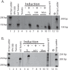
Detection of plus-sense PSTVd and PSTVd circles in total RNA from induced yeast by use of RT-PCR. (A) RT-PCR with the inward primer set 112F/259R revealed that all PSTVd RNAs tested produced a 148-bp DNA fragment. Lanes: 2, empty yeast strain; 3, PSTVd-infected tomato; 4, healthy tomato; 5 to 10, RNAs from yeast strain YPH500 transformed with plasmids containing the PSTVd constructs listed in Fig. 1. The positive control was an in vitro transcript of a PSTVd monomer (lane 11), and lane 12 contains a transcript-free RT-PCR control. RNAs were extracted from yeast grown in galactose-containing induction medium (+) or noninducing dextrose medium (−). The molecular size markers are from a 50-bp DNA ladder. (B) RT-PCR with the outward primer set 2F/259R revealed the presence of circular PSTVd RNA as a 240-bp (tomato) or 248-bp (yeast) DNA duplex. The lane order is the same as in panel A, with the exception of the infected and healthy tomato samples. The different migration behaviors of the marker bands in lanes 1 and 13 are due to an electrophoretic “smile.” Black lines indicate deleted lanes; all lanes in one panel were run in the same gel.
FIG 4
FIG 4
Northern blot analysis of total RNA from yeast expressing PSTVd. Total RNAs from healthy tomato (lane 1), infected tomato (lane 2), empty YPH500 (lane 3), and YPH500 transformed with the indicated constructs (lanes 4 to 9) were separated in a 5% PAA gel and blotted onto a positively charged nylon membrane. Induction conditions were provided by use of dextrose (−) and galactose (+) media. In vitro transcripts corresponding to the mon and TL constructs were used as positive controls (lanes 10 and 11). The ribozymes of the monomer IVT (lane 10) (see Fig. 1A) did not cleave to completion, resulting in four bands: HH-PSTVd-HP (547 nt; yellow asterisk), PSTVd-HP (482 nt; green asterisk), HH-PSTVd (424 nt; blue asterisk), and linear PSTVd (359 nt; red asterisk). The blot was probed with an α-32P-labeled minus-sense PSTVd transcript. Linear and circular PSTVd RNAs are marked. For a loading control, the yeast actin gene (1,537 nt) was probed in a second hybridization. Actin migrates above circular PSTVd. Red arrows for lanes 5 and 7 indicate primary transcripts from the in vivo transcripts prior to any yeast cleavage/ligation events (in vivo) for TL and triH. An open circle denotes circular products resulting from processing of the TL construct in vivo (lane 5). A red arrow for lane 9 marks the linear monomer-length band of the monomer. Black lines indicate deleted lanes; all lanes in one panel were run in the same gel.
FIG 5
FIG 5
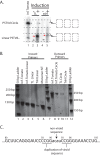
RT-PCR of gel-excised linear and circular PSTVd RNAs, Northern blot confirmation, and sequence data. A denaturing 5% polyacrylamide gel with duplication of five lanes was run and cut in half. (A) The left half was used for a Northern blot. Lane 1, positive control (infected tomato); lanes 2 and 3, yeast total RNA from the TL construct; lanes 4 and 5, yeast total RNA from the triH construct. Red arrows indicate primary transcripts, open arrows indicate cleavage products of viroid RNA, and circles designate circular PSTVd products. (B) Regions indicated by scissors and dashed boxes in panel A were excised from the right half of the gel, and the RNAs were extracted and used as templates for RT-PCR. For lanes 2 to 6, we used the inward primer set 112F/259R to reveal plus-sense PSTVd; for lanes 8 to 11, we used the outward primer set 2F/259R to reveal circular PSTVd RNA. Lanes 2, 6, and 8 are positive controls from RT-PCRs with RNAs that were not gel extracted; lanes 3 and 9 show gel-extracted linear/circular RNAs from infected tomato. The template RNAs for lanes 4 and 10 and lanes 5 and 11 were the extracted linear/circular bands from induced TL and triH RNAs, respectively, from the gel in panel A. Black lines indicate deleted lanes; all lanes in one panel were run in the same gel. (C) Sequence results for gel-extracted circles from TL in yeast. The lowercase letters represent a nonviroid vector sequence, and the bars indicate duplications of the viroid sequence.
FIG 6
FIG 6
Northern blot analysis of total RNAs from PSTVd-infected wild-type and RAT1 mutant YPH500 yeast strains. Total RNA controls included healthy tomato (lane 1), infected tomato (lane 2), empty YPH500 (lane 3), and the plasmid-free RAT1 strain (lane 4). Lane 5, galactose induction (+) of the RAT1 strain transformed with the monomer construct; lanes 6 and 7, RAT1 strain transformed with the TL construct under dextrose (−) and galactose (+) induction conditions; lanes 9 and 10, RAT1 strain transformed with the triH construct under dextrose (−) and galactose (+) induction conditions; lanes 8 and 11, YPH500 strain with galactose induction of the TL and triH transformants. In vitro transcripts corresponding to each in vivo construct were used as positive loading controls (lanes 12 to 14). The ribozymes of the monomer IVT (lane 12) (see Fig. 1A) did not cleave to completion, resulting in four bands: HH-PSTVd-HP (547 nt; yellow asterisk), PSTVd-HP (482 nt; green asterisk), HH-PSTVd (424 nt; blue asterisk), and linear PSTVd (359 nt; red asterisk). The blot was probed with an α-32P-labeled minus-sense PSTVd transcript. Linear and circular PSTVd RNAs are marked. For a loading control, the yeast Scr1 gene (522 nt) was probed in a second hybridization. Red arrows indicate primary transcripts from the in vitro transcripts or prior to any yeast cleavage/ligation events (in vivo), blue asterisks show HH-PSTVd RNA, and open circles denote circular products resulting from processing of the TL construct in vivo (lanes 7 and 8). Black lines indicate deleted lanes; all lanes were run in the same gel.
FIG 7
FIG 7
Determination of RNA 5′ ends for viroid species detectable in vivo. Total RNA was prepared from a yeast RAT1-107 mutant expressing the PSTVd TL and triH constructs. Untreated (25 μg) and MnCl2-treated (12.5 μg) RNAs were then analyzed by primer extension. In vitro transcripts (0.5 pmol) of TL and triH (lanes 1 and 8) were used as controls. The 5′ ends of both in vitro transcripts are identical, and transcription starts at position G80. Lanes 2 and 3, RNAs from plasmid-free RAT1-107 mutant, without and with MnCl2 treatment; lanes 4 and 5, RNAs from triH-transformed RAT1-107 mutant, without and with MnCl2 treatment; lanes 6 and 7, RNAs from TL-transformed RAT1-107 mutant, without and with MnCl2 treatment. Position −2C (red arrow) marks the yeast transcription start site in constructs harboring a GAL1 promoter. Position G80 marks the first PSTVd base. Open arrows indicate additional 5′ ends/degradation products detectable in the MnCl2-enriched TL RNA (lane 7). The bars in lane 5 indicate the high level of additional 5′ ends/degradation products seen in the triH variant compared to those in the TL variant.
FIG 8
FIG 8
Cleavage and ligation sites for PSTVd in yeast expressing the TL construct. Letter coloring and upper/lowercase indicate the same nucleotides throughout. The lowercase red letters are vector nucleotides added during plasmid construction. (A) Sequences of the 5′ and 3′ ends of TL transcribed in yeast. Uppercase blue and red letters are wild-type PSTVd sequences. The blue arrows (95/96) indicate the known cleavage sites of PSTVd processed in potato or tomato. The downward-facing red arrow (92/93) indicates the 5′ cleavage site of processing in yeast. The lowercase black letters are the 5′ end of the delta ribozyme. The green arrow indicates cleavage by the delta ribozyme. The dotted line with the red arrow marks the site for ligation in yeast. (B) Sequences of ligation junctions in potato and yeast. A caret (∧) marks the ligation site. Sequences shown were determined by RT-PCR with gel-extracted RNA circles from yeast and tomato. The proposed ligation scheme results in an additional 8 nt in yeast (red). (C) Ligation substrate in yeast. The marked 3′ and 5′ ends are ligated to form the yeast circle. The additional 8 nt (red) comprise nt 96, the four vector nucleotides, and nt 93 to 95.

Similar articles

Cited by

References

    1. Diener TO. 1972. Viroids. Adv Virus Res 17:295–313. doi:10.1016/S0065-3527(08)60754-X. - DOI - PubMed
    1. Diener TO. 1974. Viroids: the smallest known agents of infectious disease. Annu Rev Microbiol 28:23–39. doi:10.1146/annurev.mi.28.100174.000323. - DOI - PubMed
    1. Tsagris EM, Martinez de Alba AE, Gozmanova M, Kalantidis K. 2008. Viroids. Cell Microbiol 10:2168–2179. doi:10.1111/j.1462-5822.2008.01231.x. - DOI - PubMed
    1. Flores R, Minoia S, Carbonell A, Gisel A, Delgado S, Lopez-Carrasco A, Navarro B, Di Serio F. 2015. Viroids, the simplest RNA replicons: how they manipulate their hosts for being propagated and how their hosts react for containing the infection. Virus Res 209:136–145. doi:10.1016/j.virusres.2015.02.027. - DOI - PubMed
    1. Owens RA, Hammond RW. 2009. Viroid pathogenicity: one process, many faces. Viruses 1:298–316. doi:10.3390/v1020298. - DOI - PMC - PubMed

LinkOut - more resources