. 2017 Nov 30;91(24):e00850-17.
doi: 10.1128/JVI.00850-17. Print 2017 Dec 15.

Atomic Structures of Minor Proteins VI and VII in Human Adenovirus

Affiliations

Atomic Structures of Minor Proteins VI and VII in Human Adenovirus

Xinghong Dai et al. J Virol. .

Abstract

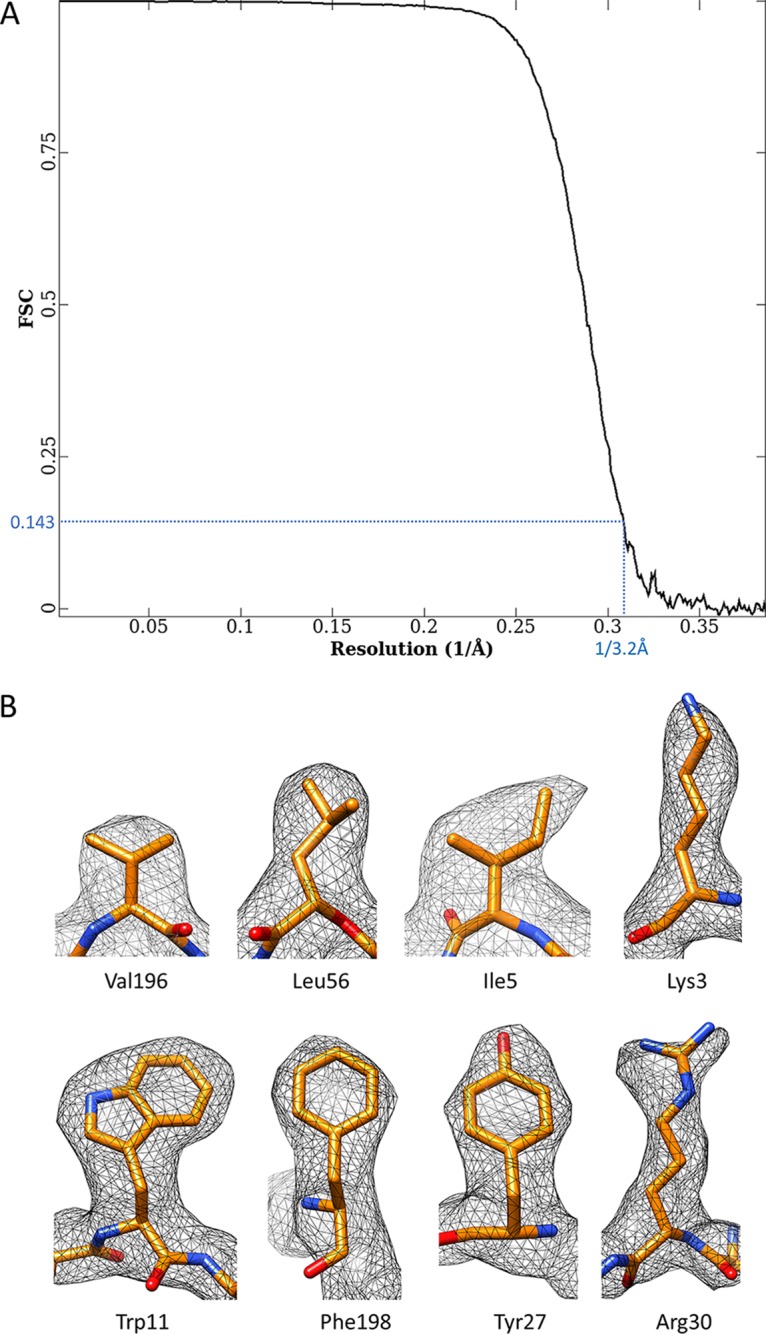
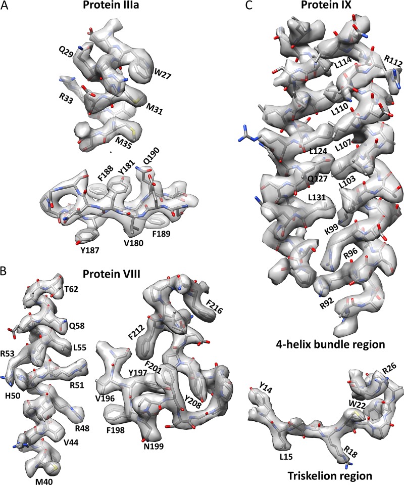
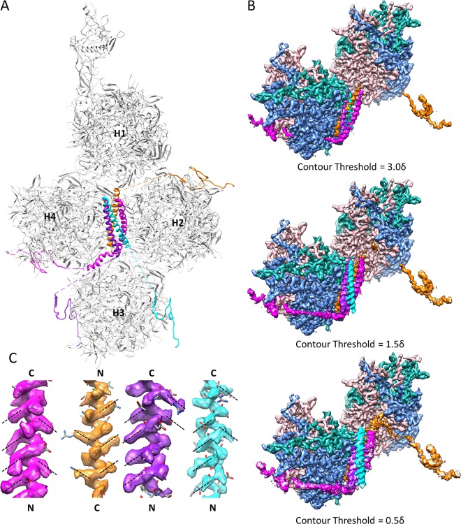
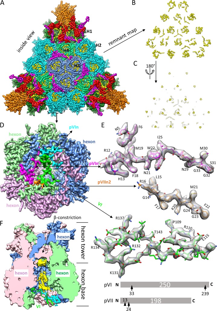
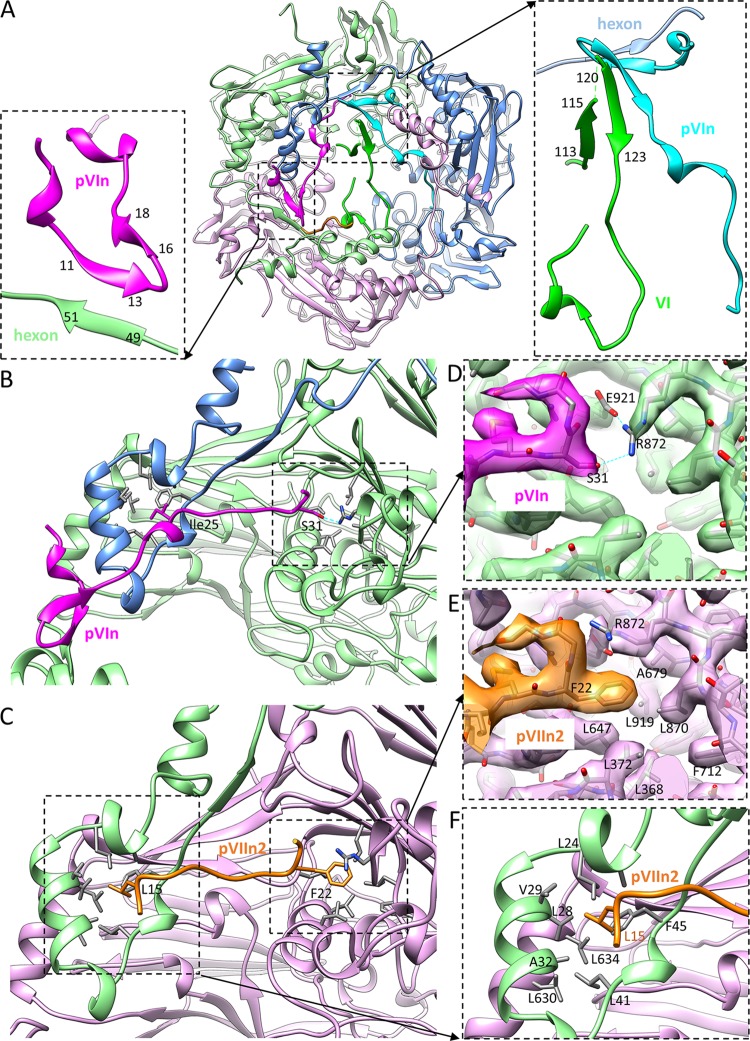
Human adenoviruses (Ad) are double-stranded DNA (dsDNA) viruses associated with infectious diseases, but they are better known as tools for gene delivery and oncolytic anticancer therapy. Atomic structures of Ad provide the basis for the development of antivirals and for engineering efforts toward more effective applications. Since 2010, atomic models of human Ad5 have been derived independently from photographic film cryo-electron microscopy (cryo-EM) and X-ray crystallography studies, but discrepancies exist concerning the assignment of cement proteins IIIa, VIII, and IX. To clarify these discrepancies, we employed the technology of direct electron counting to obtain a cryo-EM structure of human Ad5 at 3.2-Å resolution. Our improved structure unambiguously confirms our previous cryo-EM models of proteins IIIa, VIII, and IX and explains the likely cause of conflict in the crystallography models. The improved structure also allows the identification of three new components in the cavity of hexon-the cleaved N terminus of precursor protein VI (pVIn), the cleaved N terminus of precursor protein VII (pVIIn2), and mature protein VI. The binding of pVIIn2-and, by extension, that of genome-condensing pVII-to hexons is consistent with the previously proposed dsDNA genome-capsid coassembly for adenoviruses, which resembles that of single-stranded RNA (ssRNA) viruses but differs from the well-established mechanism of pumping dsDNA into a preformed protein capsid exemplified by tailed bacteriophages and herpesviruses.IMPORTANCE Adenovirus is a double-edged sword to humans: it is a widespread pathogen but can be used as a bioengineering tool for anticancer and gene therapies. The atomic structure of the virus provides the basis for antiviral and application developments, but conflicting atomic models for the important cement proteins IIIa, VIII, and IX from conventional/film cryo-EM and X-ray crystallography studies have caused confusion. Using cutting-edge cryo-EM technology with electron counting, we improved the structure of human adenovirus type 5 and confirmed our previous models of cement proteins IIIa, VIII, and IX, thus clarifying the inconsistent structures. The improved structure also reveals atomic details of membrane-lytic protein VI and genome-condensing protein VII and supports the previously proposed genome-capsid coassembly mechanism for adenoviruses.

Keywords: cement protein structure; dsDNA genome packaging; endosomal escape; genome-capsid coassembly; human adenovirus.


Figures

FIG 1
FIG 1
Distribution and structure of minor proteins IIIa, VIII, and IX in the virion of human Ad5. (A and B) Outer surface (A) and inner surface (B) views of human Ad5 capsid. The electron-counting cryo-EM density map of the human Ad5 virion at 3.2-Å resolution is differentially colored according to the identities of the penton base (P), hexons (H1 to H4), and minor proteins IX (purple) (A), IIIa (red) (B), and VIII (magenta) (B). (C to E) Atomic models of minor proteins IX, IIIa, and VIII. Each model is rainbow colored blue to red from the N terminus to the C terminus of the protein chain. The numbers denote protein sequence numbers at boundaries of the model. Three copies of the protein IX N-terminal region and four copies of the protein IX C-terminal region assemble into the triskelion and the 4-helix bundle, respectively. Note the three parallel-one antiparallel configuration of coiled-coil helices in the 4-helix bundle.
FIG 2
FIG 2
Resolution assessment of the electron-counting cryo-EM structure of the human Ad5 virion. (A) The final resolution of the density map was estimated to be 3.2 Å by the criterion FSC = 0.143 (63). (B) High-resolution features of some protein side chains in the density map of cement protein VIII. Note that subtle differences among Val, Leu, and Ile, between Phe and Tyr, and between Lys and Arg are discernible.
FIG 3
FIG 3
Validating our previous models of proteins IIIa, VIII, and IX with the improved structure of human Ad5 from electron-counting cryo-EM. Example densities (semitransparent surfaces) of proteins IIIa (A), VIII (B), and IX (C) were segmented from the 3.2-Å-resolution cryo-EM reconstruction and fitted with the corresponding atomic models (sticks) to showcase the validity of our model. Some bulky side chains are labeled. Note that only two high-quality helices of the protein IX 4-helix bundle are shown, for clarity. These two helices are antiparallel.
FIG 4
FIG 4
Cement protein IX forms the triskelions and 4-helix bundles on the outer surface of Ad5 capsid. (A) Distribution of four protein IX molecules in each asymmetric unit of Ad5 capsid. The C-terminal regions of four protein IX molecules assemble into a 4-helix bundle, while their N-terminal regions are distributed in four triskelion structures. Quasi-equivalent hexons H1 to H4 are labeled. (B) Densities connecting the N-terminal triskelion domain of protein IX to its C-terminal coiled-coil helix in the 4-helix bundle are discernible by lowering the display threshold. The density map is viewed from the same orientation as that for panel A, with the densities of hexons H2 and H3 hidden for clarity. Only two of the four triskelions and their linker densities to the 4-helix bundle are shown, for clarity. Note that the brown density links the triskelion to a helix from the top of the 4-helix bundle, while the magenta density (and the other two not shown) links the triskelion to a helix from the bottom of the 4-helix bundle. (C) Polarities of the four helices in the 4-helix bundle. Because protein side chains in an α-helix should point slightly toward the N-terminal end of the helix, the polarities of the four helices can be determined unambiguously based on the orientations of side chain densities, as denoted by dashed lines. The four helices were determined to be three parallel and one antiparallel.
FIG 5
FIG 5
Identification and modeling of proteins VI, pVIn, and pVIIn2 in the inner cavity of hexon. (A) Facet of Ad5 capsid viewed from inside the capsid. The density map is differentially colored in the same way as in Fig. 1A and B, except that previously unmodeled densities are highlighted in yellow. Note that the unmodeled densities are mainly distributed in the cavities of hexons. (B and C) A “remnant map” was calculated by removing densities of hexon, penton base, and proteins IIIa, VIII, and IX, leaving previously unmodeled densities alone. The density map in panel B is flipped over in panel C to show the cup-shaped densities hiding deeply in the cavity of each hexon, which are attributable to the disordered N- and/or C-terminal region of protein VI. (D) Zoomed-in view of hexon H2. The location of this hexon is marked with a dashed hexagon in panel A. Three subunits of the hexon trimer and the minor proteins in the cavity of the hexon are differentially colored. The hexon trimer is displayed at a threshold of 3δ (δ is the standard deviation), pVIn (cyan or magenta) and pVIIn2 (gold) at a threshold of 2.75δ, and mature VI (green) at a threshold of 1.75δ. (E) Atomic models (sticks) of pVIn, pVIIn2, and VI fitted to their corresponding cryo-EM densities (semitransparent surfaces), with some landmark residues labeled. Only one of the two equivalent copies of pVIn is shown. The schematics at the bottom show maturational cleavages in pVI and pVII. Each precursor protein is represented as a bar, with the polypeptide length (in amino acids) indicated in the center. Consensus cleavage sites are denoted by arrows, and the nonconsensus site is denoted by an arrowhead. (F) Disordered densities hidden deeply in the cavity of hexon H2. The surface presentation of the hexon was calculated by use of atomic models of the hexon trimer, and one half was removed to expose the inner cavity of the hexon. The nomenclature “hexon base,” “hexon tower,” and “β-constriction site” is used following that in the literature (5). Disordered densities (yellow) in the cavity were segmented from the cryo-EM density map and displayed at a threshold of 0.9δ. A model of protein VI (green ribbon) crossing the opening of the cavity is also shown. The two dashed lines denote that the disordered densities are speculated to be attributable to the unmodeled N- and C-terminal regions of protein VI.
FIG 6
FIG 6
Binding sites of pVIn, pVIIn2, and protein VI in the cavity of hexon. (A) Atomic model of hexon H2, corresponding to the density map shown in Fig. 5D. The two insets show zoomed-in views of the boxed regions, showing β-augmentation interactions among hexon subunit, pVIn, and VI. (B and C) Overall views of the binding sites of pVIn (B) and pVIIn2 (C) in the hexon wall. (D and E) Comparison of pVIn (D) and pVIIn2 (E) C-terminal binding to two equivalent pockets in the hexon wall. The images are zoomed-in views of the boxed regions in panels B and C, respectively. Note the similar shapes of pVIn and pVIIn2 in this region, but the prominently distinguishable pVIn Ser31 and pVIIn2 Phe22 densities, as well as distinct interactions with labeled hexon residues. (F) Second binding site shared by pVIn and pVIIn2 in the capsid wall. This site of a hydrophobic pocket is bound by pVIIn2 Leu15, as shown in the boxed region in panel C and the zoomed-in view here, or by pVIn Ile25, as shown in panel B.

References

    1. Ghebremedhin B. 2014. Human adenovirus: viral pathogen with increasing importance. Eur J Microbiol Immunol 4:26–33. doi: 10.1556/EuJMI.4.2014.1.2. - DOI - PMC - PubMed
    1. Huang GH, Xu WB. 2013. Recent advance in new types of human adenovirus. Bing Du Xue Bao 29:342–348. - PubMed
    1. Appaiahgari MB, Vrati S. 2015. Adenoviruses as gene/vaccine delivery vectors: promises and pitfalls. Expert Opin Biol Ther 15:337–351. doi: 10.1517/14712598.2015.993374. - DOI - PubMed
    1. Rosewell Shaw A, Suzuki M. 2016. Recent advances in oncolytic adenovirus therapies for cancer. Current Opin Virol 21:9–15. doi: 10.1016/j.coviro.2016.06.009. - DOI - PMC - PubMed
    1. Roberts MM, White JL, Grutter MG, Burnett RM. 1986. Three-dimensional structure of the adenovirus major coat protein hexon. Science 232:1148–1151. doi: 10.1126/science.3704642. - DOI - PubMed

Publication types

Substances