. 2017 Nov 30;91(24):e01282-17.
doi: 10.1128/JVI.01282-17. Print 2017 Dec 15.

Human Metapneumovirus Induces Formation of Inclusion Bodies for Efficient Genome Replication and Transcription

Affiliations

Human Metapneumovirus Induces Formation of Inclusion Bodies for Efficient Genome Replication and Transcription

Nicolás Cifuentes-Muñoz et al. J Virol. .

Abstract

Human metapneumovirus (HMPV) causes significant upper and lower respiratory disease in all age groups worldwide. The virus possesses a negative-sense single-stranded RNA genome of approximately 13.3 kb encapsidated by multiple copies of the nucleoprotein (N), giving rise to helical nucleocapsids. In addition, copies of the phosphoprotein (P) and the large RNA polymerase (L) decorate the viral nucleocapsids. After viral attachment, endocytosis, and fusion mediated by the viral glycoproteins, HMPV nucleocapsids are released into the cell cytoplasm. To visualize the subsequent steps of genome transcription and replication, a fluorescence in situ hybridization (FISH) protocol was established to detect different viral RNA subpopulations in infected cells. The FISH probes were specific for detection of HMPV positive-sense RNA (+RNA) and viral genomic RNA (vRNA). Time course analysis of human bronchial epithelial BEAS-2B cells infected with HMPV revealed the formation of inclusion bodies (IBs) from early times postinfection. HMPV IBs were shown to be cytoplasmic sites of active transcription and replication, with the translation of viral proteins being closely associated. Inclusion body formation was consistent with an actin-dependent coalescence of multiple early replicative sites. Time course quantitative reverse transcription-PCR analysis suggested that the coalescence of inclusion bodies is a strategy to efficiently replicate and transcribe the viral genome. These results provide a better understanding of the steps following HMPV entry and have important clinical implications.IMPORTANCE Human metapneumovirus (HMPV) is a recently discovered pathogen that affects human populations of all ages worldwide. Reinfections are common throughout life, but no vaccines or antiviral treatments are currently available. In this work, a spatiotemporal analysis of HMPV replication and transcription in bronchial epithelial cell-derived immortal cells was performed. HMPV was shown to induce the formation of large cytoplasmic granules, named inclusion bodies, for genome replication and transcription. Unlike other cytoplasmic structures, such as stress granules and processing bodies, inclusion bodies are exclusively present in infected cells and contain HMPV RNA and proteins to more efficiently transcribe and replicate the viral genome. Though inclusion body formation is nuanced, it corresponds to a more generalized strategy used by different viruses, including filoviruses and rhabdoviruses, for genome transcription and replication. Thus, an understanding of inclusion body formation is crucial for the discovery of innovative therapeutic targets.

Keywords: HMPV; inclusion bodies; pneumovirus; replication.


Figures

FIG 1
FIG 1
HMPV FISH probes are specific. (A) Schematic of the FISH probes designed for the present work. Probes span half of the genome, including the genes for N, P, M, F, and M2. The two sets of probes target the vRNA and the +RNA sequences, in red and purple, respectively. (B) BSR cells were transfected to express either HMPV N and P mRNAs (top) or the HMPV full-length genome (bottom) of strain JPS02-76 under the control of the T7 promoter. At 48 h posttransfection, cells were processed for FISH to detect vRNA (red) and +RNA (purple) and for immunofluorescence to detect the P protein (green). (Insets) Enlarged images of the small squares with dotted outlines. Bars, 10 μm.
FIG 2
FIG 2
Inclusion body formation is a hallmark of HMPV infection. (A) BEAS-2B cells were infected with HMPV, fixed at 24 hpi (strain CAN97-83) or 72 hpi (strains TN9332 and TN9316), and processed for detection of vRNA (red) and the N protein (green). (Insets) Enlarged images of the small squares with dotted outlines. Bars, 10 μm. (B) Z-stack analysis of a cell infected with HMPV CAN97-83, showing an inclusion body (red) close to the cell nucleus (blue) at 24 hpi. (C) BEAS-2B cells were infected with HMPV CAN97-83, and at 24 hpi the distance between the IBs and the cell nuclei was measured using Z-stack analysis of 17 cells. The red bar indicates the average distance to the nucleus.
FIG 3
FIG 3
HMPV inclusion bodies are maintained at low numbers. (A) BEAS-2B cells were infected with rgHMPV CAN97-83 at an MOI of 1 PFU/cell, and cells were processed for FISH and immunofluorescence at 24, 48, and 72 hpi. (B) BEAS-2B cells were infected with rgHMPV CAN97-83 at different MOIs, and the cells were processed for FISH and immunofluorescence at 24 hpi. An inclusion body was counted as a vRNA-positive/N-positive perinuclear structure ≥0.5 μm in size. Student's t test was performed for statistical analysis. *, P < 0.01; ***, P < 0.0001. Red lines indicate the average number of inclusion bodies per infected cell.
FIG 4
FIG 4
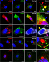
Inclusion bodies are sites of replication and transcription but not assembly. BEAS-2B cells were infected with HMPV CAN97-83 at an MOI of 3 PFU/cell and fixed at 24 hpi. FISH and immunofluorescence were performed to detect vRNA (red), +RNA (purple; green as a pseudocolor), polyadenylated RNAs (red), and the HMPV P, M, and F proteins (green). Bars, 10 μm. The squares with dotted outlines are magnified and shown as the zoom images in the right column.
FIG 5
FIG 5
Inclusion bodies do not colocalize with stress granule or P-body markers. BEAS-2B cells were infected with HMPV CAN97-83 at an MOI of 3 PFU/cell and fixed at 24 (left panels) or 48 hpi. FISH and immunofluorescence were performed to detect vRNA (red) and the SG markers G3BP1 and TIAR-1 (green) or P-body marker Ge-1 (green). (Insets) Enlarged images of the small squares with dotted outlines. Bars, 10 μm.
FIG 6
FIG 6
Rab11, β-tubulin, and actin do not colocalize in IBs. (A) BEAS-2B cells were infected with HMPV CAN97-83 at an MOI of 3 PFU/cell and fixed at 24 hpi. FISH and immunofluorescence were performed to detect vRNA (red) and Rab11 (green). Bars, 10 μm. (B) Vero cells were transfected to express GFP-Rab11 or GFP-Rab11 DN. At 24 h posttransfection, the cells were infected with HMPV CAN97-83 at different MOIs (in numbers of PFU per cell). Virus titers were calculated at 72 hpi. (C) BEAS-2B cells were infected with HMPV CAN97-83 at an MOI of 3 PFU/cell and fixed at 24 hpi. FISH and immunofluorescence were performed to detect vRNA (red) and tubulin or β-actin (green). Bars, 10 μm.
FIG 7
FIG 7
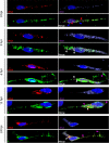
Time course analysis suggests the coalescence of inclusion bodies. BEAS-2B cells were infected with HMPV CAN97-83 at an MOI of 3 PFU/cell and fixed at 0 (3 h postinoculation), 3, 6, 12, and 24 hpi. FISH and immunofluorescence were performed to detect vRNA (red), +RNA (purple), the HMPV P protein (green), and nuclei (blue). Z-stack analysis was performed at every time postinfection.
FIG 8
FIG 8
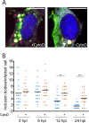
Actin polymerization is involved in inclusion body coalescence. (A) BEAS-2B cells were infected with HMPV CAN97-83 at an MOI of 3 PFU/cell, and at 3 h postinoculation, the cells were treated with DMSO or incubated with 2 μM cytochalasin D (CytoD) until 24 hpi. FISH and immunofluorescence were performed to detect vRNA (red), +RNA (purple), and the HMPV P protein (green). (B) The number of vRNA/+RNA replicative spots in at least 15 DMSO- or cytochalasin D-treated infected cells was counted at each time postinfection. Student's t test was performed for statistical analysis. **, P < 0.001; ***, P < 0.0001. Black bars indicate the average number of inclusion bodies per infected cell.
FIG 9
FIG 9
Quantitative time course analysis of HMPV transcription and replication. BEAS-2B cells were infected with HMPV CAN97-83 at an MOI of 3 PFU/cell, and at 3 h postinoculation, the cells were treated with DMSO or incubated with 2 μM cytochalasin D or 17 μM nocodazole until 24 hpi. At each time postinfection, total RNA was extracted from the cells and then quantitative RT-PCR was performed to detect P mRNA (A) or vRNA (B). The levels of expression shown are relative to GAPDH expression levels. Experiments were performed in triplicate. Student's t test was performed for statistical analysis. **, P < 0.001; *, P < 0.01.
FIG 10
FIG 10
Proposed events of HMPV transcription and replication. After attachment and endocytosis (a), viral nucleocapsids are released into the cell cytoplasm (b). Incoming particles can give rise to replicative sites (c), where vRNA (red), cRNA (green), mRNAs (blue), and viral proteins are readily synthesized. At between 6 and 12 hpi, replicative sites are more compact and start to coalesce (d) into larger inclusion bodies located close to cell nuclei (e). The synthesis of mRNA and vRNA is exponential at these times postinfection. From inclusion bodies, vRNA might be transported to assembly sites (f) through the actin cytoskeleton. The cell cytoplasm is populated by viral mRNAs. By 24 hpi, large inclusion bodies are visible at the perinuclear area (g). At this and later times postinfection, IBs remain as the only sites where transcription and replication are still active. At this point, cells are engaged in particle assembly and spread (h). Light blue circles, matrix protein; brown circles, nucleoprotein; purple circles, phosphoprotein.

References

    1. Pelletier G, Dery P, Abed Y, Boivin G. 2002. Respiratory tract reinfections by the new human metapneumovirus in an immunocompromised child. Emerg Infect Dis 8:976–978. doi: 10.3201/eid0809.020238. - DOI - PMC - PubMed
    1. van den Hoogen BG, de Jong JC, Groen J, Kuiken T, de Groot R, Fouchier RA, Osterhaus AD. 2001. A newly discovered human pneumovirus isolated from young children with respiratory tract disease. Nat Med 7:719–724. doi: 10.1038/89098. - DOI - PMC - PubMed
    1. Widmer K, Zhu Y, Williams JV, Griffin MR, Edwards KM, Talbot HK. 2012. Rates of hospitalizations for respiratory syncytial virus, human metapneumovirus, and influenza virus in older adults. J Infect Dis 206:56–62. doi: 10.1093/infdis/jis309. - DOI - PMC - PubMed
    1. Boivin G, Abed Y, Pelletier G, Ruel L, Moisan D, Cote S, Peret TC, Erdman DD, Anderson LJ. 2002. Virological features and clinical manifestations associated with human metapneumovirus: a new paramyxovirus responsible for acute respiratory-tract infections in all age groups. J Infect Dis 186:1330–1334. doi: 10.1086/344319. - DOI - PubMed
    1. Afonso CL, Amarasinghe GK, Banyai K, Bao Y, Basler CF, Bavari S, Bejerman N, Blasdell KR, Briand FX, Briese T, Bukreyev A, Calisher CH, Chandran K, Cheng J, Clawson AN, Collins PL, Dietzgen RG, Dolnik O, Domier LL, Durrwald R, Dye JM, Easton AJ, Ebihara H, Farkas SL, Freitas-Astua J, Formenty P, Fouchier RA, Fu Y, Ghedin E, Goodin MM, Hewson R, Horie M, Hyndman TH, Jiang D, Kitajima EW, Kobinger GP, Kondo H, Kurath G, Lamb RA, Lenardon S, Leroy EM, Li CX, Lin XD, Liu L, Longdon B, Marton S, Maisner A, Muhlberger E, Netesov SV, Nowotny N, et al. 2016. Taxonomy of the order Mononegavirales: update 2016. Arch Virol 161:2351–2360. doi: 10.1007/s00705-016-2880-1. - DOI - PMC - PubMed

LinkOut - more resources