. 2017 Oct 4;9(410):eaai7466.
doi: 10.1126/scitranslmed.aai7466.

Engineering a highly elastic human protein-based sealant for surgical applications

Affiliations

Engineering a highly elastic human protein-based sealant for surgical applications

Nasim Annabi et al. Sci Transl Med. .

Erratum in

Abstract

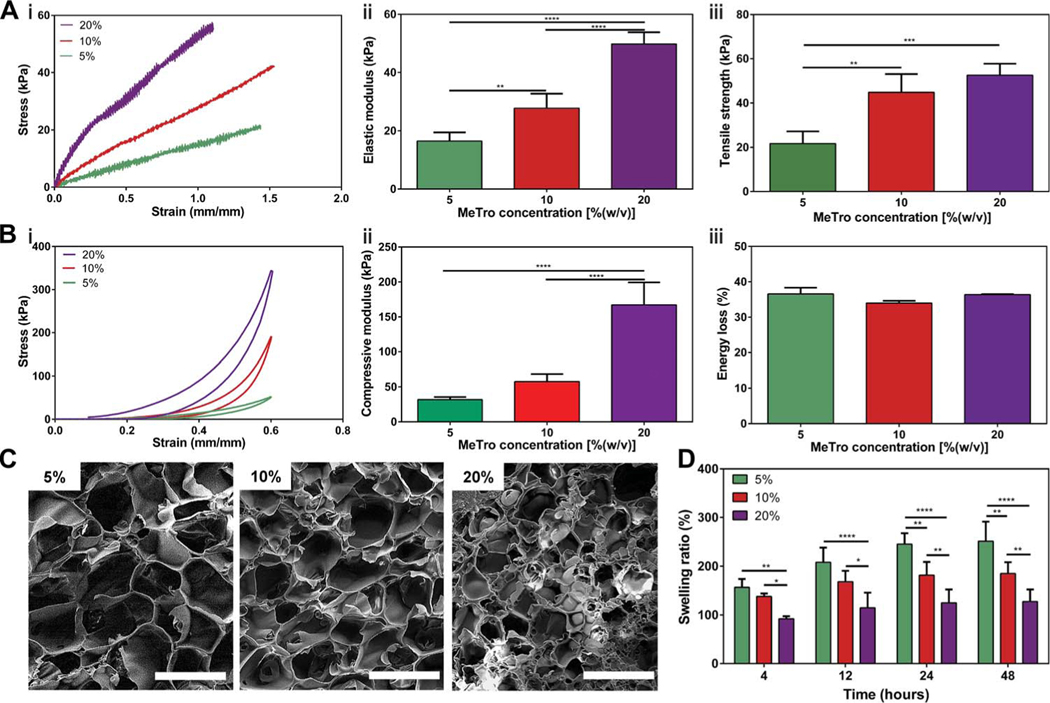
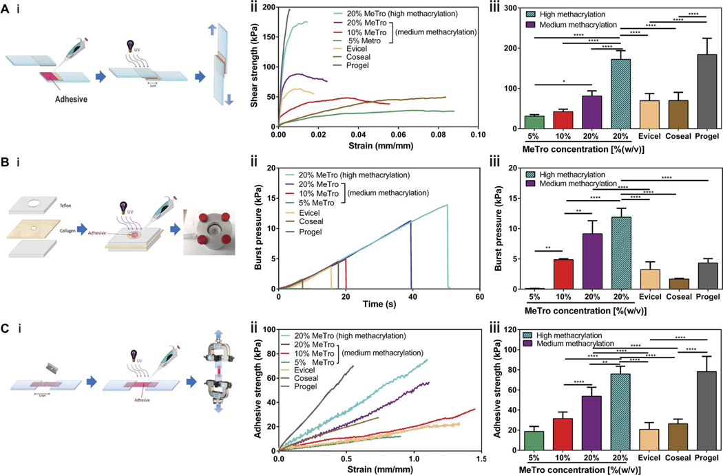
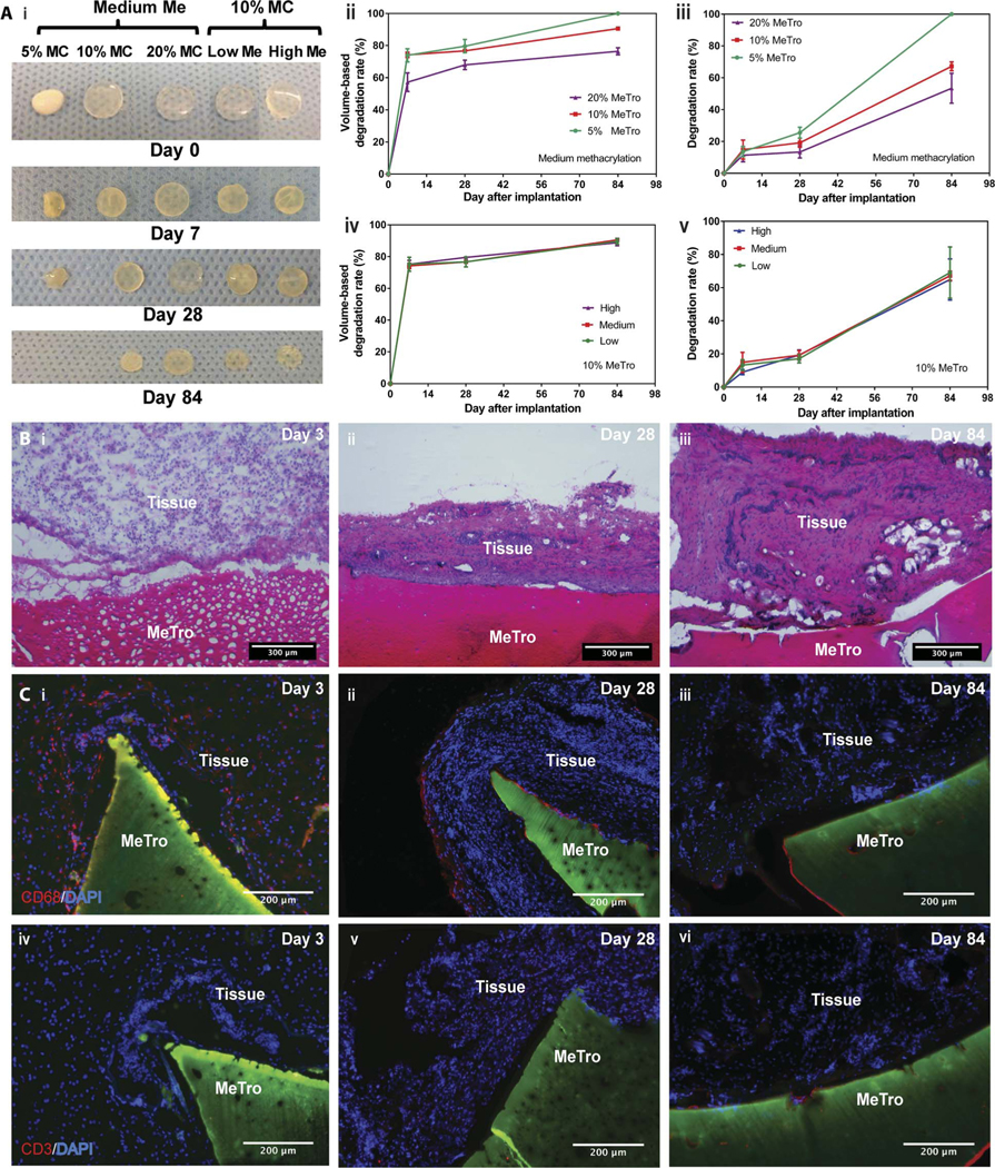
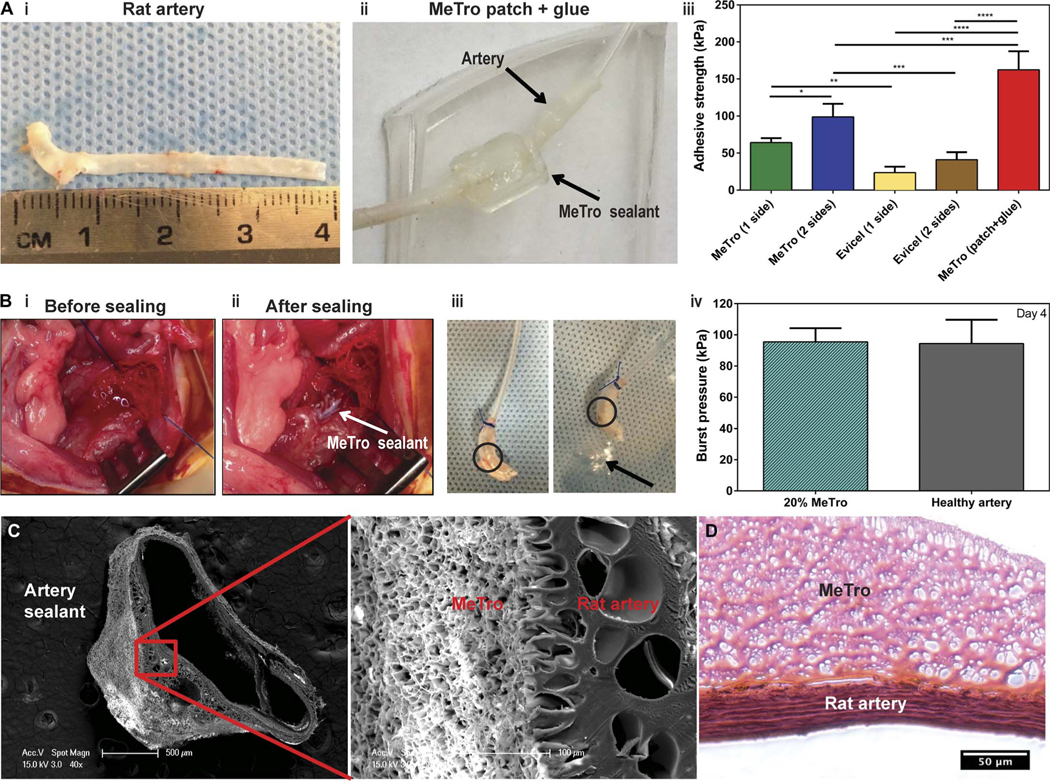
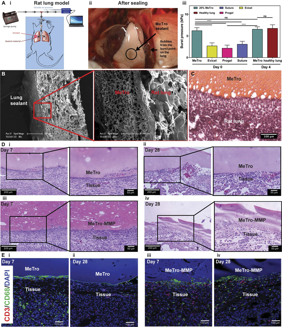
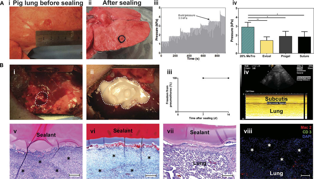
Surgical sealants have been used for sealing or reconnecting ruptured tissues but often have low adhesion, inappropriate mechanical strength, cytotoxicity concerns, and poor performance in biological environments. To address these challenges, we engineered a biocompatible and highly elastic hydrogel sealant with tunable adhesion properties by photocrosslinking the recombinant human protein tropoelastin. The subcutaneous implantation of the methacryloyl-substituted tropoelastin (MeTro) sealant in rodents demonstrated low toxicity and controlled degradation. All animals survived surgical procedures with adequate blood circulation by using MeTro in an incisional model of artery sealing in rats, and animals showed normal breathing and lung function in a model of surgically induced rat lung leakage. In vivo experiments in a porcine model demonstrated complete sealing of severely leaking lung tissue in the absence of sutures or staples, with no clinical or sonographic signs of pneumothorax during 14 days of follow-up. The engineered MeTro sealant has high potential for clinical applications because of superior adhesion and mechanical properties compared to commercially available sealants, as well as opportunity for further optimization of the degradation rate to fit desired surgical applications on different tissues.


Conflict of interest statement

Competing interests: A.S.W. has equity interest in Elastagen Pty Ltd., which supplied tropoelastin used in these studies. N.A., A.S.W., and A.K. are co-inventors on patent US9688741 held/submitted by the Brigham and Women’s Hospital, Harvard Medical School and Elastagen Pty Ltd. that covers elastic hydrogel. All other authors declare that they have no competing interests.

Figures

Fig. 1.
Fig. 1.. Physical characterizations of the MeTro sealant.
(A) Tensile tests on the MeTro hydrogel produced by using different MeTro concentrations with medium degree of methacryloyl substitution (76%) (n ≥ 3). (i) Representative tensile strain-stress curves, (ii) elastic modulus, and (iii) ultimate tensile strength. (B) Compression tests on the MeTro hydrogel produced at different MeTro concentrations with medium degree of methacryloyl substitution (n = 3). (i) Representative compression strain-stress curves, (ii) compressive modulus, and (iii) energy loss. (C) Representative SEM images of the MeTro hydrogel synthesized by (i) 5%, (ii) 10%, and (iii) 20% (w/v) MeTro concentrations at medium degree of methacryloyl substitution (scale bars, 100 μm). (D) Swelling ratios of the MeTro hydrogel in PBS at 37°C, depending on different MeTro concentrations with the medium methacryloyl substitution over 48 hours (n = 4). Data are means ± SD. P values were determined by one-way analysis of variance (ANOVA) followed by Tukey’s multiple comparisons test for (A) and (B), and two-way ANOVA for (D) (*P < 0.05, **P < 0.01, ***P < 0.001, ****P < 0.0001).
Fig. 2.
Fig. 2.. In vitro sealing properties of the MeTro sealant.
(A) Standard lap shear test to determine the shear strength of MeTro sealants (n ≥ 4) with different formulations and several commercially available sealants: Evicel (n = 5), Coseal (n = 3), and Progel (n = 4). (i) Schematic of the modified standard test method for shear test (ASTM F2255–05), (ii) representative strain-stress curves for lap shear tests, and (iii) average shear strength of MeTro sealants produced with varying MeTro concentrations and degree of methacryloyl substitution, and commercially available sealants including Evicel, Coseal, and Progel. (B) Standard burst pressure test to evaluate the burst pressure of the MeTro sealant (n ≥ 4) with different formulations [except 5% Metro (n = 2)] and several commercially available sealants: Evicel (n = 4), Coseal (n = 3), and Progel (n = 5). (i) Schematic of the modified standard test method for the burst pressure (ASTM F2392–04), (ii) representative strain-stress curves for burst pressure test, and (iii) average burst pressure of the MeTro sealants with variable MeTro concentrations and degree of methacryloyl substitution, and commercially available sealants. (C) Standard wound closure using porcine skin as the biological substrate to test the adhesion strength of the MeTro sealant (n ≥ 4) with different formulations and commercially available sealants: Evicel (n = 5), Coseal (n = 3), and Progel (n = 3). (i) Schematic of the modified standard test method for adhesion strength (ASTM F2458–05), (ii) representative strain-stress curves for wound closure test, and (iii) average adhesive strength of MeTro sealants produced with variable MeTro concentrations and degree of methacryloyl substitution, and their comparison with commercially available sealants. Data are means ± SD. P values were determined by one-way ANOVA followed by Tukey’s multiple comparisons test (*P < 0.05, **P < 0.01, ****P < 0.0001).
Fig. 3.
Fig. 3.. In vivo biocompatibility and degradation of the MeTro sealant using a rat subcutaneous model.
(A) Evaluation of the in vivo degradation of MeTro sealants on days 0, 7, 28, and 84 of implantation (n = 4). (i) Representative images of the MeTro hydrogel implants with different extents of methacryloyl substitution (Me) and MeTro concentrations (MC). In vivo MeTro degradation based on volume loss of the implant for (ii) varying MeTro concentrations with medium methacryloyl substitution and (iv) varying extents of methacryloyl substitution at 10% (w/v) MeTro concentration. In vivo MeTro degradation based on weight loss of the implant for (iii) variable MeTro concentration with medium methacryloyl substitution and (v) variable degree of methacryloyl substitution at 10% (w/v) MeTro concentration. The in vivo degradation profile of MeTro hydrogels shows significant volume loss by day 7 and almost constant weight loss afterward until day 84. (B) Histology images of MeTro with the surrounding tissue stained with H&E after (i) 3 days, (ii) 28 days, and (iii) 84 days of implantation in subcutaneous tissue of a rat using 20% MeTroconcentration and medium methacryloyl substitution (scale bars, 300 μm). H&E reveals insignificant amount of inflammatory cells. (C) Immunostaining of subcutaneously implanted MeTro hydrogels showing macrophage (CD68) only at (i) day 3 but disappeared at days (ii) 28 and (iii) 84, and resulting in no local lymphocyte infiltration (CD3) at days (iv) 3, (v) 28, and (vi) 84 (scale bars, 200 μm). Green color in (C) and (D) represents the autofluorescent MeTro gel, red color represents the lymphocytes, and blue color represents the nuclei (DAPI).
Fig. 4.
Fig. 4.. Ex vivo and in vivo function of the MeTro sealant using rat incision model of arteries.
(A) Ex vivo wound closure test using explanted rat aorta as a biological substrate for tissue adhesion. (i) An explanted aorta from a rat (about 4 cm in length). (ii) A patch generated using the MeTro hydrogel and wrapped the artery tube segments. The connecting anastomosis points were further glued with MeTro. Arrows present the rat artery and MeTro sealant. (iii) Adhesive strength of the MeTro sealant applied on one side (n = 4) and both sides (n = 3) of the artery in comparison with Evicel applied on one side (n = 4) and both sides (n = 4). The MeTro patch (n = 2) further improved the adhesive strength. (B) In vivo tests on rat arteries sealed by MeTro (n = 3). Operative sites (i) before and (ii) after sealing. White arrow presents the MeTro sealant on the artery. (iii) Image of a sealed artery pressurized with air, demonstrating that MeTro could adhere to the outer arterial surface and seal the incision, but the artery burst in another area. Circles present the MeTro seals. Arrow indicates bubbles from the burst point on the artery instead of the MeTro sealing site. (iv) Burst pressure values of artery sealed by MeTro with 20% concentration and high methacryloyl substitution after 4 days compared to a healthy artery as a control. Representative SEM (C) and H&E-stained (D) images from the interface between the explanted rat artery and the MeTro sealant. Tight interfaces between the MeTro hydrogel and the tissues indicate strong bonding and interlocking at the interfaces. Data are means ± SD. P values were determined by one-way ANOVA followed by Tukey’s multiple comparisons test for (A) and (B) (*P < 0.05, **P < 0.01, ***P < 0.001, ****P < 0.0001).
Fig. 5.
Fig. 5.. In vivo function of MeTro sealants using rat incision model of lungs.
(A) In vivo tests on rat lungs sealed by MeTro. (i) Schematic diagram showing the experimental setup. (ii) Image of the sealed lung pressurized with air, demonstrating that MeTro could adhere to the lung surface and seal the incision, but the lung tissue burst from other areas (the source of the bubbles) when pressures of >5.5 kPa were applied. (iii) Burst pressure values for lungs sealed with the MeTro sealant on day 0 (n = 4) and day 4 (n = 3) as compared to lungs sealed by Evicel (n = 3), Progel (n = 6), and suture only (n = 6) as well as healthy lung (n = 3) on day 0. Representative (B) SEM and (C) H&E-stained images from the interface between the explanted rat lung and the MeTro sealant. Tight interfaces between the MeTro hydrogel and the tissues indicate strong bonding and interlocking at the interfaces. (D) H&E staining of MeTro sections at days (i) 7 and (ii) 28 and MeTro-MMP sections at days (iii) 7 and (iv) 28 after sealing. (E) Fluorescent immunohistochemical analysis of the MeTro and MeTro-MMP sealants, showing no significant local lymphocyte infiltration (CD3) at days (i) 7 and (ii) 28 for MeTro samples and at days (iii) 7 and (iv) 28 for MeTro-MMP samples. Also, the images exhibit remarkable macrophage (CD68) expression at day 7 (i and iii) but reduced expression at day 28 for MeTro and remained the same for MeTro-MMP samples (ii and iv). Green, red, and blue colors in (E) represent the macrophages, lymphocytes, and cell nuclei (DAPI), respectively. Data are means ± SD. P values were determined by one-way ANOVA followed by Tukey’s multiple comparisons test for (A) (**P < 0.01, ***P < 0.001).
Fig. 6.
Fig. 6.. Ex vivo and in vivo test to evaluate the sealing capability of the MeTro sealant using a porcine lung incision model.
(A) Ex vivo porcine lung burst pressure testing. (i) Image from the superficial wound created in deflated lung before sealing, (ii) wound covered with the MeTro sealant, (iii) representative graph depicting the incremental increase in pressure during ventilation, and (iv) average burst pressures for MeTro (n = 5), Evicel (n = 3), Progel (n = 5), and a suture control (n = 3). MeTro shows the highest burst pressure. (B) In vivo sealing capacity of MeTro using a porcine lung incision model (n = 3). (i and ii) A right lung lobe is exposed through a small lateral thoracotomy, and a standardized defect is created [broken line in (B, i)] and sealed by applying and photocrosslinking the MeTro (20% MeTro concentration with high methacryloyl substitution) sealant [broken line in (B, ii)]. (iii and iv) At postoperative days 7 and 14, freedom from pneumothorax was confirmed by sonography, as displayed in a representative ultrasound image at day 14, indicating the absence of any relevant amount of air in the intercostal space. (v) Representative histological sections of the lung leakage site after 14 days revealed sufficient formation of wound healing tissue [asterisks in (v); H&E staining] including a stable collagenous (blue) defect cover [asterisks in (vi); Masson’s trichrome staining]. (vii) H&E staining of the attachment of MeTro sealant to noninjured lung tissue around the defect site. (viii) Immunohistological staining of the defect area and the formed wound healing tissue (asterisks) did not show relevant macrophage (Mac2) or lymphocyte (CD3) infiltration. Scale bars, 200 μm (v to vii) and 100 μm (viii). Data are means ± SD (*P < 0.05 and **P < 0.01).

References

    1. Annabi N, Tamayol A, Shin SR, Ghaemmaghami AM, Peppas NA, Khademhosseini A, Surgical materials: Current challenges and nano-enabled solutions. Nano Today 9, 574–589 (2014). - PMC - PubMed
    1. Smith TO, Sexton D, Mann C, Donell S, Sutures versus staples for skin closure in orthopaedic surgery: Meta-analysis. BMJ 340, c1199 (2010). - PMC - PubMed
    1. Sanders L, Nagatomi J, Clinical applications of surgical adhesives and sealants. Crit. Rev. Biomed. Eng. 42, 271–292 (2014). - PMC - PubMed
    1. Lins RD, Gomes RC, Santos KS, Silva PV, Silva RT, Ramos IA, Use of cyanoacrylate in the coaptation of edges of surgical wounds. An. Bras. Dermatol. 87, 871–876 (2012). - PMC - PubMed
    1. Burdick JA, Murphy WL, Moving from static to dynamic complexity in hydrogel design. Nat. Commun. 3, 1269 (2012). - PubMed