Review
. 2017 Oct;207(2):389-412.
doi: 10.1534/genetics.117.199968.

Gene Tagging Strategies To Assess Protein Expression, Localization, and Function in Drosophila

Affiliations
Review

Gene Tagging Strategies To Assess Protein Expression, Localization, and Function in Drosophila

Oguz Kanca et al. Genetics. 2017 Oct.

Erratum in

Abstract

Analysis of gene function in complex organisms relies extensively on tools to detect the cellular and subcellular localization of gene products, especially proteins. Typically, immunostaining with antibodies provides these data. However, due to cost, time, and labor limitations, generating specific antibodies against all proteins of a complex organism is not feasible. Furthermore, antibodies do not enable live imaging studies of protein dynamics. Hence, tagging genes with standardized immunoepitopes or fluorescent tags that permit live imaging has become popular. Importantly, tagging genes present in large genomic clones or at their endogenous locus often reports proper expression, subcellular localization, and dynamics of the encoded protein. Moreover, these tagging approaches allow the generation of elegant protein removal strategies, standardization of visualization protocols, and permit protein interaction studies using mass spectrometry. Here, we summarize available genomic resources and techniques to tag genes and discuss relevant applications that are rarely, if at all, possible with antibodies.

Keywords: Drosophila; Flybook; gene tagging; genome engineering; techniques and resources; transgenesis.


Figures

Figure 1
Figure 1
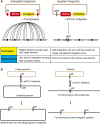
Overview of transgenesis strategies. (A) Comparison of nontargeted insertion to targeted insertion. During P-element transformation, the transposase integrates the plasmid containing transposon ends into the genome in an untargeted way. Multiple insertion sites can be obtained with the same injection. In φC31-mediated transgenesis, the plasmid is inserted into a specific target locus in the genome. IR, inverted repeats. (B) In the natural use of integrase, a phage genome-containing attP site is integrated into the attB site in the bacterial genome. This creates attR and attL sites that can no longer be used by the φC31-integrase enzyme as a substrate. In fly transformation, integrase enzyme integrates the plasmid-containing attB site in an attP site previously inserted in the fly genome by nontargeted insertion strategies.
Figure 2
Figure 2
Schematics of transgenic constructs with different options. (A) Upstream activating sequence (UAS)-cDNA strategy. (B) Genomic strategy. Advantages and disadvantages of possible choices are explained below the corresponding parts of the construct. Dotted lines indicate splicing, yellow lines indicate coding introns, and black lines indicate noncoding regions. SV40, Simian virus 40.
Figure 3
Figure 3
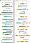
Overview of recombineering principle and its applications. (A) The basic principle of recombineering: a linear DNA piece containing homology arms (HA-L and HA-R) and a selection marker is inserted precisely into a circular target DNA. (B) Molecular mechanism of recombineering: 5′ to 3′ exonuclease activity and single-strand annealing mediated by the Red/ET system. (C) Applications of recombineering: simple scar-containing variant for C- and N-terminal tagging followed by Flp-induced removal of the selection cassette vs. two-step recombineering for seamless integration of a point mutation (red asterisk). (B) was modified from Sharan et al. (2009).
Figure 4
Figure 4
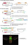
Fosmid library preparation, tagging, and transgenesis workflow. (A) FlyFos design and library construction. (B) Two-step fosmid tagging strategy, first generating the pretagged TRG fosmid clone library and second the sGFP-tagged TRG fosmid clone library. (C) Transgenesis step to build the fly TRG strain library. Note the expression of the dsRed marker in the adult eyes and the sGFP in the muscles, in particular the flight muscles, of this strain (tagged myosin heavy chain-sGFP protein, fTRG500; we acknowledge the help of Nicholas Gompel for acquiring the fly image). sGFP, superfolder GFP; TRG, TransgeneOme.
Figure 5
Figure 5
Overview of fly TransgeneOme strain library. (A) Number of available fly strains with the respective tags. (B) Functional verification by genetic rescue. (C) Collection of expression patterns in the adult fly thorax for Prm-sGFP (fTRG475, localizes at sarcomeric M-line in flight muscles), Adar-sGFP (fTRG570, localizes in nuclei of all cells), Hts-sGFP (fTRG585, expressed in motor neurons and present at the cortex of the gut epithelium) and Ergic53 (fTRG158, localizes in vesicles in the gut epithelium). GFP is shown in green and actin with phalloidin in red. Size bars are 100 µm in thorax and 10 µm in the enlarged boxes. We acknowledge the help of Christiane Barz for acquiring the images.
Figure 6
Figure 6
Overview of MiMIC (Minos-Mediated Integration Cassette) lines. (A) Schematics of MiMIC construct. MIR, Minos Inverted Repeats; SA, Splice Acceptor; 3 × STOP, stop codons in all possible open reading frames; yellow+, mini yellow dominant selection marker. (B) Schematic of genomic region with or without MiMIC. Dotted lines indicate splicing. (C) MiMIC collection numbers.
Figure 7
Figure 7
CRIMIC scheme. Coding intron sequence is cut by Cas9-gRNA complex. The sequences on either side of the cut site are used as 1-kb homology arms to integrate the SIC. Two possible SICs from published literature are included as examples. gRNA, guide RNA; RFP, red fluorescent protein; SIC, Swappable Insertion Cassette.
Figure 8
Figure 8
RMCE conversion scheme. (A) Conversion scheme of a MiMIC in the coding intron of a gene to tag the gene with GFP. Before conversion, MiMIC causes truncation of mRNA and protein. After conversion, mature mRNA will contain the artificial exon and the protein product will contain an internal GFP tag. (B) Explanation of possible open reading frames of a gene and how an RMCE cassette should be designed accordingly. N’s stand for nucleotides in the codons. (C) Different RMCE conversion strategies by crossing of fly strains. 3 × STOP, stop codons in all possible open reading frames; GSFTF, a multi-tag containing EGFP-FlAsH-StrepII-3XFlag and L flexible linkers; MiMIC, Minos-Mediated Integration Cassette; MIR, Minos Inverted Repeats; RMCE, Recombination-Mediated Cassette Exchange; SA, Splice Acceptor; yellow+, mini yellow dominant selection marker.
Figure 9
Figure 9
Schematic mode of action for protein removal tools. (A) deGradFP construct binds to GFP-tagged proteins constitutively through peptide binder and recruits the ubiquitination machinery to the target. Ubiquitinated protein is often degraded by the proteasome. (B) Auxin-induced degradation (AID) works by the TIR1 binding to AID in an Auxin-dependent manner. In the absence of Auxin, TIR does not bind to AID-tagged protein. Auxin causes rapid TIP1 binding to AID-tagged protein, which recruits the ubiquitination machinery. Ubiquitinated protein is often degraded by the proteasome. YFP, yellow fluorescent protein.

References

    1. Aleksic J., Lazic R., Müller I., Russell S. R., Adryan B., 2009. Biases in Drosophila melanogaster protein trap screens. BMC Genomics 10: 249. - PMC - PubMed
    1. Baena-Lopez L. A., Alexandre C., Mitchell A., Pasakarnis L., Vincent J.-P., 2013. Accelerated homologous recombination and subsequent genome modification in Drosophila. Development 140: 4818–4825. - PMC - PubMed
    1. Banaszynski L. A., Chen L.-C., Maynard-Smith L. A., Ooi A. G. L., Wandless T. J., 2006. A rapid, reversible, and tunable method to regulate protein function in living cells using synthetic small molecules. Cell 126: 995–1004. - PMC - PubMed
    1. Bassett A. R., Liu J.-L., 2014. CRISPR/Cas9 and genome editing in Drosophila. J. Genet. Genomics 41: 7–19. - PubMed
    1. Bateman J. R., Lee A. M., Wu C.-T., 2006. Site-specific transformation of Drosophila via phiC31 integrase-mediated cassette exchange. Genetics 173: 769–777. - PMC - PubMed

Publication types

MeSH terms

LinkOut - more resources