. 2017 Oct 5;2(19):e94298.
doi: 10.1172/jci.insight.94298.

DOCK8 enforces immunological tolerance by promoting IL-2 signaling and immune synapse formation in Tregs

Affiliations

DOCK8 enforces immunological tolerance by promoting IL-2 signaling and immune synapse formation in Tregs

Erin Janssen et al. JCI Insight. .

Abstract

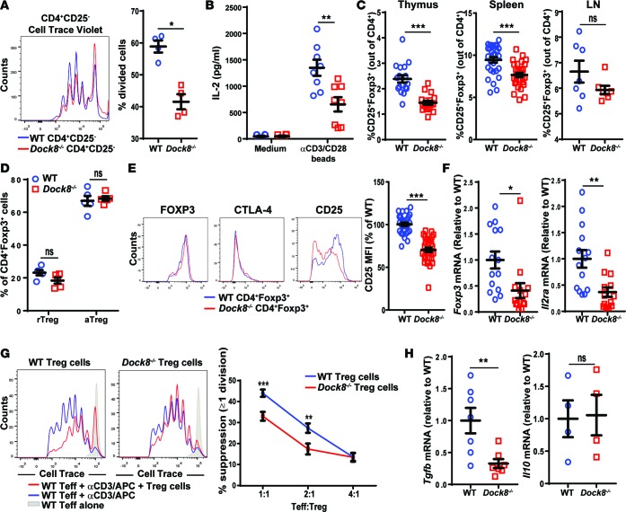
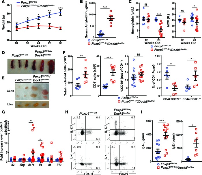
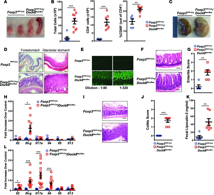
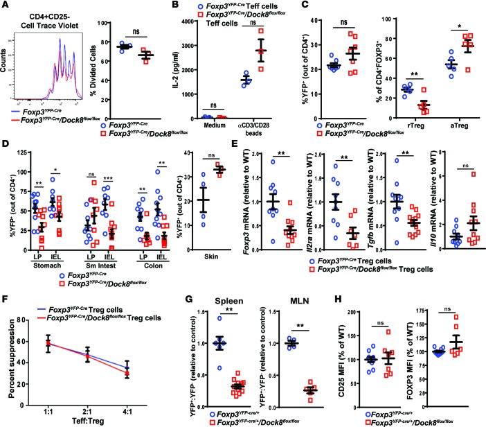
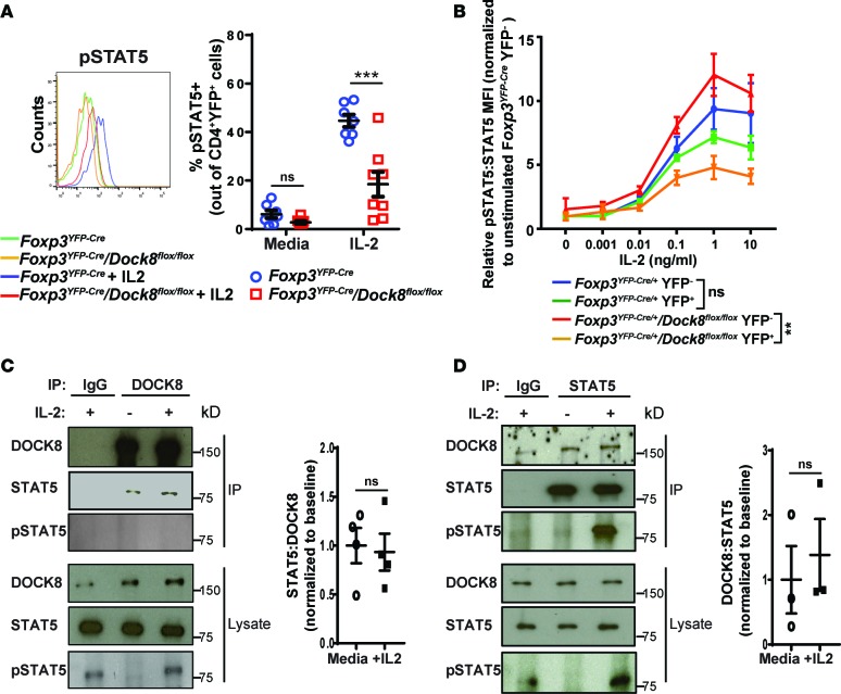
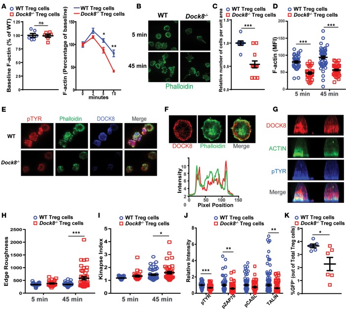
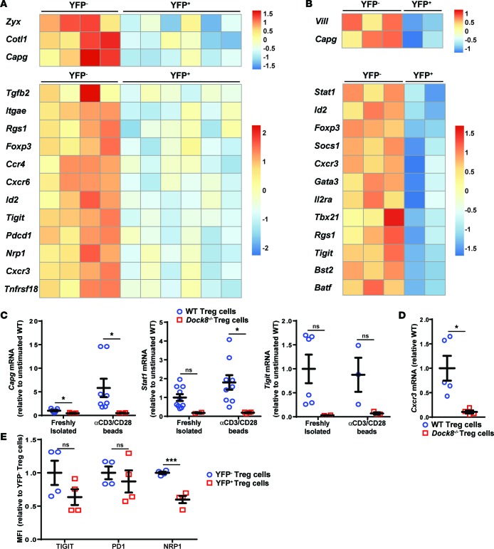
Patients deficient in the guanine nucleotide exchange factor DOCK8 have decreased numbers and impaired in vitro function of Tregs and make autoantibodies, but they seldom develop autoimmunity. We show that, similarly, Dock8-/- mice have decreased numbers and impaired in vitro function of Tregs but do not develop autoimmunity. In contrast, mice with selective DOCK8 deficiency in Tregs develop lymphoproliferation, autoantibodies, and gastrointestinal inflammation, despite a normal percentage and in vitro function of Tregs, suggesting that deficient T effector cell function might protect DOCK8-deficient patients from autoimmunity. We demonstrate that DOCK8 associates with STAT5 and is important for IL-2-driven STAT5 phosphorylation in Tregs. DOCK8 localizes within the lamellar actin ring of the Treg immune synapse (IS). Dock8-/- Tregs have abnormal TCR-driven actin dynamics, decreased adhesiveness, an altered gene expression profile, an unstable IS with decreased recruitment of signaling molecules, and impaired transendocytosis of the costimulatory molecule CD86. These data suggest that DOCK8 enforces immunological tolerance by promoting IL-2 signaling, TCR-driven actin dynamics, and the IS in Tregs.


Conflict of interest statement

Conflict of interest: The authors have declared that no conflict of interest exists.

Figures

Figure 1
Figure 1. Dock8–/– mice have reduced Treg percentages and in vitro Treg-suppressive ability.
(A and B) Proliferation measured by Cell Trace Violet dilution (A) and IL-2 secretion in culture supernatants (B) by CD4+CD25 Teffs isolated from the spleens of Dock8–/– and WT mice cultured for 3 days with anti-CD3+anti-CD28–coated beads. (C) Percentage of CD25+FOXP3+ Tregs among CD4+ cells in the thymuses, spleens, and LNs of Dock8–/– and WT mice. n = 17 mice from each group for the thymus, n = 31 mice from each group for the spleen, n = 7 mice from each group for the LN. (D) Percentages of CD44CD62Lhi rTregs and CD44+CD62Llo aTregs of total CD4+FOXP3+ cells in the spleens of Dock8–/– and WT mice. n = 5 mice from each group. (E) Representative FACS plots of intracellular FOXP3 and CTLA-4 and surface CD25 expression gating on CD4+FOXP3+ splenocytes (left). Quantitative analysis of surface CD25 expression by splenic CD4+FOXP3+ cells from Dock8–/– mice and WT controls. n = 29 mice from each group. (F) qPCR analysis of Foxp3 and Il2ra mRNA levels in FACS-sorted CD4+CD25+CD39+ Tregs from Dock8–/– and WT mice. Results are expressed as fold increase relative to the WT control ratio of the mRNA of interest/b2microglobulin. (G) Suppression of the proliferation of CD4+CD25 Teffs by CD4+CD25+CD39+ Tregs from Dock8–/– mice and WT controls. Teff proliferation was measured by FACS analysis of Cell Trace Violet dilution. The left panel is a representative experiment; the right panel shows the pooled results. (H) qPCR analysis of Tgfb and Il10 mRNA expression by sorted CD4+CD25+CD39+ Tregs from Dock8–/– mice and WT controls. Symbols represent individual mice, and error bars represent mean and SEM. Results in A, B, and FH are representative of 3 independent experiments. t test, NS P > 0.05, *P < 0.05, **P < 0.01, ***P < 0.001.
Figure 2
Figure 2. Mice with Treg-specific DOCK8 deficiency develop weight loss, splenomegaly, and lymphadenopathy.
(A) Weights between age 10 and 30 weeks in Foxp3YFP–Cre/Dock8flox/flox mice and Foxp3YFP–Cre controls. n = 11 Foxp3YFP–Cre/Dock8flox/flox and 10 Foxp3YFP–Cre mice. (B) Serum amyloid P levels in Foxp3YFP–Cre/Dock8flox/flox mice and Foxp3YFP–Cre controls at 28 weeks of age. (C) Hemoglobin concentration and mean red blood cell volume (MCV) in Foxp3YFP–Cre/Dock8flox/flox mice and Foxp3YFP–Cre controls at 12 and 28 weeks of age. (D and E) Representative photographs of the spleens (D), cervical LNs (CLN), and inguinal LNs (ILN) (E) from 30-week-old Foxp3YFP–Cre/Dock8flox/flox mice and Foxp3YFP–Cre controls. (F) Nucleated cells counts, CD4+ cell counts, percentage of CD69+ cells among CD4+ cells, and percentage of CD4+CD44CD62Lhi naive and CD4+CD44+CD62Llo memory T cells among CD4+FOXP3 cells in the spleens of 30-week-old Foxp3YFP–Cre/Dock8flox/flox mice and Foxp3YFP–Cre controls. (G) Cytokine mRNA expression in pooled CLNs and ILNs from 30-week-old Foxp3YFP–Cre/Dock8flox/flox mice and Foxp3YFP–Cre controls. Results of the qPCR analysis were expressed as fold increase relative to control of the cytokine mRNA/b2microglobulin mRNA ratio. (H) Representative FACS plots showing intracellular cytokine expression after gating on CD4+ T cells stimulated for 3.5 hours with phorbol 12,13-dibutyrate and ionomycin. (I) Serum IgA and IgE levels in 28-week-old Foxp3YFP–Cre/Dock8flox/flox mice and Foxp3YFP–Cre controls. Symbols represent individual mice, and error bars in AC, F, G, and I represent mean and SEM. t test, NS P > 0.05, *P < 0.05, **P < 0.01, ***P < 0.001.
Figure 3
Figure 3. Mice with a Treg-specific DOCK8 deficiency develop gastrointestinal inflammation.
(A) Representative photographs of the MLNs from 30-week-old Foxp3YFP–Cre/Dock8flox/flox mice and Foxp3YFP–Cre controls. (B) Total cellularity, number of CD4+ T cells, and percentage of activated CD69+ T cells of CD4+ T cells in MLNs from 30-week-old Foxp3YFP–Cre/Dock8flox/flox mice and Foxp3YFP–Cre controls. (C and D) Representative photographs of the stomachs (C) and representative H&E-stained sections of the forestomach and glandular stomach (original magnification, ×10) (D) from 30-week-old Foxp3YFP–Cre/Dock8flox/flox mice and Foxp3YFP–Cre controls. Scale bar: 100 μm (D). (E) Representative images (original magnification, ×20) of indirect immunofluorescent staining of stomach sections for IgG using dilutions of serum from a 28-week-old Foxp3YFP–Cre/Dock8flox/flox mouse and Foxp3YFP–Cre controls. (F and G) Representative H&E-stained sections (original magnification, ×20) (F) and enteritis pathology score (G) of the small intestine (SI) from 30-week-old Foxp3YFP–Cre/Dock8flox/flox mice and Foxp3YFP–Cre controls. Scale bar: 100 μm. (H) qPCR analysis of cytokine mRNA expression in the SI of 30-week-old Foxp3YFP–Cre/Dock8flox/flox mice and Foxp3YFP–Cre controls. Results are expressed as fold increase of the cytokine mRNA/b2microglobulin mRNA ratio relative to control. (I and J) Representative H&E-stained sections (original magnification, ×20) (I) and colitis pathology score (J) of the mid colon from 30-week-old Foxp3YFP–Cre/Dock8flox/flox mice and Foxp3YFP–Cre controls. (K) Fecal lipocalin-2 concentrations in the stools of 30-week-old Foxp3YFP–Cre/Dock8flox/flox mice and Foxp3YFP–Cre controls. (L) qPCR analysis of cytokine mRNA expression in the colons from 30-week-old Foxp3YFP–Cre/Dock8flox/flox mice and Foxp3YFP–Cre controls. Results are expressed as fold increase of the cytokine mRNA/b2microglobulin mRNA ratio relative to control. Symbols represent individual mice, and error bars in B, G, H, J, and K represent mean and SEM. t test, *P < 0.05, **P < 0.01, ***P < 0.001.
Figure 4
Figure 4. Tregs from mice with a Treg-specific DOCK8 deficiency have impaired fitness.
(A and B) Proliferation measured by Cell Trace Violet dilution (A) and IL-2 secretion in culture supernatants (B) by CD4+CD25 Teffs isolated from the spleens of Foxp3YFP–Cre/Dock8flox/flox and Foxp3YFP–Cre control mice cultured for 3 days with anti-CD3+anti-CD28–coated beads. (C) Percentage of YFP+ Tregs of CD4+ T cells and percentage of CD44CD62Lhi rTregs and CD44+CD62Llo aTregs among total Tregs in spleens of 30-week-old Foxp3YFP–Cre/Dock8flox/flox mice and Foxp3YFP–Cre controls. (D) Percentage of YFP+ cells of total CD4+ cells in the intraepithelial lymphocytes (IEL) and LP of the stomachs and colons and in the skin from Foxp3YFP–Cre/Dock8flox/flox mice and Foxp3YFP–Cre controls. (E) qPCR analysis of Foxp3, Il2ra, Tgfb, and Il10 mRNA levels in FACS-sorted CD4+CD25+YFP+ Tregs from Foxp3YFP–Cre/Dock8flox/flox mice and Foxp3YFP–Cre controls. Results are expressed as fold increase relative to the ratio of the mRNA of interest/b2microglobulin in Foxp3YFP–Cre controls. (F) Suppression of the proliferation of CD4+CD25 Teffs stimulated with mitomycin C–treated APCs and soluble anti-CD3 mAb at a Teff/Treg ratio of 1:1. Teff proliferation was measured by FACS analysis of Cell Trace Violet dilution. (G) YFP+/YFP cell ratio in the spleens and MLNs of Foxp3YFP–Cre/+/Dock8flox/flox females and Foxp3YFP–Cre/+ controls normalized to a ratio of 1.0 in the controls. (H) Quantitative analysis of surface CD25 and intracellular FOXP3 protein expression by splenic CD4+YFP+ cells from Foxp3YFP–Cre/+/Dock8flox/flox females and Foxp3YFP–Cre/+ controls. Results in A, B, and F are representative of 3 independent experiments. Symbols represent individual mice, and error bars represent mean and SEM. t test, NS P > 0.05, *P < 0.05, **P < 0.01, ***P < 0.001.
Figure 5
Figure 5. DOCK8-deficient Tregs have decreased IL-2-driven STAT5 phosphorylation, and DOCK8 associated with STAT5.
(A) Representative FACS analysis of pSTAT5 staining (left) and percentage of pSTAT5+ cells (right) in splenic CD4+YFP+ Tregs from Foxp3YFP–Cre/Dock8flox/flox mice and Foxp3YFP–Cre controls stimulated for 15 minutes with 10 ng/ml mouse IL-2. (B) Relative pSTAT5 content in CD4+FOXP3+YFP and CD4+FOXP3+YFP+ Tregs from Foxp3YFP–Cre/+/Dock8flox/flox female mice and Foxp3YFP–Cre/+ controls at baseline and following stimulation with increasing concentrations of IL-2 for 15 minutes. The graph plots the relative pSTAT5/STAT5 MFI ratio normalized to the Foxp3YFP–Cre/+ YFP Treg baseline ratio. (C) Representative immunoblot of STAT5, pSTAT5, and DOCK8 in DOCK8 immunoprecipitates and cell lysates from WT T cells, and quantification of 4 individual experiments, showing the ratio of immunoprecipitated STAT5/DOCK8. Results were normalized to media alone samples. (D) Representative immunoblot of STAT5, pSTAT5, and DOCK8 in STAT5 immunoprecipitates and cell lysates from WT T cells, and quantification of 3 individual experiments, showing the ratio of immunoprecipitated DOCK8/STAT5. Results were normalized to media-alone samples. Results in A and B are representative of 3 independent experiments using 2–3 mice per group in each. Error bars represent the mean and SEM. Significance was determined by unpaired t test in A, C, and D, while ANOVA was used to compare the curves in B. ns P > 0.05, ***P < 0.001.
Figure 6
Figure 6. DOCK8-deficient Tregs have abnormal shape and F-actin kinetics and an unstable immune synapse.
(A) Baseline F-actin content of CD4+CD25+ Tregs from Dock8–/– and WT mice, and effect of CD3 crosslinking on the F-actin content of Tregs from Dock8–/– and WT mice. Results are expressed as the change in the MFI of F-actin from the baseline (time 0). (B) Representative images of CD4+CD25+ Tregs from Dock8–/– mice and WT controls plated on anti-CD3– and ICAM-1–coated glass chambers and stained for F-actin by phalloidin at 5 and 45 minutes (original magnification, ×100). (C) Relative number of adherent cells per unit area at 5 minutes. (D) Quantitative analysis of F-actin staining of Tregs after 5 and 45 minutes of stimulation. (E) Localization of pTYR, F-actin, and DOCK8 in Dock8–/– and WT Tregs at 10 minutes (original magnification, ×100). (F) A representative TIRF image of DOCK8 and actin distribution across the synapse after 10 minutes of synapse formation. The graph shows line scan profiles of DOCK8 and ACTIN across the middle of the cell; the green trace represents ACTIN intensity distribution, and the red trace represents DOCK8 intensity distribution. Note that DOCK8 and ACTIN intensities coincide in the cell periphery, represented by the peaks at the beginning and end of the line scan profiles. (G) Side view of 2 representative T cells, showing the whole cell ACTIN, pTYR, and DOCK8 distribution. Cells stained with DOCK8, pTYR, and ACTIN were imaged using confocal microscopy, and the images show a side view of maximum intensity projection of the confocal images. (H) Quantification of cell edge roughness determined by “shape descriptors” utility of ImageJ at 5 and 45 minutes. (I) Measurement of immune synapse instability, as denoted by the kinapse index (transient interactions index > 1) at 5 and 45 minutes. (J) Relative staining intensity of pTYR, phospho-ZAP70 (pZAP70), and TALIN at 10 minutes. (K) Transendocytosis of CD86-GFP by Tregs from Dock8–/– and WT mice cocultured with CD86-GFP–expressing CHO cells. The percentage of GFP+ Tregs of total Tregs is shown. Results in A and K are representative of 3 independent experiments. Results in BJ are representative of 2 independent experiments. Symbols represent individual measurements, and error bars represent mean and SEM. Significance was determined by unpaired t test. ns P > 0.05, *P < 0.05, **P < 0.01, ***P < 0.001.
Figure 7
Figure 7. DOCK8-deficient Tregs have decreased expression of cytoskeleton- and Treg-associated genes.
(A and B) CD4+CD25+CD39+YFP+ (DOCK8-deficient) and YFP (DOCK8-sufficient) Tregs were sorted from Foxp3YFP–Cre/+/Dock8flox/flox female mice. Tregs were cultured overnight in media alone (A) or with anti-CD3+CD28 beads (B). Heatmaps of selected genes differentially expressed in YFP+ and YFP Tregs from Foxp3YFP–Cre/+Dock8flox/flox female mice. The cutoff for significance was P < 0.05. P values were calculated using the Wald test for differential expression analysis. P values were corrected afterward for multiple testing. Expression of genes is centered and scaled by row to highlight differences in each gene sample. Each column represents an individual mouse. (C and D) CD4+CD25+CD39+ Tregs were FACS sorted from Dock8–/– and WT mice. RNA was prepared from Tregs directly after isolation (C) or after 24-hour culture with anti-CD3+CD28 beads (C and D). qPCR results are expressed as fold increase of mRNA of interest/b2microglobulin ratio relative to the unstimulated WT Tregs. (E) MFI of surface marker staining on YFP DOCK8-sufficient and YFP+ DOCK8-deficient CD4+CD25+CD39+ Tregs from Foxp3YFP–Cre/+/Dock8flox/flox female mice. Symbols represent individual mice. Bars in CE represent the mean and SEM. t test, NS P > 0.05, *P < 0.05, ***P < 0.001.

References

    1. Goldrath AW, Bevan MJ. Selecting and maintaining a diverse T-cell repertoire. Nature. 1999;402(6759):255–262. doi: 10.1038/46218. - DOI - PubMed
    1. Laufer TM, Fan L, Glimcher LH. Self-reactive T cells selected on thymic cortical epithelium are polyclonal and are pathogenic in vivo. J Immunol. 1999;162(9):5078–5084. - PubMed
    1. Sakaguchi S, Yamaguchi T, Nomura T, Ono M. Regulatory T cells and immune tolerance. Cell. 2008;133(5):775–787. doi: 10.1016/j.cell.2008.05.009. - DOI - PubMed
    1. Waisman A, Lukas D, Clausen BE, Yogev N. Dendritic cells as gatekeepers of tolerance. Semin Immunopathol. 2017;39(2):153–163. doi: 10.1007/s00281-016-0583-z. - DOI - PubMed
    1. Mahmud SA, et al. Costimulation via the tumor-necrosis factor receptor superfamily couples TCR signal strength to the thymic differentiation of regulatory T cells. Nat Immunol. 2014;15(5):473–481. doi: 10.1038/ni.2849. - DOI - PMC - PubMed

Publication types

MeSH terms