Multicenter Study
. 2017 Oct 5;2(19):e95367.
doi: 10.1172/jci.insight.95367.

Monoclonal IgG in MGUS and multiple myeloma targets infectious pathogens

Affiliations
Multicenter Study

Monoclonal IgG in MGUS and multiple myeloma targets infectious pathogens

Adrien Bosseboeuf et al. JCI Insight. .

Abstract

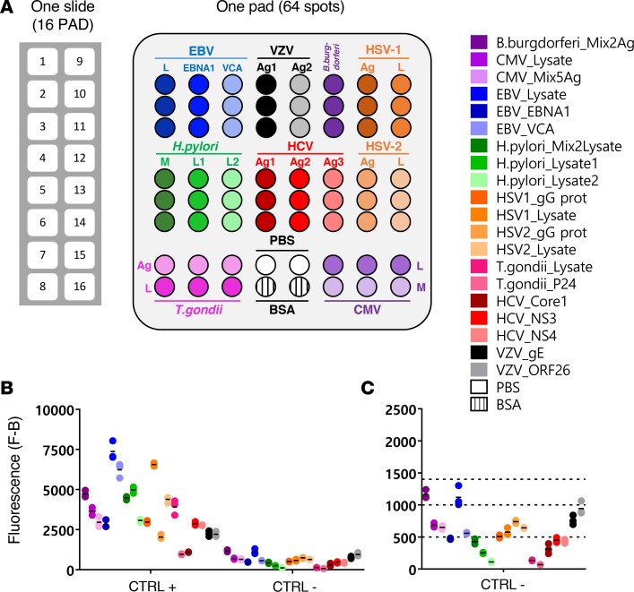
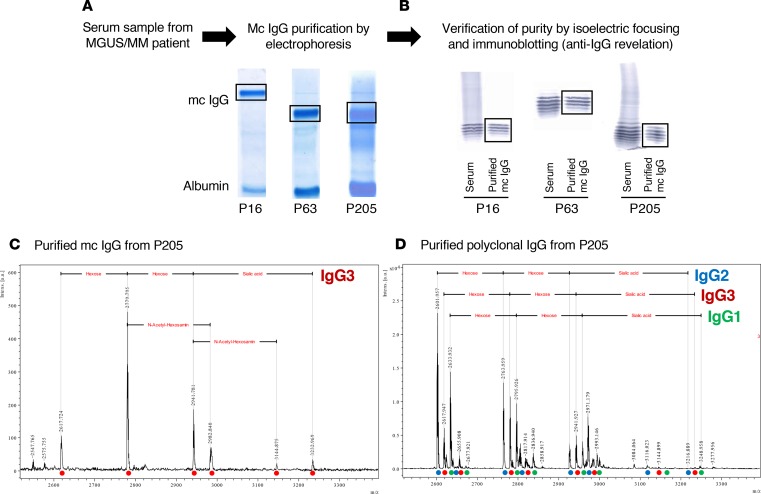
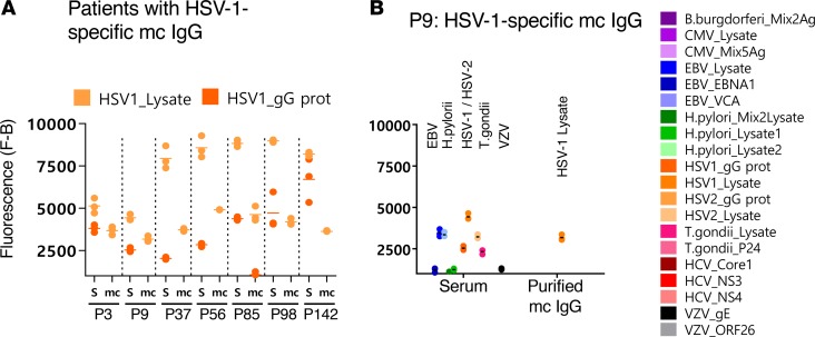
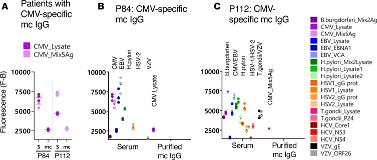
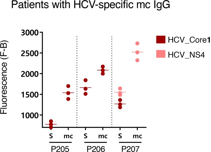
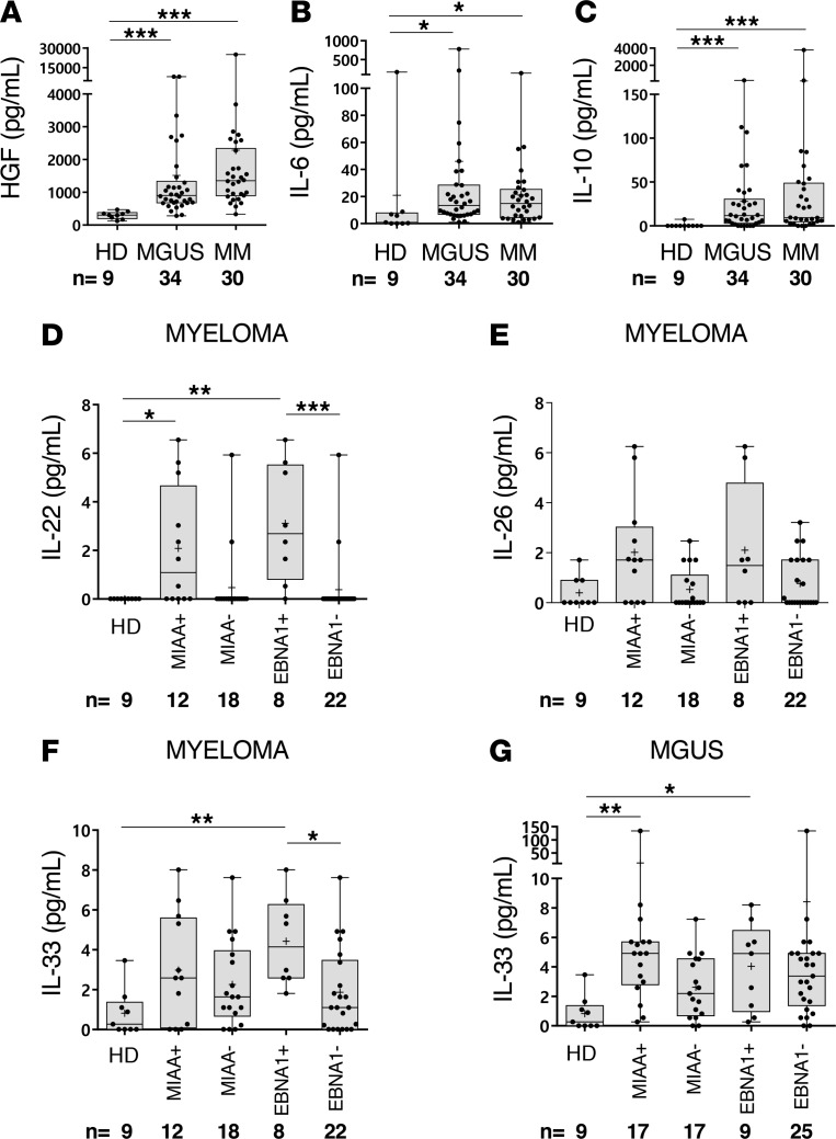
Subsets of mature B cell neoplasms are linked to infection with intracellular pathogens such as Epstein-Barr virus (EBV), hepatitis C virus (HCV), or Helicobacter pylori. However, the association between infection and the immunoglobulin-secreting (Ig-secreting) B proliferative disorders remains largely unresolved. We investigated whether the monoclonal IgG (mc IgG) produced by patients diagnosed with monoclonal gammopathy of undetermined significance (MGUS) or multiple myeloma (MM) targets infectious pathogens. Antigen specificity of purified mc IgG from a large patient cohort (n = 244) was determined using a multiplex infectious-antigen array (MIAA), which screens for reactivity to purified antigens or lysates from 9 pathogens. Purified mc IgG from 23.4% of patients (57 of 244) specifically recognized 1 pathogen in the MIAA. EBV was the most frequent target (15.6%), with 36 of 38 mc IgGs recognizing EBV nuclear antigen-1 (EBNA-1). MM patients with EBNA-1-specific mc IgG (14.0%) showed substantially greater bone marrow plasma cell infiltration and higher β2-microglobulin and inflammation/infection-linked cytokine levels compared with other smoldering myeloma/MM patients. Five other pathogens were the targets of mc IgG: herpes virus simplex-1 (2.9%), varicella zoster virus (1.6%), cytomegalovirus (0.8%), hepatitis C virus (1.2%), and H. pylori (1.2%). We conclude that a dysregulated immune response to infection may underlie disease onset and/or progression of MGUS and MM for subsets of patients.


Conflict of interest statement

Conflict of interest: The authors have declared that no conflict of interest exists.

Figures

Figure 1
Figure 1. Description of the MIAA assay.
(A) The multiplex infectious-antigen array (MIAA) assay consists of microarray slides that contain 16 identical pads; 1 pad (represented enlarged) is shown in detail, with Ag or lysate spotted in triplicate. For each patient, serum and purified monoclonal (mc) IgGs are examined in parallel using the MIAA assay; each type of sample is tested in duplicate or more (some samples were tested 6 times within 1 experiment). Fluorescence signal is used to determine the serological status of each sample (15). (B) Human serum samples containing polyclonal IgGs specific for each of the 9 infectious pathogens (= positive controls, CTRL+) were used to set up the assay and assess reproducibility. Human serum control samples that did not contain IgG specific for 1 or several pathogens (= negative controls, CTRL–) served to evaluate nonspecific binding and to determine the fluorescence threshold of specific positivity for each pathogen, Ag, or lysate (L), or mix of Ag (M). Shown is the net fluorescence intensity of the samples after subtracting background fluorescence (F–B). (C) Detail of negative controls and thresholds of positivity. Three fluorescence thresholds of specific positivity were used in all MIAA experiments: 500 for hepatitis C virus (HCV), H. pylori, and T. gondii; 1,000 for CMV, herpes simplex virus-1 (HSV-1), and HSV-2; and 1,400 for EBV, varicella zoster virus (VZV), and B. burgdorferi. Signals below these thresholds were considered to be negative. Dots may be superimposed; black horizontal bars represent the means of results obtained for a pathogen, Ag, or lysate. Positive and negative controls are run in every MIAA experiment, as internal controls.
Figure 2
Figure 2. Purification of mc IgGs, and verification of purity of the mc IgGs.
(A) For all 244 patients, monoclonal (mc) IgGs were purified as previously reported (12, 13, 15). (B) The purity of mc IgGs was verified using isoelectric focusing and immunoblotting; 3 examples (patients P16, P63, and P205) are shown. For certain patients, purified mc IgG (C) and polyclonal IgGs (D) were also studied by mass spectrometry. Only one IgG3 isotype (red circle) was observed in the mc IgG, while the polyclonal IgGs contained 3 isotypes: IgG1 (green circle), IgG2 (blue circle), and IgG3 (red circle), demonstrating the high purity of the mc IgG. The different peaks observed for each isotype correspond to the cumulative mass of the different hexoses carried by the IgG Fc fragment. Experiments were repeated twice.
Figure 3
Figure 3. EBV-specific mc IgG as determined by the MIAA assay.
For each patient, serum and purified monoclonal (mc) IgGs were incubated in parallel in the multiplex infectious-antigen array (MIAA) assay; results of hybridization are shown for patients P16 (A and B) and P63 (C and D). (A and B) For P16, serum contained IgG that recognized EBV nuclear antigen-1 (EBNA-1), EBV viral capsid antigen (VCA), herpes simplex virus-1 (HSV-1), HSV-2, and varicella zoster virus (VZV) glycoprotein E (gE), whereas the purified mc IgG recognized EBNA-1 only. (C and D) For P63, serum contained IgG that recognized EBNA-1, EBV VCA, HSV-1, HSV-2, and T. gondii, whereas the purified mc IgG recognized EBNA-1 only. (B and D) Quantification of the fluorescence signals generated by the serum and purified mc IgG of patients P16 (B) and P63 (D) as assessed with the MIAA assay. (E) Results obtained for the 36 patients found to have an EBNA-1–specific mc IgG. For each patient, the results obtained for EBV with the patient’s serum (S) and purified mc IgG are shown in dark blue (EBNA-1) or light blue (VCA). The fluorescence values shown for EBNA-1 or EBV VCA were obtained after subtraction of the threshold of specific positivity for each pathogen, protein, or lysate (1,400 for EBV). Dots may be superimposed; horizontal bars represent the means of results obtained for a pathogen, Ag, or lysate. Experiments were performed at least twice.
Figure 4
Figure 4. Results of 2 patients with EBV VCA–specific mc IgG.
For each patient, results obtained with the serum (S) and the purified monoclonal (mc) IgG are represented. (A) For patients P10 and P50, the serum contained IgG that recognized EBV nuclear antigen-1 (EBNA-1) and EBV viral capsid antigen (VCA), but the purified mc IgG recognized only EBV VCA. (B) Detail of results obtained for patient P10: serum P10 contained IgG that recognized B. burgdorferi, CMV, EBNA-1, EBV VCA, H. pylori, herpes simplex virus-2 (HSV-2), T. gondii, and varicella zoster virus (VZV), whereas the purified mc IgG recognized EBV VCA only. (C) Detail of results obtained for patient P50: serum P50 contained IgG that recognized CMV, EBNA-1, EBV VCA, HSV-2, and VZV, whereas the purified mc IgG recognized EBV VCA only. The fluorescence values shown for each pathogen, Ag, or lysate were obtained after subtraction of the threshold of specific positivity of the pathogen, Ag, or lysate (1,400 for EBV). Dots may be superimposed; horizontal bars represent the means of results obtained for a pathogen, Ag, or lysate. Experiments were performed at least twice.
Figure 5
Figure 5. HSV-1–specific mc IgG as determined by the MIAA assay.
(A) Results of the multiplex infectious-antigen array (MIAA) assay for the 7 patients found to have a herpes simplex virus-1–specific (HSV-1–specific) monoclonal (mc) IgG. For each patient, the results obtained for HSV-1 lysate and glycoprotein G (gG) with the patient’s serum (S) and with purified mc IgG (mc) are shown. (B) Detail of results obtained for patient P9: P9 serum contained IgG that recognized EBV nuclear antigen-1 (EBNA-1), EBV viral capsid antigen (VCA), CMV, HSV-1, varicella zoster virus (VZV), and T. gondii, whereas the purified mc IgG recognized only HSV-1 gG. The fluorescence values shown for each pathogen, Ag, or lysate were obtained after subtraction of the threshold of specific positivity of the pathogen, Ag, or lysate (1,000 for HSV-1). Dots may be superimposed; horizontal bars represent the means of results obtained for a pathogen, Ag, or lysate. Experiments were performed at least twice.
Figure 6
Figure 6. VZV-specific mc IgGs as determined by the MIAA assay.
(A) Results of the multiplex infectious-antigen array (MIAA) assay for the 4 patients found to have a varicella zoster virus–specific (VZV-specific) monoclonal (mc) IgG. For each patient, the results obtained for VZV glycoprotein E (gE) and ORF-26 with the patient’s serum (S) and purified mc IgG (mc) are shown. (B) Detail of results obtained for patient P109: P109 serum contained IgG that recognized CMV, EBV nuclear antigen-1 (EBNA-1), herpes simplex virus-1 (HSV-1), VZV gE, and VZV ORF-26, whereas purified mc IgG recognized only VZV gE. The fluorescence values shown for each pathogen, Ag, or lysate were obtained after subtraction of the threshold of specific positivity of the pathogen, Ag, or lysate (1,400 for VZV). Dots may be superimposed; black horizontal bars represent the means of results obtained for a pathogen, Ag, or lysate. Experiments were performed at least twice.
Figure 7
Figure 7. CMV-specific mc IgGs as determined by the MIAA assay.
(A) Results of the multiplex infectious-antigen array (MIAA) assay for the 2 patients found to have a CMV-specific monoclonal (mc) IgG. For each patient, the results obtained for the CMV lysate and a mix of 5 CMV Ags (see Methods) with the patient’s serum (S) and purified mc IgG are shown. (B) Detail of results obtained for patient P84: serum P84 contained IgG that recognized CMV, EBV nuclear antigen-1 (EBNA-1), EBV viral capsid antigen (VCA), H. pylori, herpes simplex virus-1 (HSV-1), HSV-2, and varicella zoster virus (VZV) ORF-26, whereas the purified mc IgG recognized only the CMV lysate. (C) Detail of results obtained for patient P112: serum P112 contained IgG that recognized B. burgdorferi, CMV, EBNA-1, EBV VCA, H. pylori, HSV-1, HSV-2, T. gondii, and VZV, whereas the purified mc IgG recognized only the CMV Ag mix. The fluorescence values shown for each pathogen, Ag, or lysate were obtained after subtraction of the threshold of specific positivity of the pathogen, Ag, or lysate (1,000 for CMV). Dots may be superimposed; horizontal bars represent the means of results obtained for a pathogen, Ag, or lysate. Experiments were performed at least twice.
Figure 8
Figure 8. H.pylori–specific mc IgGs as determined by the MIAA assay.
(A) Results of the multiplex infectious-antigen array (MIAA) assay for the 3 patients with a H. pylori–specific monoclonal (mc) IgG. For each patient, the results obtained for H. pylori lysate 1 and lysate 2 with the patient’s serum (S) and purified mc IgG are shown. (B) The results obtained for patient P186 are detailed: serum P186 contained IgG that recognized CMV, EBV nuclear antigen-1 (EBNA-1), EBV viral capsid protein (VCA), H. pylori, herpes simplex virus-1 (HSV-1), HSV-2, T. gondii, and varicella zoster virus (VZV) ORF-26, whereas the purified mc IgG recognized only the H. pylori lysates (dark green: mix of lysates 1 and 2; medium green: lysate 1; light green: lysate 2). Note that certain H. pylori proteins and Ags are likely present both in lysate 1 and lysate 2. The fluorescence values shown for each pathogen, Ag, or lysate were obtained after subtraction of the threshold of specific positivity of the pathogen, Ag, or lysate (500 for H. pylori). Dots may be superimposed; horizontal bars represent the mean of results obtained for a pathogen, Ag, or lysate. Experiments were performed at least twice. (C) The immunoblot assay was performed using the commercial kit Helico Blot 2.1 (MP Biomedicals), which consisted of a Western blot made from bacterial lysate of H. pylori strain ATCC 49503. The test strip contained H. pylori Ags with molecular weights of 116 kDa (CagA), 89 kDa (VacA), 65 kDa (urease B), 60 kDa (heat shock protein [HSP]), 37 kDa (H. pylori undetermined protein [UP]), 35 kDa (H. pylori UP), and 30 kDa (urease A) as separate bands. The assay was performed and interpreted according to the instructions of the manufacturers. Experiments were performed at least twice.
Figure 9
Figure 9. HCV-specific mc IgGs as determined by the MIAA assay.
Results of the multiplex infectious-antigen array (MIAA) assay for the 3 patients with an hepatitis C virus–specific (HCV-specific) monoclonal (mc) IgG. For each patient, the results obtained for HCV core, NS-3, or NS-4 proteins with the patient’s serum and purified mc IgG are shown. Serum contained IgG that recognized the HCV core (P205, P206, P207) and NS-3 (P206) or NS-4 (P205, P207), whereas the purified mc IgG recognized only the HCV core (P205, P206) or NS4 (P207). The fluorescence values shown for each HCV protein were obtained after subtraction of the threshold of specific positivity (500 for HCV). Dots may be superimposed; horizontal bars represent means of results obtained for HCV core, NS3, or NS4. Experiments were performed at least twice.
Figure 10
Figure 10. Confirmation of the specificity of EBNA-1 recognition of purified mc IgGs.
(A) Dot blotting assays with purified recombinant EBV nuclear antigen-1 (EBNA-1) were first performed in parallel with serum and purified monoclonal (mc) IgG from 3 patients: P169 and P190, patients with EBNA-1–specific mc IgG as assessed by the multiplex infectious-antigen array (MIAA) assay, as positive controls; and patient P160, whose mc IgG did not recognize EBNA-1 as assessed by the MIAA assay, as a negative control. EBNA-1 dot blotting was then performed with the purified mc IgGs of 30 additional patients with EBNA-1–specific mc IgG as assessed by the MIAA assay. (B) Dot blotting assays performed by coating relevant (RRPPPGRRPFFHPVA) or irrelevant (RRPPFGRRFFFHPVA) EBNA-1–derived peptides and studying reactivity with serum and purified mc IgGs. Amino acids of the relevant epitope are in bold and underlined. The modified amino acids in the irrelevant peptide are shown in blue. Signal was obtained only with relevant peptide, but only 2 of 24 of the purified mc IgGs that could be tested bound to the relevant peptide. The serum of patient P98 does not contain IgG specific for EBNA-1. Patient P1 is shown as an example of serum containing polyclonal IgGs that bound to the relevant peptide, while the purified mc IgG did not bind. (C) Dose-dependant inhibition of recognition of EBNA-1 protein by purified mc IgG specific for EBNA-1 in the presence of the relevant peptide in the MIAA assay. Recombinant EBNA-1 protein was spotted on the array. A purified mc IgG found to be EBNA-1 specific with the MIAA assay and to recognize the relevant EBNA-1–derived peptide RRPPPGRRPFFHPVA with the dot blot assay, was preincubated with different concentrations of this peptide for 1 hour at room temperature before adding the mc IgG to the MIAA pad. The irrelevant peptide RRPPFGRRFFFHPVA was used as a negative control. Results are means ± SD of triplicates. *P < 0.05 by ANOVA. Experiments were performed at least twice.
Figure 11
Figure 11. Confirmation of the specificity of HSV-1 recognition of purified mc IgGs.
(A, left): The purified monoclonal (mc) IgG from herpes simplex virus-1–positive (HSV-1+) index patient P56 was immobilized on Protein A/G beads and used for 4 rounds (R1–R4) of phage display peptide purification, with a CX7C library; the Fd-tet lacking peptide inserts served as a negative control. After the fourth round of selection, bacterial colonies were picked at random for sequencing. Results are expressed as mean ± SEM of duplicate wells. (A, right) Alignment of 14 related sequences, used for a BLAST alignment identifying the sequence VPALR from HSV-1 tegument protein, UL36 (in blue, aa 1498–1510). (B) Dot blotting with HSV-1 lysate and relevant (EAVLHLSEDLGGVPALRQYVP) and irrelevant (EAVLHLSEDLGGRPAERQYVP) HSV-1–derived peptides was performed for serum and purified mc IgG from the 7 patients found to have HSV-1–specific mc IgG with the multiplex infectious-antigen array (MIAA) assay. Patient P1, whose mc IgG did not recognize HSV-1 using the MIAA assay, was used as a negative control. Amino acids of the relevant epitope are in bold and underlined. The modified amino acids of the irrelevant peptide are in blue. Dot blots were performed at least twice.
Figure 12
Figure 12. Cytokine levels in serum of MGUS and SM/MM patients with infectious pathogen–specific or EBNA-1–specific mc IgG.
Six cytokines were quantified in serum using Luminex technology (Bio-Plex 200) with Bio-Plex Pro Human Cytokine Panel kits (Bio-Rad) in 3 different groups: healthy donors (HD, n = 9), monoclonal gammopathy of undetermined significance (MGUS) patients (n = 34), and a group of 4 smoldering myeloma (SM) patients and 26 multiple myeloma (MM) patients (designated as MM; n = 30). The MGUS and SM/MM groups were subdivided according to the pathogen specificity of monoclonal (mc) IgG: patients with mc IgG specific for one of the infectious pathogens from the multiplex infectious-antigen array (MIAA) assay are designated as MIAA+ patients; all others are designated as MIAA patients. Patients with EBV nuclear antigen-1–specific (EBNA-1–specific) mc IgG are designated as EBNA-1+ patients; all others are designated as EBNA-1. The cytokines measured were (A) HGF, (B) IL-6, (C) IL-10, (D) IL-22, (E) IL-26, and (F) IL-33. Cytokine levels were measured in serum from MIAA+/– and EBNA1+/– MM patients. (G) Serum IL-33 levels of MIAA+/– and EBNA1+/– MGUS patients. Note that the scales for IL-33 concentrations are different in F and G. For a given patient, results are means of technical duplicate measurements, expressed in pg/ml. Median values of groups of patients are shown as bars; means are indicated with the + symbol. Normal cytokine values in blood of healthy donors are as follows: HGF, 63–1,283 pg/ml; IL-6, < 9 pg/ml (in our group of HD, one individual had 165.5 pg/ml IL-6); IL-10, < 2 pg/ml; IL-22, 0 pg/ml; IL-26 and IL-33, normal values are not defined. *P < 0.05, **P < 0.01, and ***P < 0.001 by ANOVA.

References

    1. Kyle RA. Monoclonal gammopathy of undetermined significance. Natural history in 241 cases. Am J Med. 1978;64(5):814–826. doi: 10.1016/0002-9343(78)90522-3. - DOI - PubMed
    1. Dhodapkar MV. MGUS to myeloma: a mysterious gammopathy of underexplored significance. Blood. 2016;128(23):2599–2606. doi: 10.1182/blood-2016-09-692954. - DOI - PMC - PubMed
    1. Kyle RA, Greipp PR. Smoldering multiple myeloma. N Engl J Med. 1980;302(24):1347–1349. doi: 10.1056/NEJM198006123022405. - DOI - PubMed
    1. Rajkumar SV, et al. International Myeloma Working Group updated criteria for the diagnosis of multiple myeloma. Lancet Oncol. 2014;15(12):e538–e548. doi: 10.1016/S1470-2045(14)70442-5. - DOI - PubMed
    1. Drouet E, et al. Oligo-monoclonal immunoglobulins frequently develop during concurrent cytomegalovirus (CMV) and Epstein-Barr virus (EBV) infections in patients after renal transplantation. Clin Exp Immunol. 1999;118(3):465–472. doi: 10.1046/j.1365-2249.1999.01084.x. - DOI - PMC - PubMed

Publication types

MeSH terms