

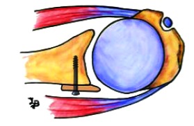
Treatment of The Posterior Unstable Shoulder
- PMID: 28979596
- PMCID: PMC5611705
- DOI: 10.2174/1874325001711010826
Treatment of The Posterior Unstable Shoulder
Abstract
Background: It is estimated that approximately 5% of glenohumeral instabilities are posterior. There are a number of controversies regarding therapeutic approaches for these patients.
Methods: We analyse the main surgery alternatives for the treatment of the posterior shoulder instability. We did a research of the publications related with posterior glenohumeral instability.
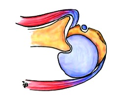
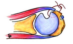
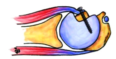
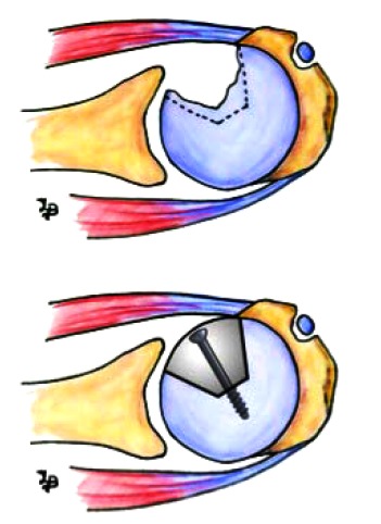
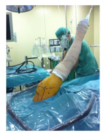
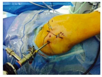
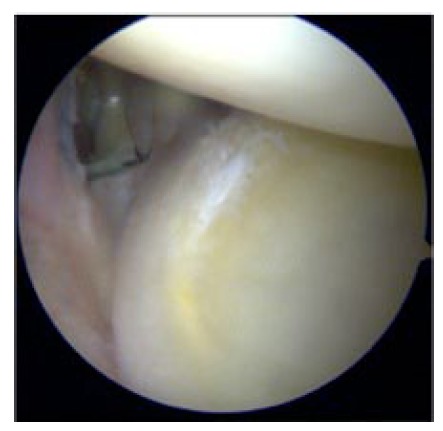
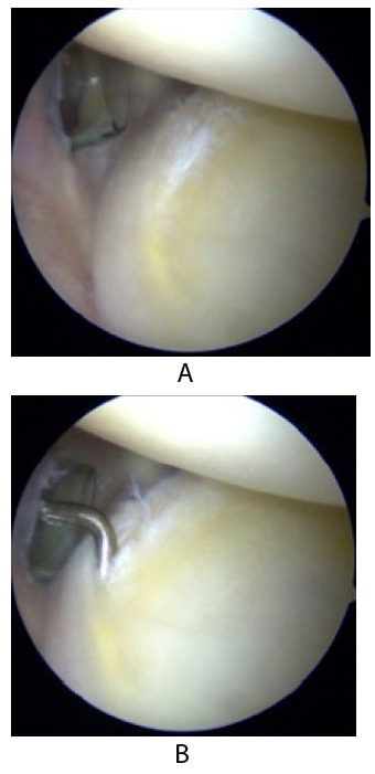
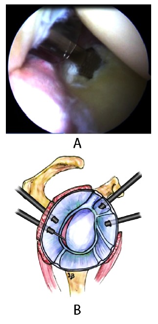
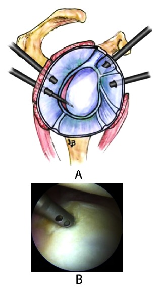
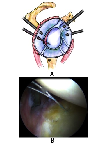
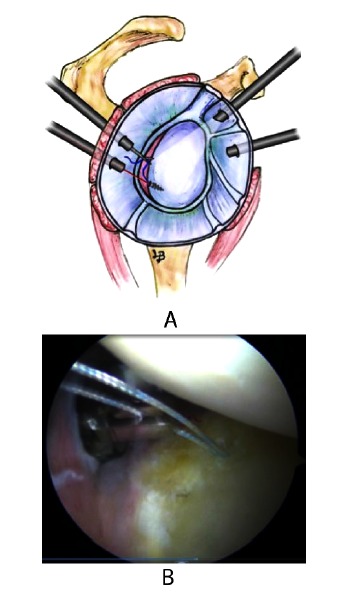
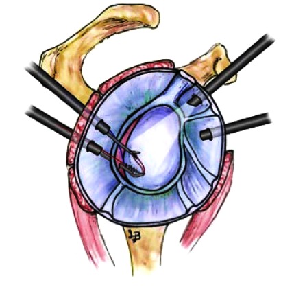
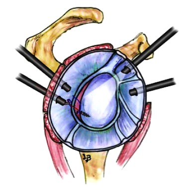
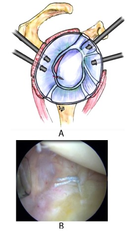
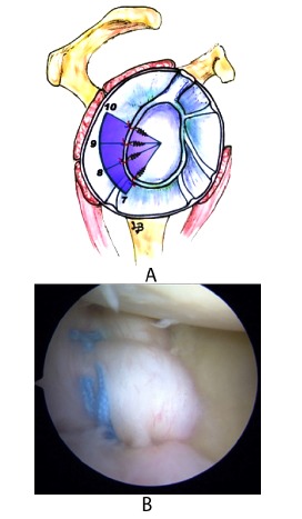
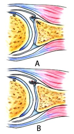
Results: There are conservative and surgical treatment options. Conservative treatment has positive results in most patients, with around 65 to 80% of cases showing recurrent posterior dislocation. There are multiple surgical techniques, both open and arthroscopic, for the treatment of posterior glenohumeral instability. There are procedures that aim to repair bone defects and others that aim to repair soft tissues and capsulolabral injuries. The treatment should be planned according to each case on an individual basis according to the patient characteristics and the injury type. Surgical treatment is indicated in patients with functional limitations arising from instability and/or pain that have not improved with rehabilitation treatment. The indications for arthroscopic treatment are recurrent posterior subluxation caused by injury of the labrum or the capsulolabral complex; recurrent posterior subluxation caused by capsuloligamentous laxity or capsular redundancy; and multidirectional instability with posterior instability as a primary component. Arthroscopic assessment will help identify potential injuries associated with posterior instability such as bone lesions or defects and lesions or defects of soft tissues. The main indications for open surgery would be in cases of Hill Sachs lesions or broad reverse Bankart lesions not accessible by arthroscopy. We indicated non-anatomical techniques (McLaughlin or its modifications) for reverse Hill-Sachs lesions with impairment of the articular surface between 20% and 50%. Disimpaction of the fracture and placement of bone graft (allograft or autograft) is a suitable treatment for acute lesions that do not exceed 50% of the articular surface and with articular cartilage in good condition. Reconstruction with allograft may be useful in lesions affecting up to 50% of the humeral surface and should be considered when there is a situation of non-viable cartilage at the fracture site. For defects greater than 50% of the articular surface or in the case of dislocations over 6 months in duration where there is poor bone quality, some authors advocate substitution techniques as a treatment of choice. The main techniques for treating glenoid bone defects are posterior bone block and posterior opening osteotomy of the glenoid.
Conclusions: The treatment of the posterior glenohumeral instability has to be individualized based on the patient´s injuries, medical history, clinical exam and goals. The most important complications in the treatment of posterior glenohumeral instability are recurrent instability, avascular necrosis and osteoarthritis.
Keywords: Glenohumeral instability; Instability; Posterior instability; Shoulder; Shoulder arthroscopic; Shoulder surgery.
Figures
References
-
- OBrien S.J., Pagnani M.J., Fealy S., McGlynn S.R., Wilson J.B. The active compression test: A new and effective test for diagnosing labral tears and acromioclavicular joint abnormality. Am. J. Sports Med. 1998;26(5):610–613. - PubMed
-
- Rockwood C.A., Matsen F.A., Wirth M.A., Lippitt S.B. The Shoulder. London: Saunders Elsevier; 2009.
LinkOut - more resources
Full Text Sources
Other Literature Sources