. 2017 Sep 13;7(10):1499-1514.
doi: 10.1002/2211-5463.12281. eCollection 2017 Oct.

The periplasmic binding protein NrtT affects xantham gum production and pathogenesis in Xanthomonas citri

Affiliations

The periplasmic binding protein NrtT affects xantham gum production and pathogenesis in Xanthomonas citri

Aline Sampaio et al. FEBS Open Bio. .

Abstract

In Xanthomonas citri, the bacterium that causes citrus canker, three ATP-binding cassette (ABC) transporters are known to be dedicated to the uptake of sulfur compounds. In this work, using functional, biophysical and structural methods, we showed that NrtT, a periplasmic component of the ABC transporter NrtCB, is an alkanesulfonate-binding protein and that the deletion of the nrtT gene affected xantham gum synthesis, adhesion and biofilm production, similarly to the phenotype obtained in the X. citri ssuA-knockout strain, in which the alkanesulfonate-binding protein SsuA is absent. Although NrtA and SsuA share similar ligands, the function of these proteins is not complementary. These results emphasize that organic-sulfur sources are directly involved with bacterial infection in vivo and are needed for pathogenesis in X. citri.

Keywords: ABC transporter; NrtT; Xanthomonas citri; alkanesulfonates/taurine; pathogenesis; periplasmic binding protein.


Figures

Figure 1
Figure 1
Genetic organization of gene nrt in Xanthomonas citri in comparison with Xanthomonas campestris, Pseudomonas aeruginosa and Escherichia coli. (A) Promoter regions (black boxes) predicted in the cluster of X. citri by the softberry bprom program. (B) Genetic organization of operons putatively involved with the transport and metabolism of mineral and organic ions and nitrate/nitrite/taurine. Orthologues and genes belonging to the same operon are represented in same colors, using the KEGG code and name. The percentage amino acid sequence identity is shown for each protein in comparison with X. citri orthologues.
Figure 2
Figure 2
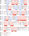
Multiple sequence and structure alignment of Xanthomonas citri NrtT model with proteins identified in the blastp against PDB. The alignment was performed by the promals3d program 48. The α‐helices and β‐sheets are colored in red (arrow) and blue (cylinders), respectively. Residues that interact with the ligands in the PDB structures are showed in bold underlined. The same was indicated for NrtT. The first three letter code refers to the PDB data bank. 2G29, nitrate‐binding protein NrtA from Synechocystis sp. PCC 6803; 2I48, bicarbonate‐binding protein CmpA from Synechocystis sp. PCC 6803; 2X26, sulfonate‐binding protein SsuA from Escherichia coli; 3E4R, sulfonate‐binding protein SsuA from X. citri; 3UIF, sulfonate‐binding protein SsuA from Methylobacillus flagelatus KT; NrtT, nitrate/alkanesulfonate/taurine‐binding protein NrtT from X. citri.
Figure 3
Figure 3
Comparison of the ligand‐binding pockets of NrtT and other proteins. (A) Surface electrostatic potential and residues from the ligand‐binding pockets of the five proteins identified after NrtT blast against the PDB evidencing that NrtT is closely related to the alkanesulfonate‐binding proteins. Charges are shown in red (negative) and blue (positive) and residues that interact with the ligands in the structures are shown as yellow sticks. Residues from NrtT pocket were predicted based on the structural alignment with the sulfonate‐binding proteins. Proteins name are showed with the PDB code and the ligands (except for NrtT and Xanthomonas citri SsuA/1). The percentages correspond to the query coverage and the amino acid sequence identity after alignment. 2G29, nitrate‐binding protein NrtA from Synechocystis sp. PCC 6803 bound to nitrate (blue stick); 2I48, bicarbonate‐binding protein CmpA from Synechocystis sp. PCC 6803 bound to bicarbonate (green stick); 2X26, alkanesulfonate‐binding protein SsuA from Escherichia coli; 3E4R, alkanesulfonate‐binding protein SsuA/2 from X. citri bound to HEPES (gray sticks); 3UIF, sulfonate‐binding protein SsuA from Methylobacillus flagelatus KT; NrtT, nitrate/alkanesulfonate/taurine‐binding protein NrtT from X. citri. (B) Comparison of the electrostatic potential at the pocket entrance of NrtT with the X. citri alkanesulfonate‐binding proteins SsuA/1 (Xac0849, model) and SsuA/2 (Xac3198, PDB code 3E4R) The proteins are shown as external surface and the black line shows the conserved profile of charges.
Figure 4
Figure 4
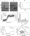
Production of Xanthomonas citri NrtT and biophysical analyses in the presence of putative ligands. (A) Production of NrtT from Escherichia coli BL21(DE3) cells. (I) 15% SDS/PAGE of NrtT samples obtained in different steps of expression, purification and cleavage with SUMO protease. (II) Western blot using antibodies against NrtT. M, molecular mass marker; 1, insoluble fraction of the E. coli BL21 (DE3) induced cells; 2, soluble fraction; 3, size‐exclusion chromatography sample of NrtT before cleavage with SUMO‐protease; 4, sample after cleavage with SUMO‐protease; 5, concentrated sample used for biophysical and structural assays. The arrows indicate the estimated molecular mass for SUMO‐NrtT (NC) and the estimated molecular mass of the NrtT after cleavage with SUMO‐protease (CL). (III) DLS plot showing that after SUMO protease cleavage NrtT was 89% monodisperse. (B) Thermal denaturation curve of NrtT in the presence of different molecules evidencing a gain of thermal stability in the presence of MOPS. (C) Plots of the different melting temperatures identified after thermal denaturation of NrtT in the absence and presence of different putative ligands. (D) Circular dichroism spectra of NrtT in the absence and presence of MOPS and taurine showing the α–β profile expected for periplasmic binding proteins and slight structural changes. (E) Intrinsic fluorescence of tryptophans from NrtT in the absence and presence of MOPS and taurine. The inset shows the NrtT structural model in cartoon with the two tryptophans (W72 and W134) shown as gray spheres. DLS, dynamic light scattering.
Figure 5
Figure 5
SAXS analysis of NrtT in the absence and presence of MOPS. (A) Experimental scattering curve and the best intensity fitting obtained from SAXS data. The inset shows the normalized pair‐distance distribution function P(r) showing a small difference between NrtT and NrtT + MOPS. (B) Kratky plot for the samples NrtT and NrtT + MOPS evidencing that in presence of MOPS NrtT is more globular. (C) Porod–Debye plot showing the interaction between NrtT and MOPS causes loss of flexibility. (D,E) Ab initio envelopes generated from the three‐dimensional structure model of NrtT and fitting of SAXS data by the rigid body optimization for NrtT (D) and NrtT + MOPS (E).
Figure 6
Figure 6
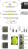
The role assessment of nrtT in the colonies’ phenotype, adhesion and biofilme formation. (A) Illustrative scheme of the construction of mutant (Xac::nrtT) and complementary (Xac::nrtTc) strains. (I) chromosomal deletion of the nrtT gene was obtained after electroporation of the suicide plasmid p.nrtT in Xanthomonas citri 306 strain and recombination for insertion at the Bam HI site of the nrtT gene of a 3 kbp fragment encoding spectinomycin and streptomycin resistances. (II) Final genome organization of the Xac::nrtT mutant. (B) Confimation of the mutant clones. (I) Products of the PCR reaction for amplification of the 3 kb Spr/Smr cassette. 1, molecular mass marker; 2, PCR product using X. citri wild‐type DNA as template; 3–5, PCR product using putative mutant DNA as template, evidencing the amplification of a 3 kb fragment. (II) Western blot of the wild‐type and Xac::nrtT mutant 1 using the anti‐NrtT antibody. 1 and 2, total cellular extracts of X. citri and Xac::nrtT cells. (C) Scheme of the construction of the complementary strain Xac::nrtTc. (D) Morphology and color of the X. citri (Xac), Xac::nrtT and Xac::nrtTc colonies after growth in LB broth plates for 24 h. Colonies from mutant strains showed decreased size and attenuation of the color, which was partly reestablished by the complementary strain. (E) Xantham gum production by wild‐type and mutant strain in LB broth. A significant reduction of 26% was observed in the mutant strain (P < 0.05). (F) Biofilm production on abiotic surface (polypropylene plates) by X. citri and Xac::nrtT strains showing significant reduction in the mutant strain (P < 0.01). (G) Analysis of adhesion and biofilm formation on surface of Citrus sinensis leaves after 24 h of the cultures growth evidencing significant reduction in the mutant strain.
Figure 7
Figure 7
Role assessment of nrtT during in vitro growth. Growth curves of Xanthomonas citri and Xac::nrtT strains in (A) LB, (B) XAM1 and (C) XAM1 modified with 3.0 mm MOPS or 3.0 mm taurine replacing the ammonium sulfate to give XAM1a and XAM1b, respectively. Cells were growth at 30 °C, 250 r.p.m. for 72 h. The absorbance at 600 nm was measured every 24 h. The experiment was carried out in triplicate and the results represent the mean and standard deviation.
Figure 8
Figure 8
In vivo role assessment of nrtT. (A) Growth curves of the strains obtained after recovering the cells and plating in LB broth medium for 24 h. Sectors of a Citrus sinensis leaf inoculated with a syringe containing 100 μL of normalized CFU·mL −1 of the strains Xanthomonas citri, Xac::nrtT and Xac::nrtTc. Three sectors from three distinct leaves were processed for each strain for replicates. (B) Development of the canker symptoms in Citrus sinensis leaves monitored during 12 days. With the lack of nrtT gene, X. citri mutant strain produced evident chlorosis and attenuated pustules when compared with the wild‐type strain. Complementation of the nrtT mutation with pKX33.pnrtT was not evidenced in these experiments corroborating the hypothesis of deletion of the full transporter by polar mutation. Leaves were photographed using a microscope at ×100 magnification.

References

    1. da Silva ACR, Ferro JA, Reinach FC, Farah CS, Furlan LR, Quaggio RB, Monteiro‐Vitorello CB, Van Sluys MA, Almeida NF, Alves LM et al (2002) Comparison of the genomes of two Xanthomonas pathogens with differing host specificities. Nature 414, 459–463. - PubMed
    1. Gottwald TR, Graham JH and Schubert TS (2002) Citrus canker: the pathogen and its impact. Plant Health Prog 10, 1–34.
    1. Civerolo E (1984) Bacterial canker disease of citrus. J Rio Gd Val Hortic Soc 37, 127–146.
    1. Brunings A and Gabriel D (2003) Xanthomonas citri: breaking the surface. Mol Plant Pathol 4, 141–157. - PubMed
    1. Graham JH, Gottwald TR, Cubero J and Achor AD (2004) Xanthomonas axonopodis pv. citri: factors affecting successful eradication of citrus canker. Mol Plant Pathol 5, 1–15. - PubMed

LinkOut - more resources