. 2017 Oct 1;58(12):5030-5042.
doi: 10.1167/iovs.16-21402.

Therapeutic Effects of a Novel Agonist of Peroxisome Proliferator-Activated Receptor Alpha for the Treatment of Diabetic Retinopathy

Affiliations

Therapeutic Effects of a Novel Agonist of Peroxisome Proliferator-Activated Receptor Alpha for the Treatment of Diabetic Retinopathy

Guotao Deng et al. Invest Ophthalmol Vis Sci. .

Abstract

Purpose: Clinical studies have shown that peroxisome proliferator-activated receptor alpha (PPARα) agonist fenofibrate has therapeutic effects on diabetic retinopathy (DR). The purpose of this study was to identify a novel PPARα agonist and to evaluate its beneficial effects on DR.

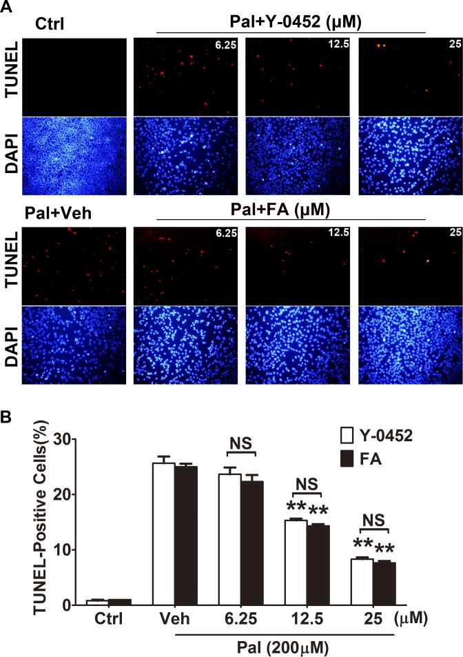
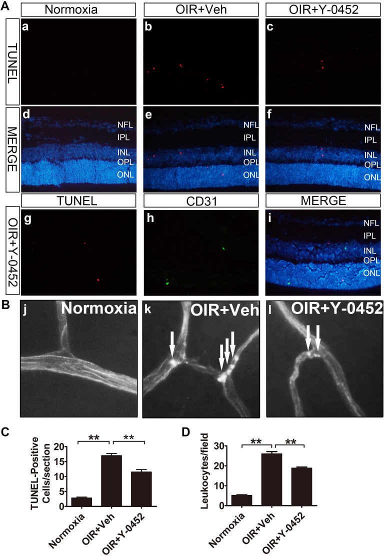
Methods: The transcriptional activity of PPARα was measured by a luciferase-based promoter assay. TUNEL was used to evaluate apoptosis in retinal precursor cells (R28). Diabetes was induced in rats by injection of streptozotocin. Retinal inflammation was examined using leukostasis assay, and retinal vascular leakage was measured using permeability assay. Retinal function was measured using electroretinogram (ERG) recording, and retinal apoptosis was quantified using the cell death ELISA. The anti-angiogenic effect was evaluated in the oxygen-induced retinopathy (OIR) model.

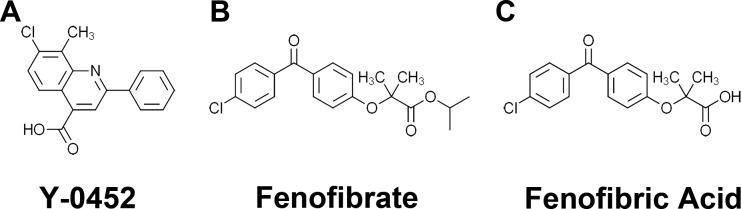
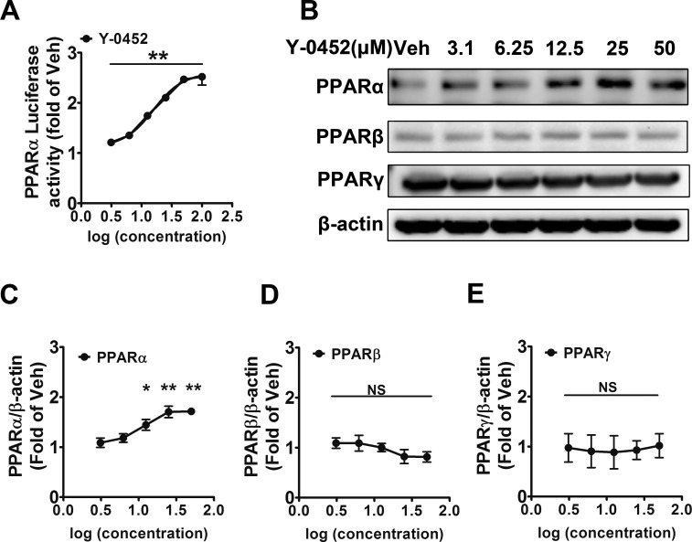
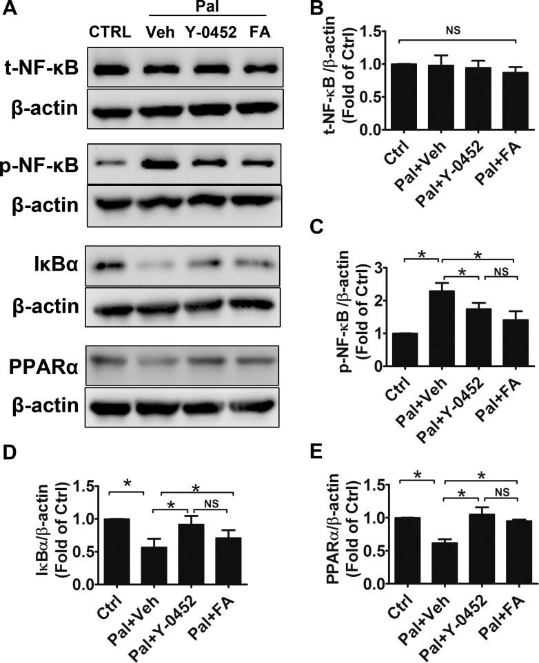
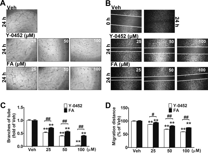
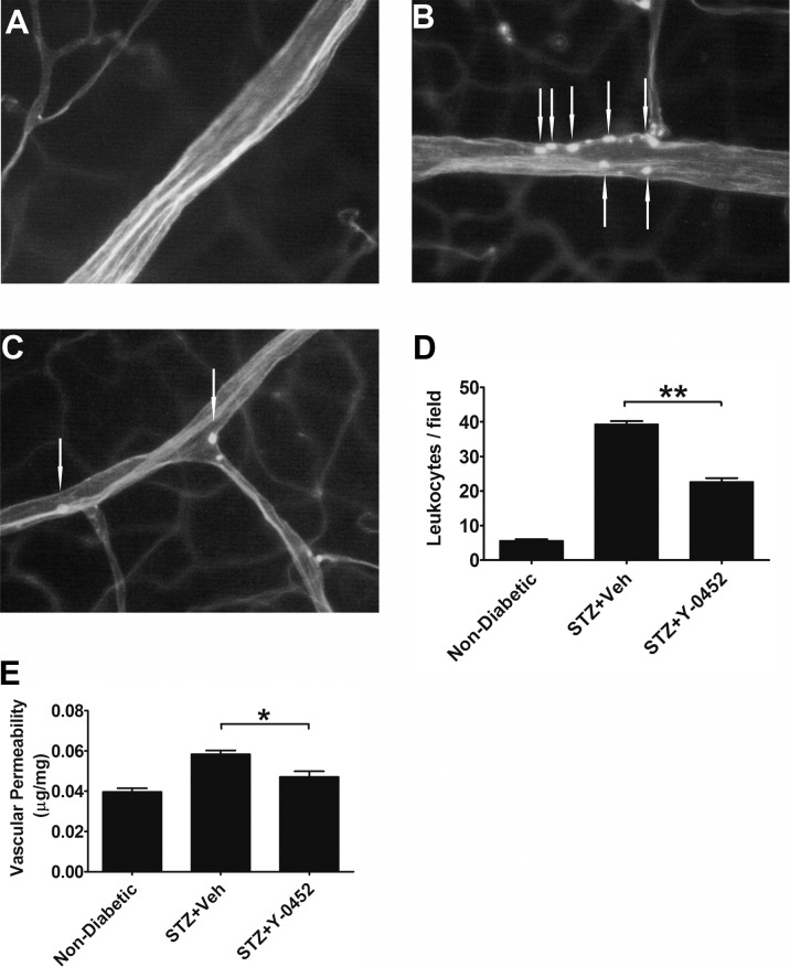
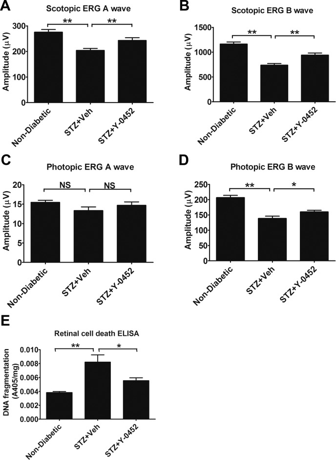
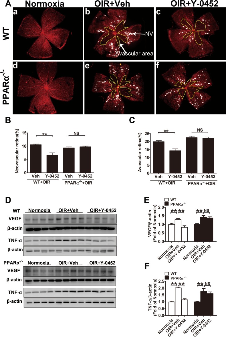
Results: A compound, 7-chloro-8-methyl-2-phenylquinoline-4-carboxylic acid (Y-0452), with a chemical structure distinct from existing PPARα agonists, activated PPARα transcriptional activity and upregulated PPARα expression. Y-0452 significantly inhibited human retinal capillary endothelial cell migration and tube formation. The compound also protected R28 cells against apoptosis and inhibited NF-κB signaling in R28 cells exposed to palmitate. In diabetic rats, Y-0452 ameliorated leukostasis and vascular leakage in the retina. In addition, Y-0452 preserved the retinal function and reduced retinal cell death in diabetic rats. Y-0452 also alleviated retinal neovascularization in the OIR model.

Conclusions: Y-0452 is a novel PPARα agonist and has therapeutic potential for DR.


Figures

Figure 1
Figure 1
Chemical structures of Y-0452 (MW: 297), 7-chloro-8-methyl -2-phenylquinoline-4-carboxylic acid (A), fenofibrate (B) and fenofibric acid (C).
Figure 2
Figure 2
Activation of PPARα and upregulation of PPARα expression by Y-0452. (A) Induction of luciferase reporter under the control of PPRE by Y-0452. The PPARα reporter Combo cells were treated with Y-0452 at various concentrations (3.12, 6.25, 12.5, 25, 50, and 100 μM) for 36 hours, and then luciferase activities were measured (n = 3, **P < 0.01 versus vehicle). (B) R28 cells were treated with Y-0452 at indicated concentrations for 24 hours. PPAR protein levels were measured by Western blot analysis with β-actin as loading control. (C–E) Densitometry quantification of PPARα, PPARβ, and PPARγ levels (n = 3, *P < 0.05, **P < 0.01, versus vehicle, 1-way ANOVA).
Figure 3
Figure 3
The protective effect of Y-0452 on oxidative stress–induced apoptosis in retinal precursor cells (R28). R28 cells were treated with Y-0452 and Feno-FA (FA) at various concentrations for 4 hours, and 200 μmol/L palmitate (Pal) was added and incubated with the cells for another 24 hours. (A) Representative images of TUNEL-positive cells (red) and total cells (blue, DAPI staining). (B) Quantification of TUNEL-positive cells, presented as percentages of total cells (n = 3, **P < 0.01, 2-way ANOVA).
Figure 4
Figure 4
Regulation of inflammation factors by Y-0452. R28 cells were treated with 25 μmol/L Y-0452 for 4 hours and then co-incubated with 200 μmol/L palmitate (Pal) for another 24 hours. (A) Levels of total NF-κB (t-NF-κB), phosphorylated NF-κB (p-NF-κB), IκBα, and PPARα were determined by Western blotting. β-actin was used as an internal control. Representative blots were shown from three independent experiments. (B–E) Densitometry quantification of t-NF-κB, p-NF-κB, IκBα, and PPARα levels (n = 3, *P<0.05 versus vehicle, 2-way ANOVA).
Figure 5
Figure 5
Inhibitory effects of Y-0452 on endothelial cell migration and tube formation. (A, C) HRCECs were treated with Y-0452 and Feno-FA (FA) at indicated concentrations for 16 hours and seeded onto Matrigel containing different concentrations of Y-0452 or Feno-FA. After 6 hours of incubation, branch numbers were counted in five random fields under a microscope. (B, D) HRCECs were seeded in a 24-well plate and cultured in growth medium until 80% confluency. A straight line across the center of the well was scratched. After washing with PBS, cells were incubated with different concentrations of Y-0452 and Feno-FA for 24 hours. Images were captured, and cell migration distances were quantified using ImageJ software and expressed as percentages of vehicle control (n = 3, **P < 0.01 versus vehicle, #P < 0.05 versus Feno-FA, ##P < 0.01 versus Feno-FA, 2-way ANOVA).
Figure 6
Figure 6
Effects of Y-0452 on retinal vascular inflammation and leakage in diabetic rats. Rats with 1 month of STZ-induced diabetes received daily intraperitoneal injections of Y-0452 (10 mg/kg/d) or the same volume of the vehicle (Veh) as a control for 3 weeks. Retinal vascular endothelial cells and adherent leukocytes were stained with FITC–concanavalin-A and visualized under a fluorescence microscope. (A–C) Representative images of retinal adherent leukocytes in nondiabetic and STZ-induced diabetic rats ([A] nondiabetic, [B] STZ+Veh, [C] STZ+Y-0452). (D) Quantification of retinal adherent leukocytes was performed from ×40 magnification images. Adherent leukocytes were counted in six random fields per retina (white arrows indicate adherent leukocytes). (E) Retinal vascular leakage was measured using Evans blue as a tracer and normalized by total retinal protein concentrations (n = 6; *P < 0.05, **P < 0.01, 2-way ANOVA).
Figure 7
Figure 7
Effects of Y-0452 on ERG responses and retinal cell death in STZ-induced diabetic rats. Rats with 2 months of diabetes and treated with Y-0452 for 3 weeks were used for ERG recording and retinal DNA fragmentation ELISA. (A–D) Scotopic and photopic ERG A and B wave amplitudes in the indicated groups (n = 6; *P < 0.05, **P < 0.01). (E) Quantification of retinal DNA fragmentation, reflective of total retinal apoptosis, in the retina of nondiabetic and STZ-induced diabetic rats treated with Y-0452 and vehicle (n = 6; *P < 0.05, **P < 0.01, 2-way ANOVA).
Figure 8
Figure 8
Effects of Y-0452 on retinal apoptosis and inflammation in OIR mice. WT OIR mice were intraperitoneally injected with Y-0452 (10 mg/kg/d) from P12 to P16 or the same volume of vehicle. (A) Representative images of retinal apoptotic cells in OIR mice (normoxia: [a, d], OIR+Veh: [b, e], OIR+Y-0452: [c, f], costaining with anti-CD31: [g–i]). Retinal apoptotic cells were labeled by TUNEL (red), and nuclei stained with DAPI (blue). CD31 (green) cells were co-immunostained with TUNEL (red) and DAPI (blue) in OIR retinal sections. (B) Representative images of retinal adherent leukocytes in OIR mice (white arrows indicate adherent leukocytes). (C) Quantification of retinal TUNEL-positive cells in OIR mice from retinal sections (n = 6; *P < 0.05, **P < 0.01 versus vehicle, 1-way ANOVA). (D) Quantification of retinal adherent leukocytes in OIR mice was performed from ×40 magnification images. Adherent leukocytes were counted in six random fields per retina (n = 6; *P < 0.05, **P < 0.01 versus vehicle, 2-way ANOVA).
Figure 9
Figure 9
Effect of Y-0452 on retinal neovascularization in OIR mice. WT and PPARα−/− OIR mice were intraperitoneally injected with Y-0452 (10 mg/kg/d) from P12 to P16 or the same volume of vehicle. The retinas were fixed and stained with Isolectin B4. Areas of retinal neovascularization and vaso-obliteration were quantified under a fluorescence microscope. (A) Representative images of retinal neovascularization and avascular areas in WT (a–c) and PPARα−/− OIR mice (d–f). The white dot–marked area indicates neovascular retina and the yellow line–marked area indicates avascular area. (B, C) Quantification of retinal neovascularization and avascular areas. (D) Retinal VEGF and TNF-α levels were measured by Western blot analysis. (E, F) Quantification of VEGF levels in WT and PPARα−/− OIR mice (n = 8; *P < 0.05, **P < 0.01, 2-way ANOVA).

Similar articles

Cited by

References

    1. Fong DS, Sharza M, Chen W, Paschal JF, Ariyasu RG, Lee PP. . Vision loss among diabetics in a group model Health Maintenance Organization (HMO). Am J Ophthalmol. 2002; 133: 236– 241. - PubMed
    1. Deliyanti D, Zhang Y, Khong F,et al. . FT011, a novel cardiorenal protective drug, reduces inflammation, gliosis and vascular injury in rats with diabetic retinopathy. PLoS One. 2015; 10: e0134392. - PMC - PubMed
    1. Hu Y, Chen Y, Ding L,et al. . Pathogenic role of diabetes-induced PPAR-alpha down-regulation in microvascular dysfunction. Proc Natl Acad Sci U S A. 2013; 110: 15401– 15406. - PMC - PubMed
    1. Xie TY, Yan W, Lou J, Chen XY. . Effect of ozone on vascular endothelial growth factor (VEGF) and related inflammatory cytokines in rats with diabetic retinopathy. Genet Mol Res. 2016; 15: 1– 11. - PubMed
    1. Gao X, Li Y, Wang H, Li C, Ding J. . Inhibition of HIF-1alpha decreases expression of pro-inflammatory IL-6 and TNF-alpha in diabetic retinopathy [published online ahead of print June 11, 2016]. Acta Ophthalmol. doi:10.1111/aos.13096. - DOI - PubMed

Publication types

MeSH terms