Review
. 2017 Dec;8(6):549-556.
doi: 10.1007/s13244-017-0576-z. Epub 2017 Oct 4.

MRI for adenomyosis: a pictorial review

Affiliations
Review

MRI for adenomyosis: a pictorial review

Lisa Agostinho et al. Insights Imaging. 2017 Dec.

Abstract

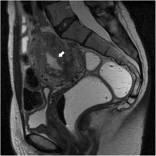
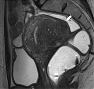
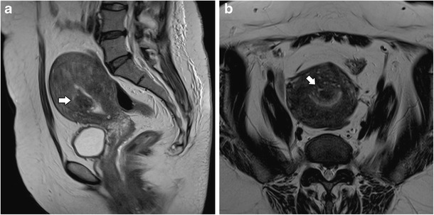
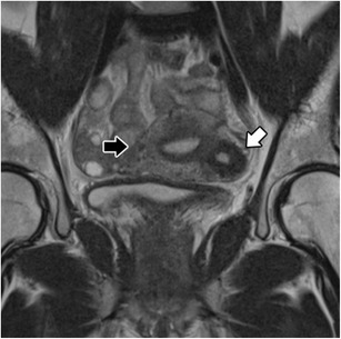
Adenomyosis is defined as the presence of ectopic endometrial glands and stroma within the myometrium. It is a disease of the inner myometrium and results from infiltration of the basal endometrium into the underlying myometrium. Transvaginal ultrasonography (TVUS) and magnetic resonance imaging (MRI) are the main radiologic tools for this condition. A thickness of the junctional zone of at least 12 mm is the most frequent MRI criterion in establishing the presence of adenomyosis. Adenomyosis can appear as a diffuse or focal form. Adenomyosis is often associated with hormone-dependent lesions such as leiomyoma, deep pelvic endometriosis and endometrial hyperplasia/polyps. Herein, we illustrate the MRI findings of adenomyosis and associated conditions, focusing on their imaging pitfalls.

Teaching points: • Adenomyosis is defined as the presence of ectopic endometrium within the myometrium. • MRI is an accurate tool for the diagnosis of adenomyosis and associated conditions. • Adenomyosis can be diffuse or focal. • The most established MRI finding is thickening of junctional zone exceeding 12 mm. • High-signal intensity myometrial foci on T2- or T1-weighted images are also characteristic.

Keywords: Adenomyosis · Uterus · Female urogenital diseases · Magnetic resonance imaging · Diagnostic imaging.


Figures

Fig. 1
Fig. 1
Diffuse adenomyosis: Sagittal T2-weighed image; thickening of the junctional zone forming an ill-defined area of low signal intensity, with punctate high-intensity myometrial foci (white arrow)
Fig. 2
Fig. 2
Focal adenomyosis: Sagittal T2-weighed image; focal asymmetric thickening of the junctional zone forming an ill-defined area of low signal intensity (black arrow)
Fig. 3
Fig. 3
Focal adenomyosis: a Axial T2- and b Axial T1 3D FS-weighted images, showing embedded bright foci on T2- and T1 3D FS-weighted images representing haemorrhagic foci (white arrows)
Fig. 4
Fig. 4
Postmenopausal uterus: Sagittal T2-weighted images; the junctional zone is not measurable (asterisk)
Fig. 5
Fig. 5
Uterine contractions mimicking adenomyosis: a and b Sagittal T2-weighted images; hypointense bands perpendicular to the junctional zone that modify after a few minutes (white arrows), and representing physiologic uterine contractions
Fig. 6
Fig. 6
Pseudo-widening of the endometrium: Sagittal T2-weighted images; thickened junctional zone with striated high-signal intensity areas radiating from the endometrium toward the myometrium (white arrow), an appearance that simulates invasion by endometrial carcinoma
Fig. 7
Fig. 7
Adenomyoma: Sagittal T2-weighted image; circumscribed intra-myometrial hypointense mass with ill-defined margins and minimal mass effect with high-signal foci (white arrow)
Fig. 8
Fig. 8
Polypoid adenomyoma: a Sagittal T2- and b Axial T2-weighted images; projection of junctional zone into the endometrial cavity with nodular morphology and ill-defined borders (white arrows)
Fig. 9
Fig. 9
Isolated or juvenile cystic adenomyoma: Coronal T2-weighted images; nodular uterine lesion with a central cavity with hyperintense signal (white arrow), without connection to the endometrial cavity in an otherwise normal uterus (black arrow)
Fig. 10
Fig. 10
Swiss cheese appearance in adenomyosis: a Axial T1 3D FS- and b Sagittal T2-weighted images; poor definition of the endometrial junctional zone with exuberant glandular myometrial cysts, myometrial nodules and linear striations (white arrows)
Fig. 11
Fig. 11
Leiomyoma: Sagittal T2-weighted image; heterogeneous and hypointense mass with well-defined borders, with mass effect on adjacent tissues (asterisk) representing leiomyoma. There are also features suggestive of adenomyosis
Fig. 12
Fig. 12
Diffuse adenomyosis and leiomyomas: a Axial T2 and b Sagittal T2-weighted images; diffuse thickening of the junctional zone (white arrow) in relation to diffuse adenomyosis and multiple hypointense masses representing leiomyomas (asterisks)
Fig. 13
Fig. 13
Adenomyosis and endometriosis: a and b Sagittal T2-weighted images; broadened junctional zone forming an ill-defined area of low signal intensity, with punctate high-intensity myometrial foci indicating adenomyosis (thin white arrow); endometriotic nodule in the bladder wall (white arrow); endometrioma in the left ovary (black arrow)
Fig. 14
Fig. 14
Subserosal endometriosis: Coronal T2-weighted image; subserosal ill-defined mass of low signal intensity, with high-intensity myometrial foci, in the left uterine wall from pelvic deep endometriosis. Leiomyomas are also seen (asterisks)
Fig. 15
Fig. 15
Diffuse adenomyosis and endometrial polyps: a and b Sagittal T2- and c Coronal contrast-enhanced T1 3D FS-weighted imagens; ill-defined thickening of the junctional zone in relation to adenomyosis (black arrow) and hypointense nodular formations in the endometrial cavity representing small endometrial polyps (white arrows)

References

    1. Siegler AM, Camilien L. Adenomyosis. J Reprod Med. 1994;39:841–853. - PubMed
    1. Benagiano G, Habiba M, Brosens I. The pathophysiology of uterine adenomyosis: an update. Fertil Steril. 2012;98:572–579. doi: 10.1016/j.fertnstert.2012.06.044. - DOI - PubMed
    1. McElin T, Bird C. Adenomyosis of the uterus. Obstet Gynecol Annu. 1974;3:425–441. - PubMed
    1. Vercellini P, Parazzini F, Oldani S, Panazza S, Bramante T, Crosignani PG. Adenomyosis at hysterectomy: a study on frequency distribution and patient characteristics. Hum Reprod. 1995;10:1160–1162. doi: 10.1093/oxfordjournals.humrep.a136111. - DOI - PubMed
    1. Templeman C, Marshall SF, Ursin G. Adenomyosis and endometriosis in the California teachers study. Fertil Steril. 2008;90:415–424. doi: 10.1016/j.fertnstert.2007.06.027. - DOI - PMC - PubMed