. 2017 Dec;26(12):2426-2437.
doi: 10.1002/pro.3317. Epub 2017 Oct 30.

Sampling and energy evaluation challenges in ligand binding protein design

Affiliations

Sampling and energy evaluation challenges in ligand binding protein design

Jiayi Dou et al. Protein Sci. 2017 Dec.

Abstract

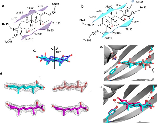
The steroid hormone 17α-hydroxylprogesterone (17-OHP) is a biomarker for congenital adrenal hyperplasia and hence there is considerable interest in development of sensors for this compound. We used computational protein design to generate protein models with binding sites for 17-OHP containing an extended, nonpolar, shape-complementary binding pocket for the four-ring core of the compound, and hydrogen bonding residues at the base of the pocket to interact with carbonyl and hydroxyl groups at the more polar end of the ligand. Eight of 16 designed proteins experimentally tested bind 17-OHP with micromolar affinity. A co-crystal structure of one of the designs revealed that 17-OHP is rotated 180° around a pseudo-two-fold axis in the compound and displays multiple binding modes within the pocket, while still interacting with all of the designed residues in the engineered site. Subsequent rounds of mutagenesis and binding selection improved the ligand affinity to nanomolar range, while appearing to constrain the ligand to a single bound conformation that maintains the same "flipped" orientation relative to the original design. We trace the discrepancy in the design calculations to two sources: first, a failure to model subtle backbone changes which alter the distribution of sidechain rotameric states and second, an underestimation of the energetic cost of desolvating the carbonyl and hydroxyl groups of the ligand. The difference between design model and crystal structure thus arises from both sampling limitations and energy function inaccuracies that are exacerbated by the near two-fold symmetry of the molecule.

Keywords: 17-hydroxylprogesterone; Computational protein design; hydrophobic small molecules; ligand binding design.


Figures

Figure 1
Figure 1
Computational design method and OHP9 design model. (a) Computational design method for binding 17‐OHP. PatchDock9 was used to place three conformers of 17‐OHP into all 257 crystal structures of NTF2‐like proteins from PDB based on shape complementarity; pocket residues were redesigned by Rosetta to fulfill the ligand hydrogen bonding and hydrophobic packing interactions. (b) OHP9 design model. Six mutations introduced by Rosetta design are highlighted in purple. 17‐OHP is shown as gray sticks with the more polar end inside the protein pocket. (c) Yeast display flow cytometry results. Under the same labeling condition, scaffold protein RV0760 does not bind 17‐OHP while design protein OHP9 shows a clear binding signal. Negative control is OHP9 cells labeled with streptavidin‐PE and FITC‐conjugated anti‐cMyc antibody without biotinylated 17‐OHP compound.
Figure 2
Figure 2
Distinct binding conformations revealed in the crystal structure of OHP9. (a) 2D representation of designed interactions around 17‐OHP. Hydrogen bonding interactions are highlighted as dashed gray lines between protein sidechains and 17‐OHP. Hydrophobic packing interactions are represented as purple shades. (b) 2D representation of interactions in OHP9 crystal chain A in the same fashion as in a. In addition, a water molecule is represented as a gray dot. Observed intraprotein hydrogen bond between Thr15 and Trp23 is highlighted. Atom O1, O2, O3, C12, and C16 of 17‐OHP are labeled explicitly for direct comparison. (c) Superimposed ligands upon aligned protein backbones. 17‐OHP in design model is shown in purple. The same ligand in crystal chain A is in cyan and its orientation deviates by a 180° rotation. (d) 2Fo‐Fc density maps of four ligand copies in OHP9 crystal: chain A in cyan; chain B and D in magenta; chain C in salmon. (e and f) Superimposed ligands upon protein alignment. In comparison with the bound ligand in crystal chain A(cyan), chain C ligand(salmon) reveals the second ligand binding configuration where 17‐OHP flips long the longer axis with two methyl groups pointing up; (f) Chain B and D(magenta) reveal the third binding configuration with an additional water molecule between Tyr108 and 17‐OHP.
Figure 3
Figure 3
Computational analysis of observed discrepancies in OHP9 design model. (a) Computed sidechain rotamer distribution for Trp23 in OHP9 design model backbone and OHP9 crystal backbone. Each colored grid represents one conformational state of Trp23 with sidechain χ1 and χ2 angles indicated by X and Y axes, respectively. Colorimetric scale is based on Boltzmann probability23 calculated from Rosetta energy term, where blue representing high‐probability(low‐energy) states and red representing low‐probability(high‐energy) states. The designed Trp23 rotamer is indicated by a purple window (χ1 ∼ −170°, χ2 ∼ 30°), and crystalized Trp23 rotamer by a cyan window (χ1 ∼ −170°, χ2 ∼ 90°). (b) Change of Cα‐Cβ vector in molecular dynamics simulation that captures the Trp23‐Thr15 hydrogen bond. Design model shown in purple serves as the starting point for simulation; representative MD model after a short simulation shown in gray closely matches the conformations in crystal structure(cyan). (c and d) Docked ligand conformations using Vina15 and Glide14. OHP9 design model in purple on the upper panel where Vina ligand (pink) and Glide ligand (yellow) are superimposed with the design ligand(purple) for comparison; (d) OHP9 crystal chain A in cyan on the lower panel where Vina ligand (pink) and Glide ligand(yellow) overlay with the chain A ligand(cyan). Ligand hydrogen bonds are highlighted by dashed gray lines. (e) Ligand energy landscapes generated by Rosetta ligand docking. OHP9 design model was used as the input conformation for the docking simulation summarized on the upper panel, where the purple color circles the design ligand configuration; cyan circle is close to the crystal chain A ligand configuration; gray color circles the ligand configuration that is 180° rotated from chain C ligand (with two polar groups inside the protein pocket); For the lower‐panel docking landscape, crystal chain A was used as the input docking conformation where salmon circle represents the crystal chain C ligand configuration. The same colors are used for indicating design(purple) and chain A(cyan) ligand configurations.
Figure 4
Figure 4
Binding fitness landscape of OHP9. The effect of each amino acid substitution(Y axis) at selected protein positions(X axis) is assessed by enrichment in the binding population ( ΔEx: enrichment value Supplementary Figure S4). Colored grids represent single mutant substitutes, where red and blue indicate high enrichment and depletion, respectively, after three rounds of selection for better binding (Supplementary Figure S4 and S5). The initial OHP9 amino acid at each position is indicated by its one‐letter amino acid code in the white box. (a) Designed interacting residues in OHP9 are highly conserved during the affinity selection. Few or no substitutions are enriched shown in the colored data matrix. Residue positions are mapped on to the OHP9 structure, where design ligand(purple) and crystal chain A ligand(cyan) are superimposed, and their hydrogen bonds are indicated by dashed lines in the same color. (b) Periphery beneficial mutations that seemingly conflict with the design model (red sidechains) can be partially explained by the crystal chain B and D conformation (gray sidechains). (c) Beneficial mutations in close vicinity to the ligand were included for constructing a pocket‐only combinatorial library of OHP9. The nine positions are mapped onto the OHP9 structure. (d) Top enriched substitutions, mostly in the periphery region of the protein, were included for making a general combinatorial library. In total, 15 positions were mutated all around the protein.
Figure 5
Figure 5
Evolved variants of OHP9: OHP9_1A and OHP9_1C. (a) Sequence alignment of OHP9, OHP9_1A and OHP9_1C. Black windows mark the positions mutated in OHP9_1A and OHP9_1C. (b) Equilibrium dissociation constants of OHP9, OHP9_1A, and OHP9_1C determined by fluorescence polarization assays. (c) 2Fo–Fc density maps of six ligand copies in OHP9_1C crystal. (d) The converged ligand binding configuration in OHP9_1C. Mutations inside the binding site are labeled and highlighted by magenta sticks. Water molecules are shown as red spheres and the sodium ion as a purple sphere. (e) Periphery mutations in OHP9_1C in magenta sticks mapped onto the crystal structure. Hydrogen bonds are represented by gray dashed lines.

References

    1. Speiser PW, White PC (2003) Congenital adrenal hyperplasia. N Engl J Med 349:776–788. - PubMed
    1. White PC (2009) Neonatal screening for congenital adrenal hyperplasia. Nat Rev Endocrinol 5:490–498. - PubMed
    1. Tinberg CE, Khare SD, Dou J, Doyle L, Nelson JW, Schena A, Jankowski W, Kalodimos CG, Johnsson K, Stoddard BL, Baker D (2013) Computational design of ligand‐binding proteins with high affinity and selectivity. Nature 501:212–216. - PMC - PubMed
    1. Griss R, Schena A, Reymond L, Patiny L, Werner D, Tinberg CE, Baker D, Johnsson K (2014) Bioluminescent sensor proteins for point‐of‐care therapeutic drug monitoring. Nat Chem Biol 10:598–603. - PubMed
    1. Feng J, Jester BW, Tinberg CE, Mandell DJ, Antunes MS, Chari R, Morey KJ, Rios X, Medford JI, Church GM, Fields S, Baker D, (2015) A general strategy to construct small molecule biosensors in eukaryotes. eLife 4:e10606. - PMC - PubMed