. 2017 Oct 5;8(10):e3076.
doi: 10.1038/cddis.2017.474.

Calcium/calmodulin-dependent kinase kinase 2 regulates hematopoietic stem and progenitor cell regeneration

Affiliations

Calcium/calmodulin-dependent kinase kinase 2 regulates hematopoietic stem and progenitor cell regeneration

Luigi Racioppi et al. Cell Death Dis. .

Abstract

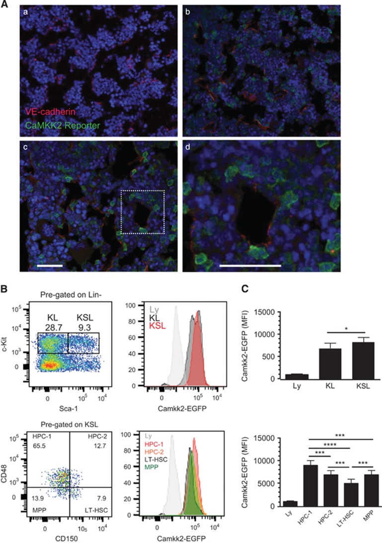
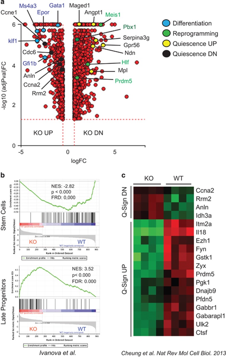
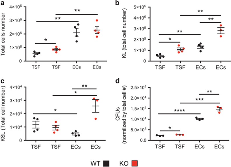
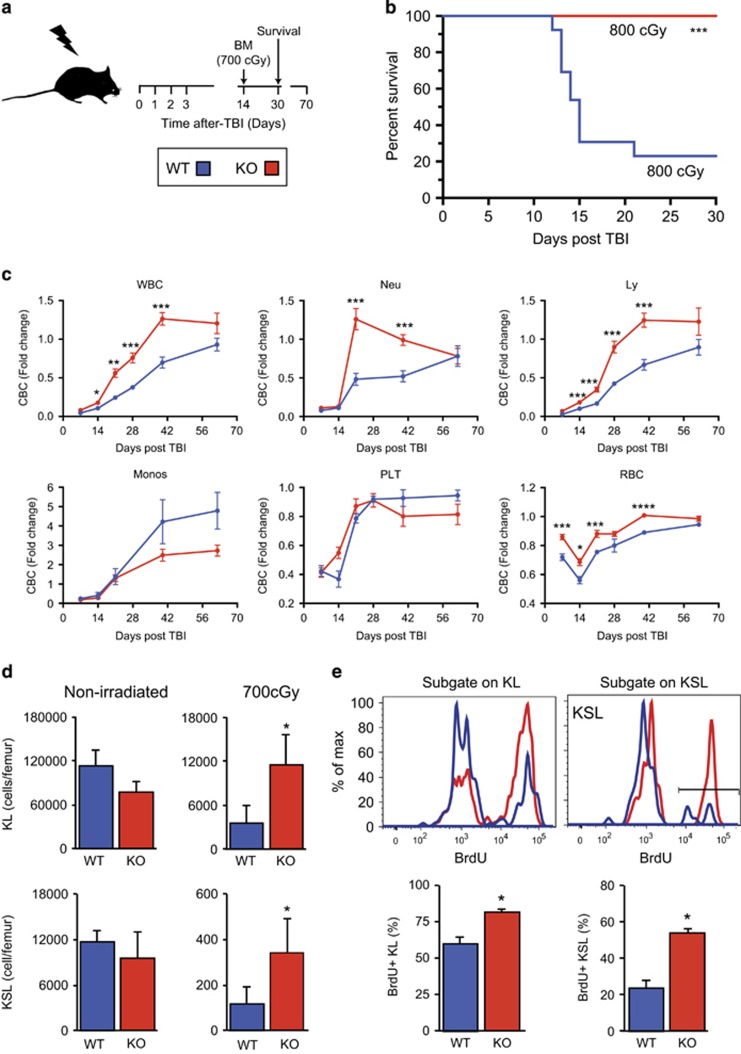
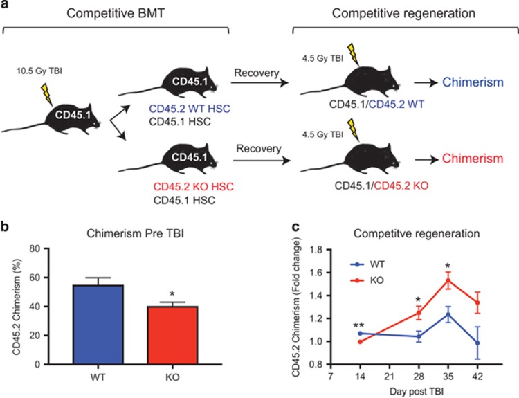
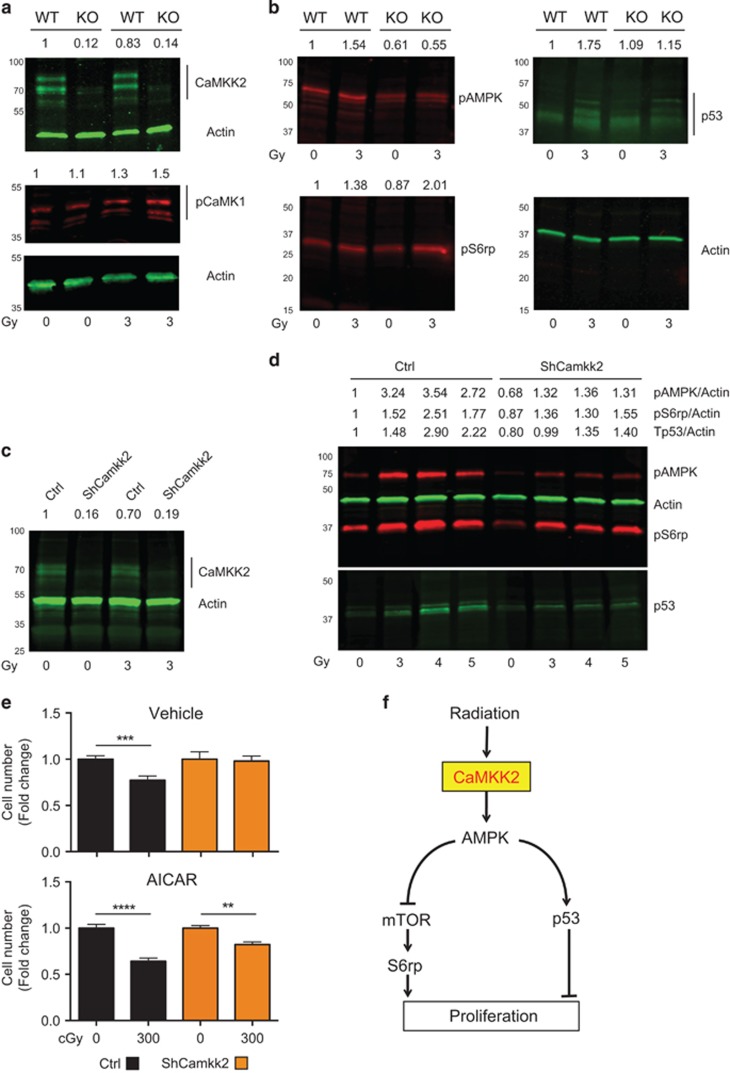
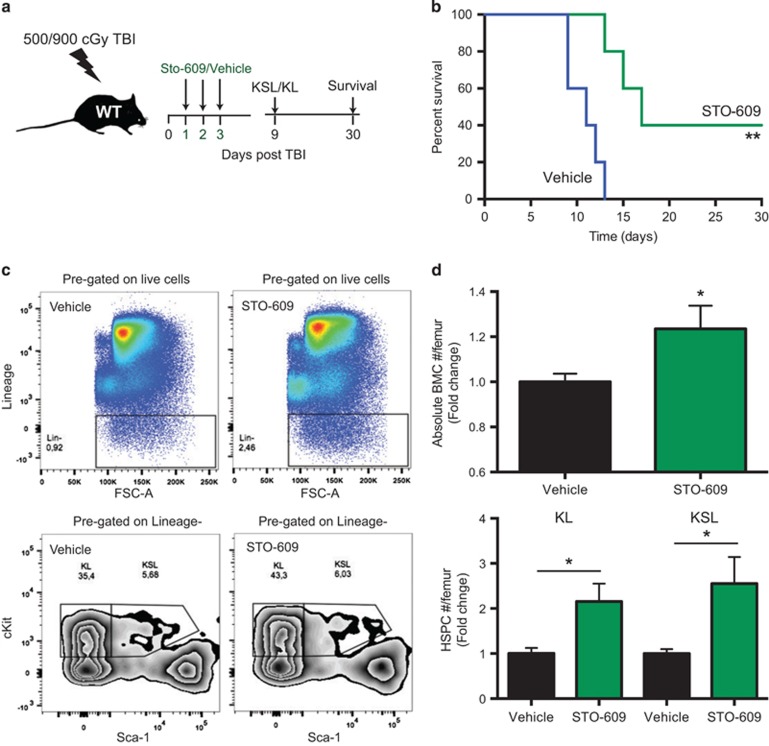
Hematopoietic stem and progenitor cells (HSPCs) are predominantly quiescent in adults, but proliferate in response to bone marrow (BM) injury. Here, we show that deletion of Ca2+/calmodulin (CaM)-dependent protein kinase kinase 2 (CaMKK2) promotes HSPC regeneration and hematopoietic recovery following radiation injury. Using Camkk2-enhanced green fluorescent protein (EGFP) reporter mice, we found that Camkk2 expression is developmentally regulated in HSPC. Deletion of Camkk2 in HSPC results in a significant downregulation of genes affiliated with the quiescent signature. Accordingly, HSPC from Camkk2 null mice have a high proliferative capability when stimulated in vitro in the presence of BM-derived endothelial cells. In addition, Camkk2 null mice are more resistant to radiation injury and show accelerated hematopoietic recovery, enhanced HSPC regeneration and ultimately a prolonged survival following sublethal or lethal total body irradiation. Mechanistically, we propose that CaMKK2 regulates the HSPC response to hematopoietic damage by coupling radiation signaling to activation of the anti-proliferative AMP-activated protein kinase. Finally, we demonstrated that systemic administration of the small molecule CaMKK2 inhibitor, STO-609, to irradiated mice enhanced HSPC recovery and improved survival. These findings identify CaMKK2 as an important regulator of HSPC regeneration and demonstrate CaMKK2 inhibition is a novel approach to promoting hematopoietic recovery after BM injury.


Conflict of interest statement

The authors declare no conflict of interest.

Figures

Figure 1
Figure 1
Camkk2 expression is enriched in primitive HSPCs in vivo. (A) Femurs were harvested from control and Camkk2-EGFP reporter mice and sectioned for immunofluorescent staining with VE-cadherin and anti-GFP antibodies (Aa, Ab; low magnification, Ac and insets high magnification) (n=3 per genotype). (B) BM cells from control and Camkk2-EGFP reporter mice were isolated, stained to identify HSPC subsets and analyzed by flow cytometry (top and bottom panels). (C) The reporter expression is shown relative to lymphocyte EGFP intensity, which is considered CaMKK2 negative. The relative EGFP expression is quantitated in the right panels (n=6 mice/genotype). Bars graph reports mean±S.E.M. *, *** and **** refer to P-values < 0.05, 0.005 and 0.001, respectively
Figure 2
Figure 2
Camkk2 regulates transcriptional program of hematopoietic stem cells. (a) Volcano plot comparison of DEGs in KSL cells isolated from BM of Camkk2 null and control mice (WT and KO, respectively). Genes downregulated or upregulated in KO compared with WT are indicated as KO DN and KO UP, respectively. Color dots indicate genes involved in the regulation of differentiation or reprogramming of mature hematopoietic cells to HSC. (b) GSEA of microarray data shows that gene signatures for genes enriched in hematopoietic stem cells are significantly downregulated in KO KSL (upper). In contrast, genes enriched in late progenitors are significantly upregulated in KO KSL (lower). (c) Loss of Camkk2 downregulates the quiescent gene signature in stem cells. Heatmap represents DEGs in quiescent stem cell signature. Q-Sign DN and UP indicate genes downregulated and upregulated in quiescent stem cells. The color key of heatmaps indicates row-wise scaled RPKM values (z-score)
Figure 3
Figure 3
Camkk2 null hematopoietic stem cells have increased proliferation in vitro. KSL cells were sorted from WT and Camkk2 null mice and cultured with TPO, SCF and Flt-3L in the presence or absence of BM endothelial cells (TSF and ECs, respectively). Cell were harvested and analyzed on day 7. (a) Total cells number. (b and c) Absolute numbers of KL and KSL cells. (d) Cells recovered at day 7 were plated in methylcellulose media for colony formation and colonies (CFUs). Graphs report total CFUs normalized by total cell expansion. The experiment was replicated twice. Bars graph reports mean±S.E.M. *P<0.05, **P<0.01, ***P<0.005. **** P<0.001
Figure 4
Figure 4
Camkk2 null mice have improved survival and accelerated hematopoietic recovery following TBI. (a) Scheme of TBI. Mice were TBI and monitored for survival, blood cell count (CBC) and BM recovery. (b) Survival of WT and Camkk2 null mice (WT and KO, respectively) irradiated with 800cGy (n=14 mice per genotype). The blue lines indicate control and the red lines indicate Camkk2 null mice. (c) Hematopoietic recovery in WT and KO mice sublethally irradiated with 700cGy TBI and bled for CBC analysis of total WBCs, platelets (PLT), RBCs, neutrophils (NE), monocytes (Mo) and lymphocytes (Ly) (n=6 and 9 mice for WT and Camkk2 null mice, respectively). (d) WT and KO mice (n=10 per group) were irradiated with 700cGy TBI and euthanized 14 days after irradiation. WT and KO non-irradiated mice were used as controls (n=6 mice per group). Upper and lower bar graphs report mean ±S.E.M. of KL and KSL, respectively. (e) BrdU incorporation in KL and KSL cells in vivo during regeneration. WT and KO mice were irradiated with 700cGy TBI, and after 14 days were pulsed with BrdU in vivo for 2 h before killing. Dot plots of KL and KSL cells and BrdU incorporation on day 14 after radiation (top panels). BrdU staining FACS profiles in KL and KSL subsets (upper panels). Bars graph reports mean±S.E.M. The percentage of BrdU+ cells is shown in lower graphs (bottom panels; n=6 per genotype). *P<0.05, **P<0.01, ***P<0.005, ****P<0.001
Figure 5
Figure 5
Camkk2 null HSC have a cell-intrinsic enhanced regenerative capability in vivo. KSL CD34- cells were isolated from WT and Camkk2 null mice (WT and KO, respectively) and transplanted in lethally irradiated recipient mice with CD45.1 competitor BM. The recipient mice receiving WT or KO KSL CD34- cells were monitored for 4 months. Subsequently, mice showing comparable percentages of WT or KO donor CD45.2 cells were irradiated with 450cGy TBI and bled weekly after irradiation (n= 6 per group). (a) Scheme of the experiment. (b) CD45.2 chimerism in mice reconstituted with WT or KO KSL CD34- before receiving 4.5Gy TBI. (c) Donor CD45.2 chimerism was monitored by flow cytometry and the results are expressed as fold change over the basal level (pre-TBI). Bars graph reports mean±S.E.M. *P<0.05, **P<0.01
Figure 6
Figure 6
CaMKK2 couples radiation signaling with the AMPK anti-proliferative pathway. HSPC (KL+KSL) were isolated from WT and Camkk2 null (KO) mice and irradiated in vitro with 400cGy or left non-irradiated. Cells were then cultured for 1-h in regular medium. Protein expression was normalized by actin and expressed as fold change over basal (non-irradiated WT HSPC), and is reported on the top of each lane. (a) Immunoblots of CaMKK2, phospho-CaMK1 (pCaMK1) and actin. (b) Immunoblots of Tp53, phosphorylated AMPK and S6rp (pAMPK and pS6rp, respectively). (c-e) M1 myeloid progenitor cells were transduced with lentiviral vectors expressing a short hairpin sequence for silencing Camkk2 or a control sequence (ShCamkk2 and Ctrl, respectively). Ctrl and ShCamkk2 M1 cells were then irradiated or left non-irradiated. One-hour after irradiation, M1 cell protein expression was assessed by immunoblotting. (c) Expression of CaMKK2 and actin. (d) Immunoblots of pAMPK, pS6rp, actin and Tp53 of M1 cells irradiated with increasing doses of radiation or left non-irradiated. (e) M1 cells transduced with Ctrl and ShCamkk2 lentiviral vectors were 300cGy irradiated and cultured for 24 h in regular medium in the presence or absence of AICAR (100 μM), a cell permeable AMPK agonist (Top and lower, respectively). Cell number was determined using a colorimetric assay, and the results are expressed as fold change of non-irradiated cells cultured in the absence of AICAR. Bars graph reports mean±S.E.M. The experiments included in this figures were replicated at least three times. (f) Modeling the radiation-induced CaMKK2-dependent signal pathway. **P<0.01,***P<0.005, ****P<0.001
Figure 7
Figure 7
Pharmacologic inhibition of CaMKK2 enhances hematopoietic regeneration in vivo. Treatment with STO-609, a CaMKK2 inhibitor, mitigates the acute hematopoietic radiation syndrome. (a) Scheme of irradiation and STO-609 treatment in wild type mice. (b) Survival of 900cGy TBI wild-type mice treated with vehicle or STO-609 (n=10 per group). Wild-type mice were irradiated with 500cGy TBI and were then treated with STO-609 or vehicle after 24 h. Nine days after TBI, the mice were killed and bones were removed. The BM cells were counted and stained to identify HPSC (BMC). (c) Representative staining and gating strategy. (d) Absolute number of BMC and HSPC (upper and lower graph, respectively). The results are expressed as the fold changes over the absolute number of cells recovered form vehicle-treated wild type mice, and are normalized for one femur. Two independent experiments have been combined (total number of mice=9 per group). Bars graph reports mean±S.E.M. *P-value <0.05, **P-value <0.01

Similar articles

Cited by

References

    1. Zhang J, Niu C, Ye L, Huang H, He X, Tong WG et al. Identification of the haematopoietic stem cell niche and control of the niche size. Nature 2003; 425: 836–841. - PubMed
    1. Ding L, Saunders TL, Enikolopov G, Morrison SJ. Endothelial and perivascular cells maintain haematopoietic stem cells. Nature 2012; 481: 457–462. - PMC - PubMed
    1. Winkler IG, Sims NA, Pettit AR, Barbier V, Nowlan B, Helwani F et al. Bone marrow macrophages maintain hematopoietic stem cell (HSC) niches and their depletion mobilizes HSCs. Blood 2010; 116: 4815–4828. - PubMed
    1. Fujisaki J, Wu J, Carlson AL, Silberstein L, Putheti P, Larocca R et al. In vivo imaging of Treg cells providing immune privilege to the haematopoietic stem-cell niche. Nature 2011; 474: 216–219. - PMC - PubMed
    1. Lucas D, Scheiermann C, Chow A, Kunisaki Y, Bruns I, Barrick C et al. Chemotherapy-induced bone marrow nerve injury impairs hematopoietic regeneration. Nat Med 2013; 19: 695–703. - PMC - PubMed

Publication types

MeSH terms