. 2017 Oct 5;8(10):e3063.
doi: 10.1038/cddis.2017.448.

Cofilin1-dependent actin dynamics control DRP1-mediated mitochondrial fission

Affiliations

Cofilin1-dependent actin dynamics control DRP1-mediated mitochondrial fission

Katharina Rehklau et al. Cell Death Dis. .

Abstract

Mitochondria form highly dynamic networks in which organelles constantly fuse and divide. The relevance of mitochondrial dynamics is evident from its implication in various human pathologies, including cancer or neurodegenerative, endocrine and cardiovascular diseases. Dynamin-related protein 1 (DRP1) is a key regulator of mitochondrial fission that oligomerizes at the mitochondrial outer membrane and hydrolyzes GTP to drive mitochondrial fragmentation. Previous studies demonstrated that DRP1 recruitment and mitochondrial fission is promoted by actin polymerization at the mitochondrial surface, controlled by the actin regulatory proteins inverted formin 2 (INF2) and Spire1C. These studies suggested the requirement of additional actin regulatory activities to control DRP1-mediated mitochondrial fission. Here we show that the actin-depolymerizing protein cofilin1, but not its close homolog actin-depolymerizing factor (ADF), is required to maintain mitochondrial morphology. Deletion of cofilin1 caused mitochondrial DRP1 accumulation and fragmentation, without altering mitochondrial function or other organelles' morphology. Mitochondrial morphology in cofilin1-deficient cells was restored upon (i) re-expression of wild-type cofilin1 or a constitutively active mutant, but not of an actin-binding-deficient mutant, (ii) pharmacological destabilization of actin filaments and (iii) genetic depletion of DRP1. Our work unraveled a novel function for cofilin1-dependent actin dynamics in mitochondrial fission, and identified cofilin1 as a negative regulator of mitochondrial DRP1 activity. We conclude that cofilin1 is required for local actin dynamics at mitochondria, where it may balance INF2/Spire1C-induced actin polymerization.


Conflict of interest statement

The authors declare no conflict of interest.

Figures

Figure 1
Figure 1
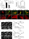
Loss of cofilin1 induced mitochondrial fragmentation in MEFs. (a) Shown are representative micrographs of a control MEF (CTR) and MEFs lacking either cofilin1 (Cfl1−/−), ADF (ADF−/−) or both ADF/cofilin proteins (Cfl1−/−/ADF−/−). Mitochondria visualized by mitochondrial-targeted YFP (mtYFP, green) and cytochrome c immunoreactivity (red) appeared fragmented in Cfl1−/−- and Cfl1−/−/ADF−/−-MEFs, but not in ADF−/−-MEFs. White boxes indicate areas shown in high magnification. Scale bars: 50 μm. (b) Relative MEF number with fragmented mitochondria was increased in Cfl1−/−- and Cfl1−/−/ADF−/−-MEFs, but not in ADF−/−-MEFs (CTR: 24.4±3.4%, n=214 cells/4 independent experiments; ADF−/−: 28.9±3.3%, n=149/4, P=0.376; Cfl1−/−: 68.6±8.0%, n=241/4, P<0.01; Cfl1−/−/ADF−/−: 70.8±7.0%, n=184/4, P<0.01). (c) Western blots demonstrating reduced cofilin1 levels upon transfection with either Cfl1-si01 or Cfl1-si03 in CTR-MEFs. Transfection of a control siRNA (scr-siRNA) did not change cofilin1 levels. β-tubulin served as a loading control. (d) Representative micrographs of Mitotracker-stained CTR-MEFs. Fragmented mitochondria were noted upon transfection of either Cfl1-si01 or Cfl1-si03, but not of scr-siRNA. White boxes indicate areas shown in high magnification. Scale bars: 50 μm. (e) Relative MEF numbers with fragmented mitochondria were increased upon transfection of either Cfl1-si01 or Cfl1-si03, but not of scr-siRNA (CTR: 6.0±1.6% scr-siRNA: 4.0±0.7% Cfl1-si01: 42.1±6.7%, P<0.01; Cfl1-si03: 62.8±11.3%, P<0.001; n>900 MEFs in three independent experiments for each condition). Columns and error bars in (b), F: mean values (MV) and standard error of the mean (S.E.M.). Open circles: values of independent experiments. **P<0.01, NS: not significant
Figure 2
Figure 2
Mitochondrial fragmentation in Cfl1−/− MEFs was caused by impaired actin dynamics. (a) Representative western blots showing actin levels in insoluble (f) and soluble (g) protein fractions from CTR-, Cfl1−/−- and ADF−/−-MEFs. Actin levels in both protein fractions were quantified to calculate the F/G-actin ratio. Compared with CTR-MEFs, the F/G-actin ratio was strongly increased in Cfl1−/−-MEFs, but not in ADF−/−-MEFs (CTR: 1.28±0.16; Cfl1−/−: 2.46±0.16, P<0.001; ADF−/−: 1.29±0.09, P=0.959; n=7 for each group). (b) Representative micrographs of Mitotracker-stained Cfl1−/−-MEFs upon expression of either GFP or various GFP-tagged cofilin1 variants: WT-Cfl1, constitutive active Cfl1-S3A, or constitutive inactive Cfl1-S3D. (c) Quantification of relative MEF numbers with fragmented mitochondria revealed that expression of either WT-Cfl1 or Cfl1-S3A, but not of GFP or Cfl1-S3D restored mitochondrial morphology in Cfl1−/−-MEFs (Cfl1−/−+GFP: 69.9±9.1; Cfl1−/−+WT-Cfl1: 12.1±5.7, P<0.01; Cfl1−/−+Cfl1-S3A: 19.1±4.4, P<0.01; Cfl1−/−+Cfl1-S3D: 76.1±10.4; n>850/3 for each condition). (d) Representative micrographs of a CTR-MEF before (−1 min) and after (+4 min, +8 min) treatment with the F-actin stabilizing drug jasplakinolide (JASP) that was added at time point 0 min. (e) Graph showing JASP-induced mitochondrial fragmentation in CTR-MEFs. For example, upon 4 min of JASP treatment, mitochondrial size was clearly reduced (57.3±7.0%, P<0.001, n=5 MEFs in five independent experiments) and it reduced to roughly 40% of basal levels after 8 min (38.9±4.3%, P<0.001). Conversely, dimethyl sulfoxide (DMSO) did not change mitochondrial morphology (8 min: 103.6±8.3%, n=5/5). (f) Representative micrographs of a Cfl1−/−-MEF before (−1 min) and after (+4 min, +8 min) treatment with the F-actin destabilizing drug cytochalasin D (CYTD) that was added at time point 0 min. (g) Graph showing CYTD-induced mitochondrial elongation in Cfl1−/−-MEFs (4 min: 142.1±7.1%, P<0.01, n=5/5; 8 min: 173.7±15.2%, P<0.01). Conversely, DMSO did not change mitochondrial morphology (8 min: 98.4±1.7%, n=4/4). White boxes in (b), (d) and (f) indicate areas shown in high magnification. Scale bars in (b), (d) and (f): 50 μm. Columns and error bars in (a) and (c): MV+S.E.M. Open circles: values of independent experiments. Squares in (e) and (g): MV+S.E.M. **P<0.01; ***P<0.001; NS: not significant
Figure 3
Figure 3
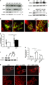
Mitochondrial fragmentation in Cfl1−/− MEFs was mediated by DRP1. (a) Western blot of two independent experiments demonstrating increased DRP1 levels in Cfl1−/−-MEFs, while TOM20 and COXII expression levels were reduced. No changes in DRP1, TOM20 or COXII expression was noted in ADF−/−-MEFs. (b) Western blot of two independent experiments demonstrating increased phosphorylation of DRP1 at S616 in Cfl1−/−-MEFs. (c) Representative western blot demonstrating reduced phosphorylation of DRP1 at S637 in Cfl1flx/flx-MEFs upon OH-TAM treatment. β-Tubulin was used as a loading control in (ac). (d) DRP1 immunoreactivity (red) in representative mtYFP-expressing (green) CTR- and Cfl1−/−-MEFs. (e) In Cfl1−/−-MEF, the Pearson's correlation of mtYFP and DRP1 was increased (CTR: 0.36±0.02; KO: 0.54±0.04; n=19, P<0.01). (f) siRNA against DRP1 efficiently depleted DRP1 in MEFs, while a scrambled control siRNA (scr-siRNA) did not alter DRP1 levels. β-Tubulin was used as a loading control. (g) Representative micrographs of a Mitotracker-stained CTR-MEF and of Cfl1−/−-MEFs upon transfection with either scr-siRNA or DRP1-siRNA. Mitochondria in Cfl1−/−-MEFs appeared fragmented upon transfection with scr-siRNA. Conversely, transfection of DRP1-siRNA (40 nM) restored mitochondrial morphology in Cfl1−/−-MEFs. (h) Quantification of relative MEF numbers with fragmented mitochondria revealed that DRP1-siRNA, but not scr-siRNA, restored mitochondrial morphology in Cfl1−/− MEFs (CTR: 23.2±2.7% Cfl1−/−+scr-siRNA: 59.8±11.5%, P<0.05; Cfl1−/−+DRP1-siRNA: 30.7±10.5, P<0.05; n>900/3 for each condition). Columns and error bars in (e) and (h): MV+S.E.M. Open circles: values of independent experiments. **P<0.01. White boxes in (d) and (g) indicate areas shown in high magnification. Scale bar in (d) and (g): 50 μm
Figure 4
Figure 4
Putative model for the role of cofilin1 in mitochondrial morphology. Previous studies revealed that actin polymerization induced by ER-anchored INF2 is relevant for mitochondrial DRP1 oligomerization and mitochondrial fission and that INF2 cooperates with mitochondrial Spire1C (not shown)., We found fragmented mitochondria and elevated mitochondrial levels of DRP1 in cofilin1-deficient MEFs. Further, we found restored mitochondrial morphology in cofilin1-deficient MEFs (i) upon expression of a constitutive active cofilin1 mutant, but not upon expression of a cofilin1 mutant that does not bind actin, (ii) upon acute F-actin destabilization (iii) and upon genetic inhibition of DRP1. Our data promote a model in which cofilin1-dependent actin dynamics acts as a negative regulator of mitochondrial DRP1 activity and mitochondrial fission. Cofilin1-dependent actin depolymerization might be required for fine-tuning actin dynamics at the mitochondrial surface by antagonizing INF2/Spire1C-mediated actin polymerization

References

    1. Orrenius S, Zhivotovsky B, Nicotera P. Regulation of cell death: the calcium-apoptosis link. Nat Rev Mol Cell Biol 2003; 4: 552–565. - PubMed
    1. Danial NN, Korsmeyer SJ. Cell death: critical control points. Cell 2004; 116: 205–219. - PubMed
    1. Bereiter-Hahn J, Vöth M, Mai S, Jendrach M. Structural implications of mitochondrial dynamics. Biotechnol J 2008; 3: 765–780. - PubMed
    1. Twig G, Liu X, Liesa M, Wikstrom JD, Molina AJ, Las G et al. Biophysical properties of mitochondrial fusion events in pancreatic beta-cells and cardiac cells unravel potential control mechanisms of its selectivity. Am J Physiol Cell Physiol 2010; 299: C477–C487. - PMC - PubMed
    1. Westermann B. Mitochondrial fusion and fission in cell life and death. Nat Rev Mol Cell Biol 2010; 11: 872–884. - PubMed