. 2017 Oct 5;8(10):256.
doi: 10.3390/genes8100256.

The Genetic Basis of Pericentral Retinitis Pigmentosa-A Form of Mild Retinitis Pigmentosa

Affiliations

The Genetic Basis of Pericentral Retinitis Pigmentosa-A Form of Mild Retinitis Pigmentosa

Jason Comander et al. Genes (Basel). .

Abstract

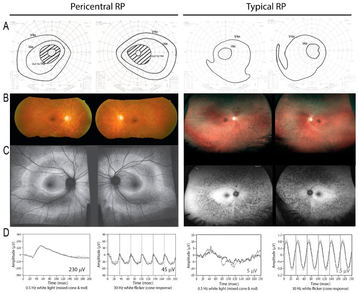
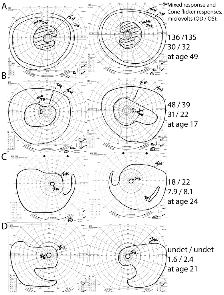
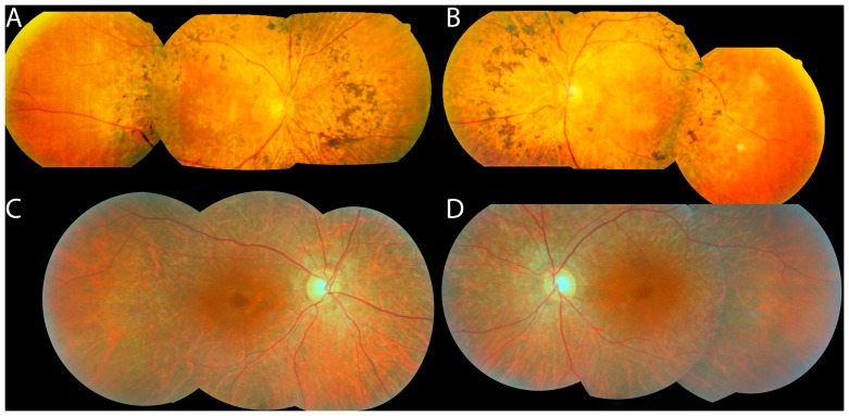
Pericentral retinitis pigmentosa (RP) is an atypical form of RP that affects the near-peripheral retina first and tends to spare the far periphery. This study was performed to further define the genetic basis of this phenotype. We identified a cohort of 43 probands with pericentral RP based on a comprehensive analysis of their retinal phenotype. Genetic analyses of DNA samples from these patients were performed using panel-based next-generation sequencing, copy number variations, and whole exome sequencing (WES). Mutations provisionally responsible for disease were found in 19 of the 43 families (44%) analyzed. These include mutations in RHO (five patients), USH2A (four patients), and PDE6B (two patients). Of 28 putatively pathogenic alleles, 15 (54%) have been previously identified in patients with more common forms of typical RP, while the remaining 13 mutations (46%) were novel. Burden testing of WES data successfully identified HGSNAT as a cause of pericentral RP in at least two patients, suggesting it is also a relatively common cause of pericentral RP. While additional sequencing might uncover new genes specifically associated with pericentral RP, the current results suggest that genetically pericentral RP is not a separate clinical entity, but rather is part of the spectrum of mild RP phenotypes.

Keywords: HGSNAT; genotype/phenotype correlations; pericentral; pericentral retinal degeneration; pericentral retinitis pigmentosa; retinitis pigmentosa; rhodopsin.


Conflict of interest statement

The authors declare no conflict of interest.

Figures

Figure 1
Figure 1
On the left, a patient (Family #14) with a heterozygous RHO mutation demonstrates a pericentral phenotype, with pericentral scotomas but preserved peripheral field to the I4e stimulus on visual field testing (A). There is retinal pigment epitheium (RPE) atrophy in the near periphery as shown by fundus photo (B) and autofluorescence imaging (C), as well as relatively preserved electroretinograms (ERGs) (D). In contrast, in typical retinitis pigmentosa (RP) (right), the peripheral response to I4e is lost (A), and the affected area is located farther away from the macula in the mid-periphery (right, B,C; note lower magnification). The ERG is more severely affected (D; note different scales; see numerical values, inset).
Figure 2
Figure 2
Intra-familial phenotypic variation in family #18. The proband (A) had pericentral RP, with near-peripheral C-shaped scotomas with preserved peripheral I4e responses, and relatively preserved full-field ERG responses. His children, in contrast, had either atypically mild RP without pericentral features (B) or typical RP (C,D). Note the constricted peripheral I4e stimulus responses and lower ERG response amplitudes in the children (BD) in comparison to the proband. undet = undetectable.
Figure 3
Figure 3
Fundus photos of patients with HGSNAT mutations: #34 (A,B) and #15 (C,D). Note that the bone spicules (A,B) or RPE depigmentation (C,D) are more posteriorly located than in typical RP, and that the fundus begins to show a more normal color peripherally (especially temporally in these photos).

References

    1. Daiger S.P. RetNet, the Retinal Information Network. [(accessed on 24 June 2017)]; Available online: http://www.sph.uth.tmc.edu/RetNet/
    1. Grover S., Fishman G.A., Brown J., Jr. Patterns of visual field progression in patients with retinitis pigmentosa. Ophthalmology. 1998;105:1069–1075. doi: 10.1016/S0161-6420(98)96009-2. - DOI - PubMed
    1. Chang S., Vaccarella L., Olatunji S., Cebulla C., Christoforidis J. Diagnostic challenges in retinitis pigmentosa: Genotypic multiplicity and phenotypic variability. Curr. Genomics. 2011;12:267–275. doi: 10.2174/138920211795860116. - DOI - PMC - PubMed
    1. Gonin J. Le scotome Annulaire Dans la Dégénérescence Pigmentaire de la Rétine. A. Maloine; Paris, France: 1901.
    1. Berson E.L., Howard J. Temporal aspects of the electroretinogram in sector retinitis pigmentosa. Arch. Ophthalmol. 1971;86:653–665. doi: 10.1001/archopht.1971.01000010655008. - DOI - PubMed

LinkOut - more resources