. 2017 Nov;117(11):2063-2078.
doi: 10.1160/TH17-01-0067. Epub 2017 Nov 30.

Extracellular Cyclophilin A Augments Platelet-Dependent Thrombosis and Thromboinflammation

Affiliations

Extracellular Cyclophilin A Augments Platelet-Dependent Thrombosis and Thromboinflammation

Saskia N I von Ungern-Sternberg et al. Thromb Haemost. 2017 Nov.

Abstract

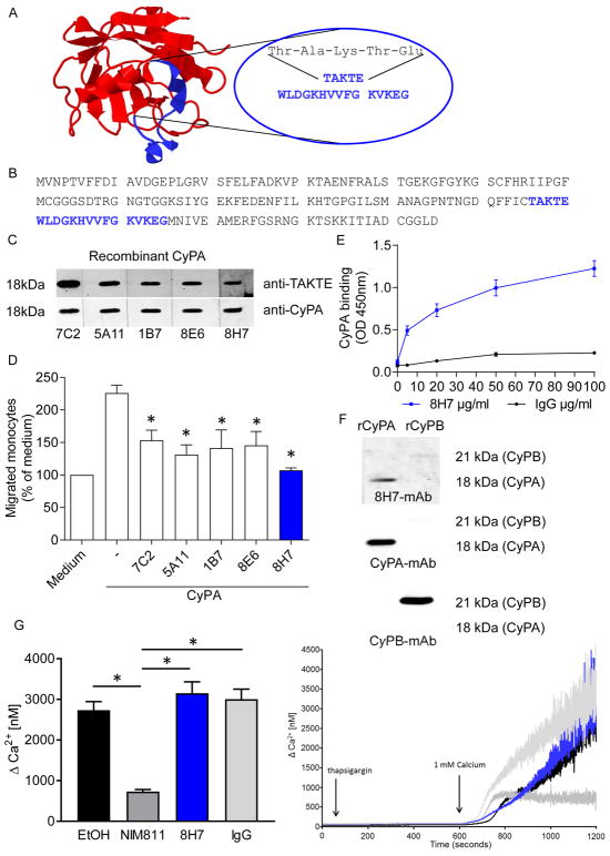
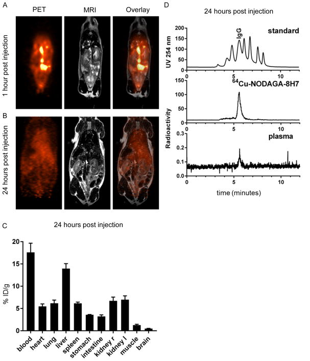
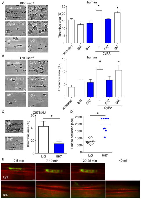
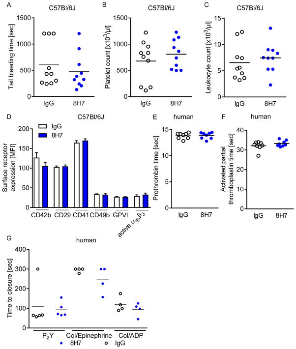
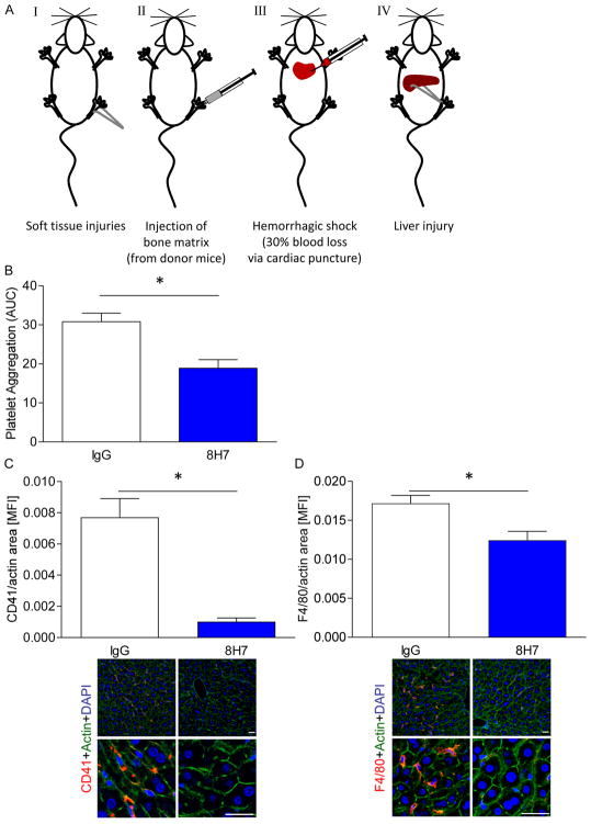
Cyclophilin A (CyPA) is involved in the pathophysiology of several inflammatory and cardiovascular diseases. To our knowledge, there is no specific inhibitor targeting extracellular CyPA without affecting other extracellular cyclophilins or intracellular CyPA functions. In this study, we developed an antibody-based inhibitor of extracellular CyPA and analysed its effects in vitro and in vivo. To generate a specific antibody, mice and rats were immunized with a peptide containing the extracellular matrix metalloproteinase inducer binding site and various antibody clones were selected and purified. At first, antibodies were tested for their binding capacity to recombinant CyPA and their functional activity. The clone 8H7-mAb was chosen for further experiments. 8H7-mAb reduced the CyPA-induced migration of inflammatory cells in vitro and in vivo. Furthermore, 8H7-mAb revealed strong antithrombotic effects by inhibiting CyPA-dependent activation of platelets and thrombus formation in vitro and in vivo. Surprisingly, 8H7-mAb did not influence in vivo tail bleeding time or in vitro whole blood coagulation parameters. Our study provides first evidence that antibody-based inhibition of extracellular CyPA inhibits thrombosis and thromboinflammation without affecting blood homeostasis. Thus, 8H7-mAb may be a promising compound for thrombi modulation in inflammatory diseases to prevent organ dysfunction.


Conflict of interest statement

Disclosure: None.

Figures

Figure 1
Figure 1. Generation and validation of a specific CyPA neutralizing antibody
5 CyPA antibodies were induced by injection of a peptide containing the EMMPRIN-binding sequence as described in material and methods. 4 mice (7C2, 5A11, 1B7, 8E6) and 1 rat (8H7) antibodies were tested for their binding capacity to CyPA and their inhibitory function. (A) Figure shows structure of CyPA (PBD-ID: 3K0N) and the epitope of the antibody highlighted in blue. (B) The CyPA amino acid sequence (blue) indicated the suggested binding region of the TAKTE-antibodies. (C) Antibodies were tested for their binding to CyPA in a SDS page using recombinant CyPA. (D) To test the function of TAKTE-antibodies in a functional assay monocyte migration was analyzed. (n=6). * means p ≤ 0.05 vs. CyPA. (E) In a modified ELISA the binding between CyPA and 8H7-mAb / IgG control was analyzed. (F) Recombinant CyPA and recombinant CyPB were loaded on a SDS page and detected with 8H7-mAb (upper panel). 8H7-mAb could only detect CyPA but not CyPB. The band size of CyPA (18 kDa) and CyPB (21 kDa) was indicated using anti-CyPA (middle panel) and anti-CyPB (lower panel) antibodies. (G) A Ca2+ measurement was performed to analyze if the 8H7-mAb antibody interfere with intracellular calcium hemostasis. The intra- and extracellular CyPA inhibitor NIM811 (200 nmol/L) and ethanol (0.2%) were used as controls (n ≤ 5). * means p ≤ 0.05.
Figure 2
Figure 2. PET/MR imaging and biodistribution analysis of radiolabeled 8H7-mAb
64Cu-NODAGA-8H7-mAb was injected i.p. and its distribution was quantified by simultaneous PET/MR imaging. (A/B) PET and MRI images of a representative animal 1 hour (A) and 24 hours (B) after i.p. injection of 64Cu-NODAGA-8H7 demonstrate systemic distribution of the antibody over time. (C) Ex vivo analysis of the organs by gamma-counting 24 hours after injection showed highest activities in blood and liver, followed by organs with good blood supply (heart, lung, spleen, kidneys). Data represent mean ± SD of three independent experiments. (D) Size standard (Vo, 669 kDa, 300 kDa, 150 kDa, 45 kDa, 17 kDa, 0.2 kDa, Vi; upper panel) and radiolabeled 8H7-mAb before injection (middle panel) are shown for reference. Representative analysis of plasma taken 24 hours after injection showed radioactivity signal corresponding to the molecular weight of 64Cu-NODAGA-8H7, demonstrating the molecular integrity of the circulating radiolabeled antibody for over 24 hours (lower panel).
Figure 3
Figure 3. Inhibition of extracellular CyPA inhibits CyPA-dependent monocyte/ endothelium adhesion and migration of monocytes/macrophages in vitro and in vivo.
(A) Human monocytes were activated with 200 nmol/L CyPA with an additional treatment of 8H7-mAb or rIgG2a. The surface expression of the integrin CD11b expression was measured using flow cytometry. * p ≤ 0.05 compared to untreated. (B) Human monocytes were treated with CyPA and 8H7-mAb or IgG control as indicated and perfused over activated HUVECs under arterial shear rates. Bar graphs show mean±SEM of adherent monocytes on activated HUVECs compared to unstimulated (n ≤ 8). * means p ≤ 0.05. (C) The migration of human monocytes was performed in a modified Boyden chamber. Human monocytes were allowed to migrate towards CyPA in presence or absence of 8H7-mAb, αCD147 or IgG control. (n≤5) * means p ≤ 0.05 vs. CyPA+IgG. (D) The infiltration of CD3+ cells into the peritoneal lavage were analyzed using flow cytometry (n≤5) * means p ≤ 0.05, with representative overlays of CD3+ cells. (E) The Ly6G+F4/80+ cells in the lavage in a CyPA-induced peritonitis model were analyzed using flow cytometry (n≤8) * means p ≤ 0.05, with representative quadrat statistic for the cells in the lavage.
Figure 4
Figure 4. Extracellular CyPA enhances platelet-activation in vitro
(A) 8H7-mAb detects CyPA in monocyte as well in platelet lysate (upper line). Afterwards the same membrane was incubated with an anti-CyPA antibody (lower line) to prove the specificity of 8H7-mAb. (B) 8H7-mAb significantly reduces the CyPA-dependent platelet activation. Representative overlays of the p-selectin expression and bar graphs show mean±SEM of P-selectin expression on platelets. * means p ≤ 0.05 vs. CyPA+IgG. (C) Monocyte-platelet aggregate (MPA) formation was analyzed by using double staining with CD42b (as platelets marker) and CD14 (as monocyte marker) as described in material and methods. The platelets were treated as indicated and incubated with monocytes for the MPA formation. Figure shows representative flow cytometry analysis. (n ≤ 6) * means p ≤ 0.05 vs. resting. Right panel shows representative quadrat statistic for the CD14+CD42+ double positive cells.
Figure 5
Figure 5. Extracellular CyPA enhances platelet-dependent thrombosis in vivo and in vitro
Human blood was preincubated with 8H7-mAb or IgG control and stimulated with 200 nmol/L CyPA. Then the blood was perfused using a shear rate of 1000 seconds−1 (A) and 1700 seconds−1 (B) over a fibrillar type I collagen coated coverslip. (n ≤ 4) * means p ≤ 0.05 vs. untreated. (C) Murine blood was perfused over a CyPA/fibrillar type I collagen coated coverslip using a shear rate of 1700 seconds−1 after i.v. injection of 8H7-mAb or IgG control (n ≤ 8) * means p ≤ 0.05, scale bar 20 μm. 8H7-mAb reduceed thrombus formation in vitro, and as the next step the in vivo thrombus formation in mesenteric arterioles was analyzed in a FeCl3 model after administration of 8H7-mAb or IgG control (D/E). Representative pictures of the in vivo thrombus formation (E) and time to occlusion for individual mice (D). * means p ≤ 0.05, scale bar 50 μm.
Figure 6
Figure 6. Blockage of extracellular CyPA through 8H7-mAb does not affect hemostasis
(A/B) 8H7-mAb treated mice did not exhibita prolonged tail bleeding time (A). After tail bleeding time the blood was collected and the number of platelets (B) and leukocytes (C) were estimated for both groups. (D) An in vivo treatment with 8H7-mAb did not change the surface expression of CD42b, CD29, CD41, CD49b, GPVI or the active form of αIIbβ3 on platelets compared to IgG control treated mice. (F/G) Every dot shows the response of one donor for the prothrombin time (E) and the activated partial thromboplastin time (F) (n=8). (G) PFA-100 measurement was performed with human blood. With the cartilages P2Y, Collagen/Epinephrine and Collage/ADP no difference could be detected.
Figure 7
Figure 7. Inhibition of extracellular CyPA limits platelet accumulation and macrophage infiltration in shock liver
The trauma/hemorrhagic shock model consists of four different parts: soft tissue injuries, injection of bone matrix, hemorrhagic shock and liver injury (AI-AIV). (B) 8H7-mAb significantly reduced platelet aggregation after trauma compared to IgG control. Moreover there were also less platelet (C) and monocyte (D) infiltration into the liver tissue compared to IgG control. Scale bar 5 μm * means p ≤ 0.05 (n ≤ 3).

References

    1. Fischer G, Wittmann-Liebold B, Lang K, et al. Cyclophilin and peptidyl-prolyl cis-trans isomerase are probably identical proteins. Nature. 1989;337(6206):476–8. - PubMed
    1. Galigniana MD, Morishima Y, Gallay PA, et al. Cyclophilin-A is bound through its peptidylprolyl isomerase domain to the cytoplasmic dynein motor protein complex. The Journal of biological chemistry. 2004;279(53):55754–9. - PubMed
    1. Rosado JA, Pariente JA, Salido GM, et al. SERCA2b activity is regulated by cyclophilins in human platelets. Arteriosclerosis, thrombosis, and vascular biology. 2010;30(3):419–25. - PubMed
    1. Kern G, Kern D, Schmid FX, et al. Reassessment of the putative chaperone function of prolyl-cis/trans-isomerases. FEBS letters. 1994;348(2):145–8. - PubMed
    1. Baker EK, Colley NJ, Zuker CS. The cyclophilin homolog NinaA functions as a chaperone, forming a stable complex in vivo with its protein target rhodopsin. The EMBO journal. 1994;13(20):4886–95. - PMC - PubMed

Publication types

MeSH terms