. 2017 Oct 17;120(4):577-590.
doi: 10.1093/aob/mcx091.

A role for LAX2 in regulating xylem development and lateral-vein symmetry in the leaf

Affiliations

A role for LAX2 in regulating xylem development and lateral-vein symmetry in the leaf

Guillermo S Moreno-Piovano et al. Ann Bot. .

Abstract

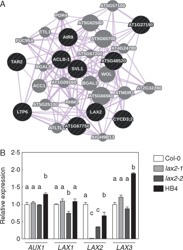
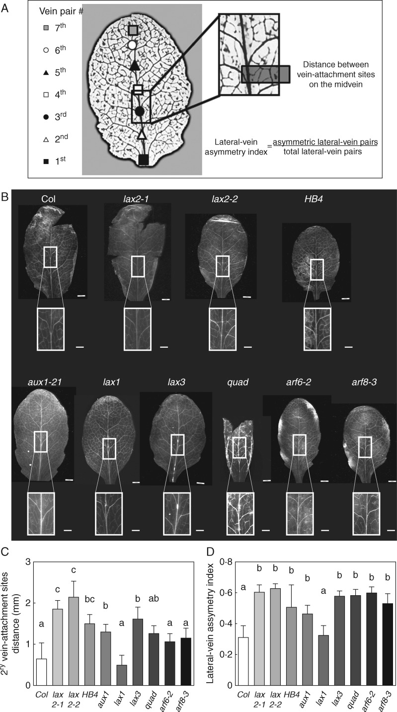
Background and aims: The symmetry of venation patterning in leaves is highly conserved within a plant species. Auxins are involved in this process and also in xylem vasculature development. Studying transgenic Arabidopsis plants ectopically expressing the sunflower transcription factor HaHB4, it was observed that there was a significant lateral-vein asymmetry in leaves and in xylem formation compared to wild type plants. To unravel the molecular mechanisms behind this phenotype, genes differentially expressed in these plants and related to auxin influx were investigated.

Methods: Candidate genes responsible for the observed phenotypes were selected using a co-expression analysis. Single and multiple mutants in auxin influx carriers were characterized by morphological, physiological and molecular techniques. The analysis was further complemented by restoring the wild type (WT) phenotype by mutant complementation studies and using transgenic soybean plants ectopically expressing HaHB4 .

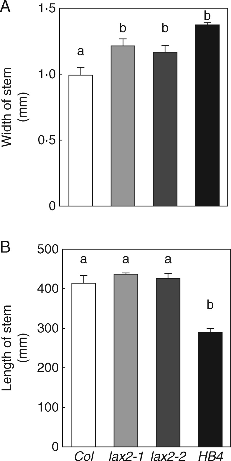
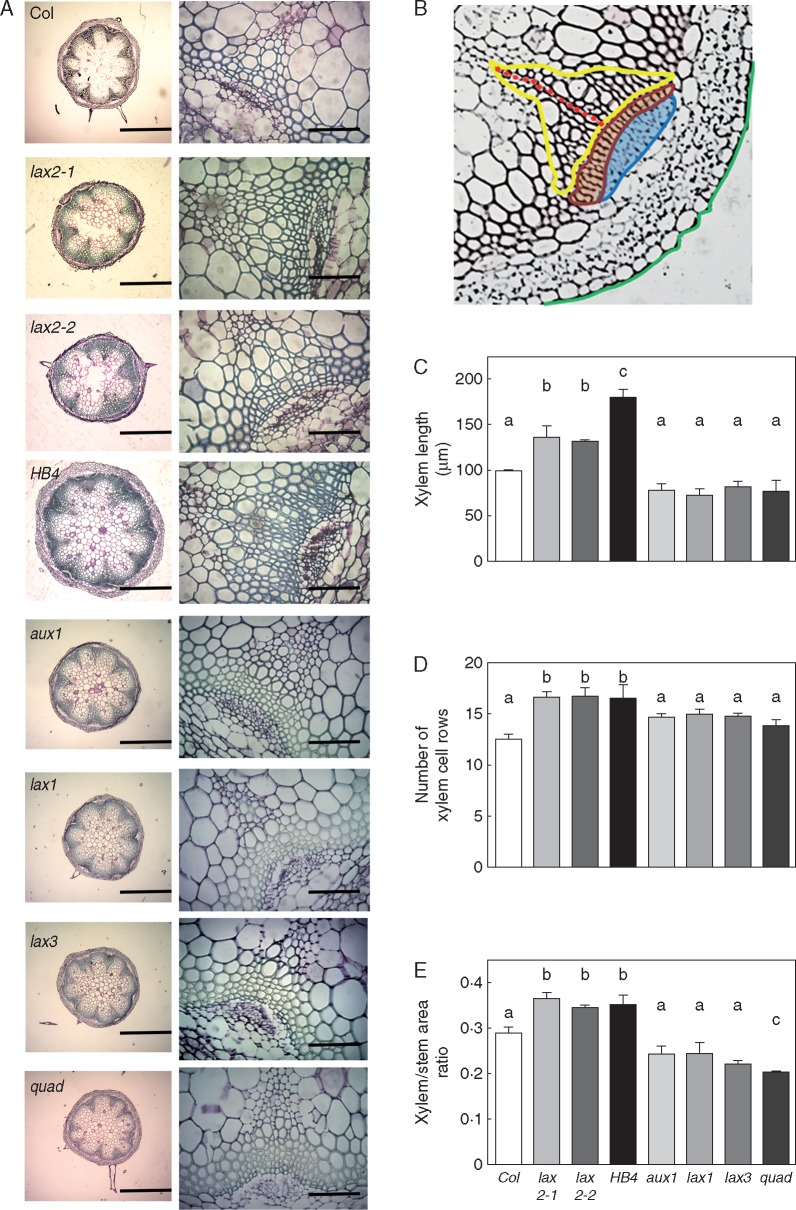
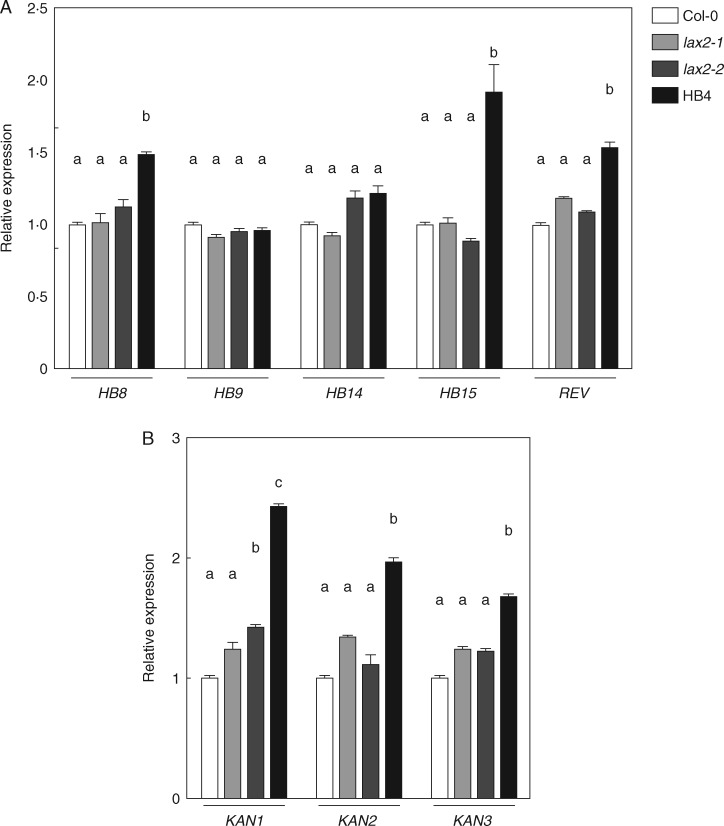
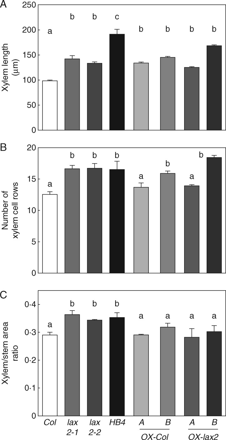
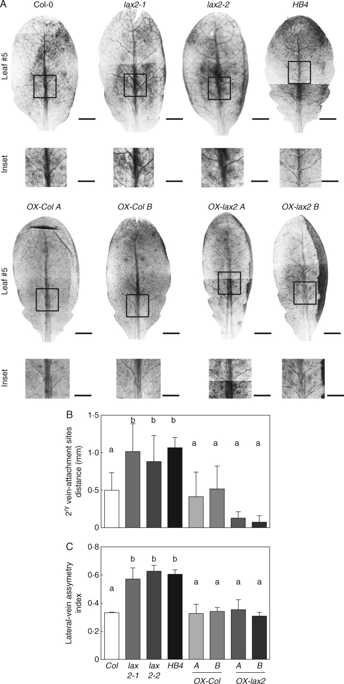
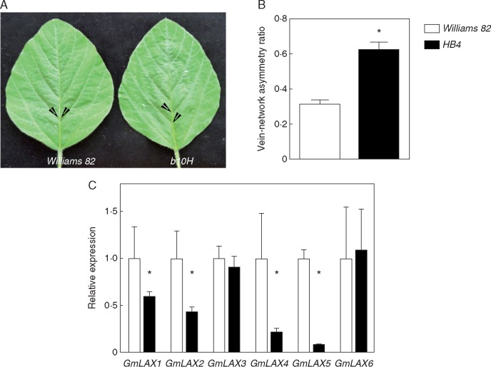
Key results: LAX2 , down-regulated in HaHB4 transgenic plants, was bioinformatically chosen as a candidate gene. The quadruple mutant aux1 lax1 lax2 lax3 and the single mutants, except lax1, presented an enhanced asymmetry in venation patterning. Additionally, the xylem vasculature of the lax2 mutant and the HaHB4 -expressing plants differed from the WT vasculature, including increased xylem length and number of xylem cell rows. Complementation of the lax2 mutant with the LAX2 gene restored both lateral-vein symmetry and xylem/stem area ratio in the stem, showing that auxin homeostasis is required to achieve normal vascular development. Interestingly, soybean plants ectopically expressing HaHB4 also showed an increased asymmetry in the venation patterning, accompanied by the repression of several GmLAX genes.

Conclusions: Auxin influx carriers have a significant role in leaf venation pattering in leaves and, in particular, LAX2 is required for normal xylem development, probablt controlling auxin homeostasis.

Keywords: Auxin influx carriers; HD-Zip I; HaHB4; LAX2; vascular patterning; venation symmetry; xylem organization.


Figures

F<sc>ig</sc>. 1.
Fig. 1.
HaHB4 represses the expression of LAX2 in Arabidopsis transgenic plants. (A) Co-expression of genes in the cluster generated using the GeneMania software package. The cluster is statistically enriched in genes related to auxin homeostasis, i.e. LAX2, TAR2 and AIR9 (P ≤ 0·05). Black nodes represent the list of query genes used in GeneMania. Grey nodes represent genes that were associated by a co-expression pattern according to GeneMania. (B) Relative transcript levels of AUX/LAX family members in rosette leaves of 20-d-old plants of WT (Col-0), lax2-1, lax2-2 and HaHB4 (displayed here as HB4) plants. AUX/LAX transcript abundance was measured and expressed relative to the level detected in Col-0 plants. Different letters indicate significant differences between means (P < 0·05, Tukey test).
F<sc>ig</sc>. 2.
Fig. 2.
Both lax2 mutants and 35:HaHB4 leaves have enhanced asymmetry on lateral-vein attachment site of the midvein resulting in leaves with increased asymmetry in venation patterning. (A) Leaf diagram showing the parameters measured on the 5th leaf lamina. (B) Illustrative photographs of leaf number 5 and the corresponding inset showing a zoom on the third vein pair of Col-0, lax2-1, lax2-2, 35S:HaHB4, aux1-21, lax1, lax3, quad, arf6-2 and arf8-3 genotypes. Whole-leaf scale bar represents 0·5 cm, whereas leaf-inset scale bar represents 1·0 mm. (C) Average distance between the two attachment sites of the lateral veins of the third pair. (D) Lateral-vein asymmetry index calculated as described in Methods. Thin bars represent s.e. Different small letters indicate significant differences (P < 0·05, Tukey test).
F<sc>ig</sc>. 3.
Fig. 3.
Both lax2 mutants and 35:HaHB4 plants exhibit a wider inflorescence stem than WT plants. Genotype effects on both main stem height (A) and width (B) of WT, lax2-1 and lax2-2 mutant alleles, and 35:HaHB4 plants. Thin bars represent s.e. Different letters indicate significant differences (P < 0·05, Tukey test).
F<sc>ig</sc>. 4.
Fig. 4.
Both lax2 mutants and 35:HaHB4 plants show an enhanced proportion of xylem in the main inflorescence stem. (A) Illustrative photographs of basal shoot cross-section of inflorescence stems together with a picture inset showing a zoom on a representative vascular bundle for analysed genotypes. Basal shoot scale bar represents 0·5 mm, whereas vascular-bundle inset scale bar represents 0·1 mm. (B) Illustration of a vascular bundle showing measured parameters as follows: yellow line is the total xylem area; red dots are xylem cell row along the main xylem axis; green dashed line illustrates a portion of the total stem area considered for xylem/stem area ratio. Procambial cells are enclosed in orange. The blue area indicates phloem cells. Column bar graphs of: (C) xylem length, (D) number of xylem cell rows and (E) xylem/stem area ratio. Vascular bundle parameters were taken from pictures of basal shoot cross-section of inflorescence stems for different genotypes, including Col-0, lax2-1, lax2-2, 35S:HaHB4, aux1-21, lax1, lax3 and quad. Thin bars represent s.e. Different letters indicate significant differences (P < 0·05, Tukey test).
F<sc>ig</sc>. 5.
Fig. 5.
Expression levels of Arabidopsis transcription factors involved in vascular development. Relative transcript levels of HD-ZIP III (A) and KANADI (B) genes were quantified by RT-qPCR using RNAs isolated from the last 3 cm of inflorescence stem excluding flower buds. Different letters indicate significant differences (P < 0·05, Tukey test).
F<sc>ig</sc>. 6.
Fig. 6.
Ectopic expression of LAX2 in Arabidopsis transgenic plants restores the xylem/stem area ratio of lax2 mutants. Column bar graphs represent (A) the xylem length, (B) the number of xylem cell rows and (C) the xylem/total stem area ratio in basal shoot cross-sections of stems. Basal shoot cross-sections of transgenic 35S:mCitrine:LAX2 stems were used to measure vascular bundle parameters. Thin bars represent s.e. Different letters indicate significant differences (P < 0·05, Tukey test).
F<sc>ig</sc>. 7.
Fig. 7.
Ectopic expression of LAX2 in Arabidopsis transgenic plants restores venation patterning in leaves of lax2 mutants. (A) Illustrative photographs of leaf number 5 and the corresponding inset showing a zoom on the third vein pair. The final image resulted from merging two photographs. Whole-leaf scale bar represents 2·0 mm, whereas leaf-inset scale bar represents 1·0 mm. (B) Average distance between the two attachment sites of the lateral veins of the third vascular pair. (C) Lateral-vein asymmetry index. Thin bars represent s.e.. Different letters indicate significant differences (P < 0·05, Tukey test).
F<sc>ig</sc>. 8.
Fig. 8.
Ectopic expression of HaHB4 in transgenic soybean leaves enhances the asymmetric formation of secondary vein pairs. (A) Illustrative photographs of the terminal foliole. White arrowheads indicate development start sites of different vein pairs. Scale bar represents 3 cm. (B) Fraction of asymmetric attachment sites compared to the total lateral vein pairs in the foliole. A total of three replicates were used to calculate the s.e. Differences were considered significant at *P < 0·05 (Student’s t-test). (C) GmLAX transcript levels detected by RT-qPCR using total RNA isolated from soybean third foliole of a fully expanded leaf. Measurements were taken on the terminal foliole from the last fully developed leaf corresponding to a 30-d-old soybean plant (V7 stage). Error bars represent the s.e. of three independent biological replicates. Statistical significance was computed by Student's t-test. *P < 0.05.

References

    1. Aloni R. 2001. Foliar and axial aspects of vascular differentiation: hypotheses and evidence. Journal of Plant Growth Regulation 20: 22–34.
    1. Aloni R. 2010. The induction of vascular tissues by auxin In: Davies PJ, ed. Plant hormones: biosynthesis, signal transduction, action! Dordrecht: Springer Netherlands, 485–518.
    1. Aloni R, Schwalm K, Langhans M, Ullrich CI.. 2003. Gradual shifts in sites of free-auxin production during leaf-primordium development and their role in vascular differentiation and leaf morphogenesis in Arabidopsis. Planta 216: 841–853. - PubMed
    1. Arcadia-Biosciences. 2015. Verdeca’s HB4 stress tolerance trait completes US Food and Drug Administration early food safety evaluation. http://www.arcadiabio.com/news/press-release/verdeca%E2%80%99s-hb4-stres...
    1. Arce AL, Raineri J, Capella M, Cabello JV, Chan RL.. 2011. Uncharacterized conserved motifs outside the HD-Zip domain in HD-Zip subfamily I transcription factors; a potential source of functional diversity. BMC Plant Biology 11: 42. - PMC - PubMed

Publication types

MeSH terms